Deep split in lower first premolar
06/01/2024
Fellow
Warning: Undefined variable $post in /home/styleendo/htdocs/styleitaliano-endodontics.org/wp-content/plugins/oxygen/component-framework/components/classes/code-block.class.php(133) : eval()'d code on line 2
Warning: Attempt to read property "ID" on null in /home/styleendo/htdocs/styleitaliano-endodontics.org/wp-content/plugins/oxygen/component-framework/components/classes/code-block.class.php(133) : eval()'d code on line 2
Knowledge of root canal morphology, proper radiographic interpretation, Cone beam CT scan interpretation, use of magnification in terms of dental operating microscope and scouting of the root canals are important in detecting the presence of the multiple canals.
CBCT in such a case is useful to know the number of canals and its location, that makes the process easier.
This case report presents a comprehensive approach to successfully treating mandibular 1st premolar with deep split.
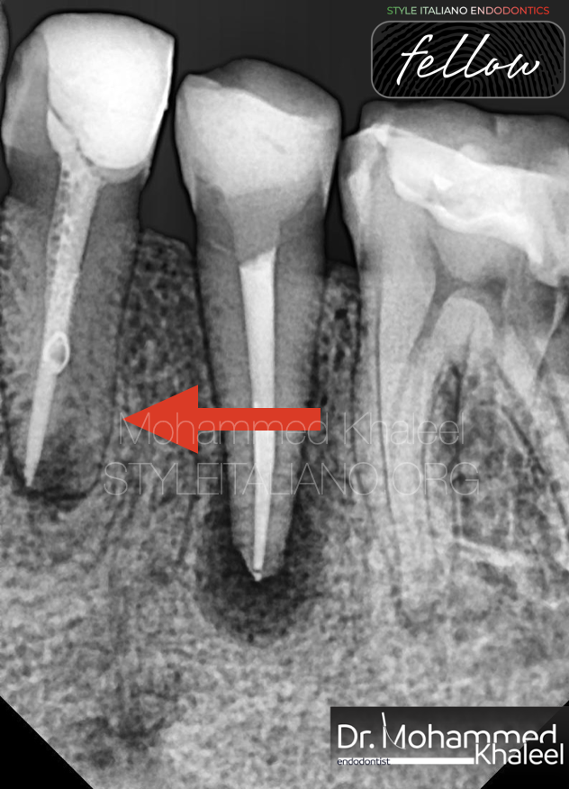
Fig. 1
A 33 years old female presented to my clinic complaining from lower left quadrant pain, the patient was generally fit.
On intra-oral examination the lower left 1St premolar was tender to percussion.
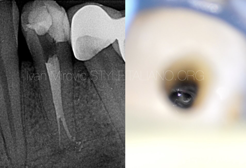
Periapical radiograph showed endodontically treated 1St premolar with periapical radiolucency.
Endodontic retreatment started immediately.
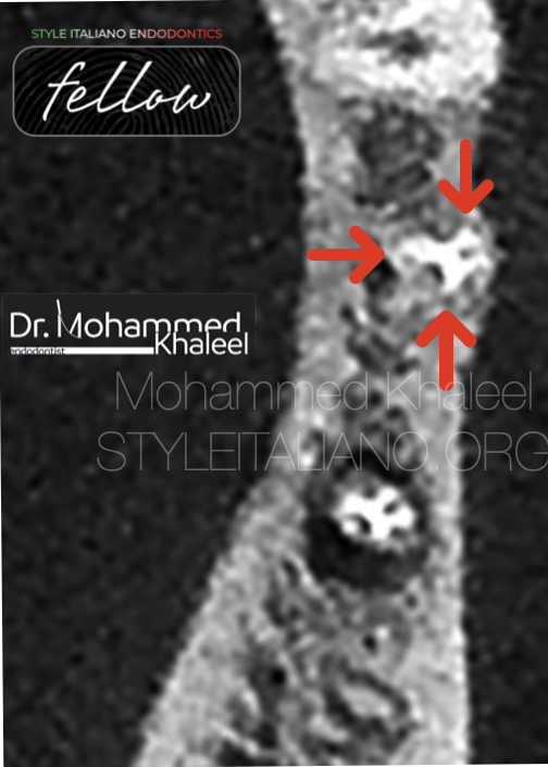
Fig. 2
the CBCT scan showed the presence of three root canals
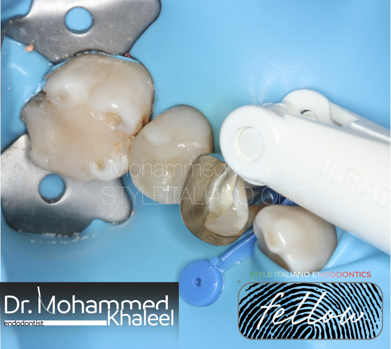
Fig. 3
Rubber dam isolation
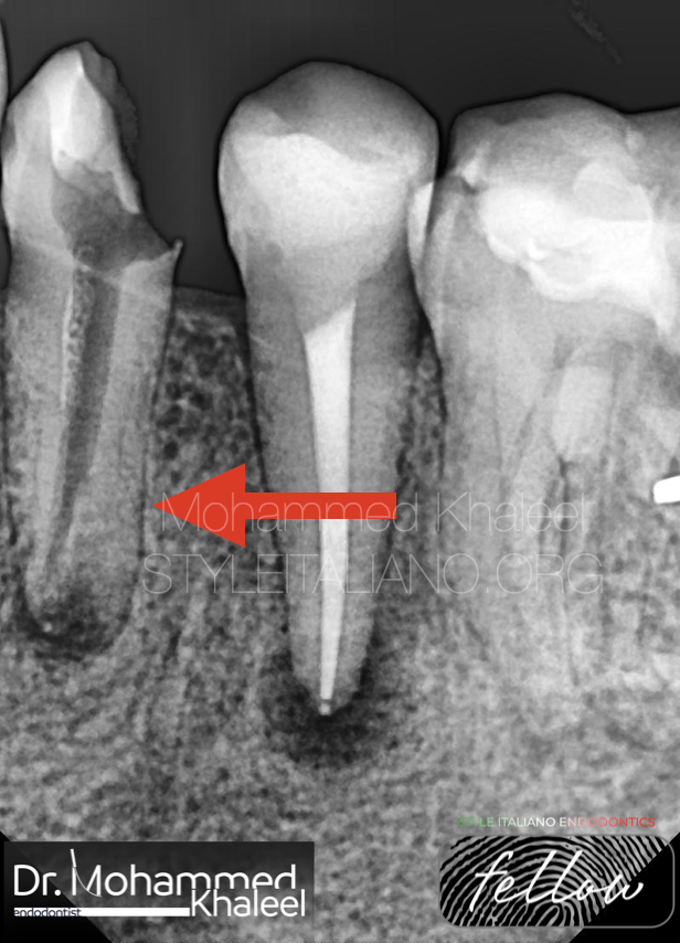
Fig. 4
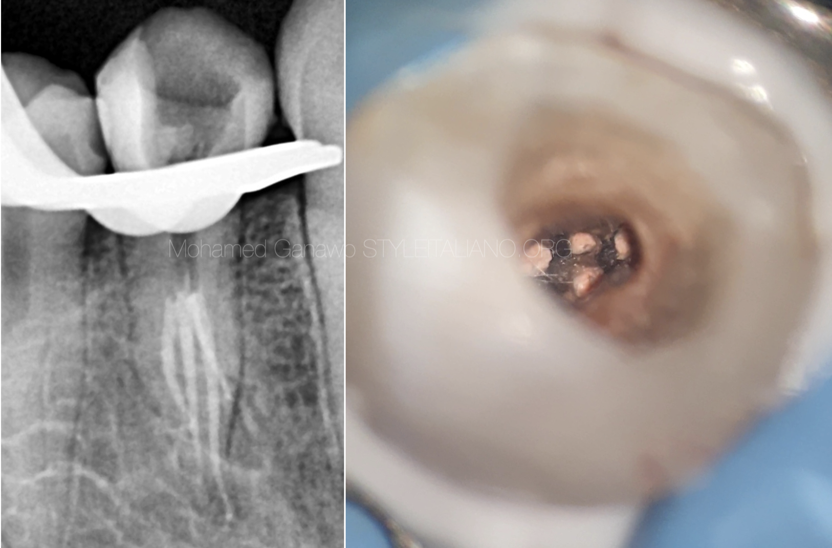
Mechanical removal of gutta percha by 25%06 rotary file with NaOCl 5.25% irrigation.
To gain sufficient access to the canals, the access was modified using ultrasonic tips acteon ET18 D.
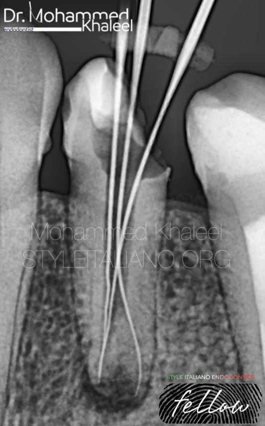
Fig. 5
Location the canals, Then instrumentation done by 20%0.4 then 25%0.4 with NaOCl 5.25% .
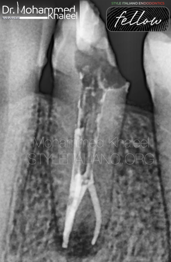
Fig. 6
Obturating the canals till the level of the canal orifices
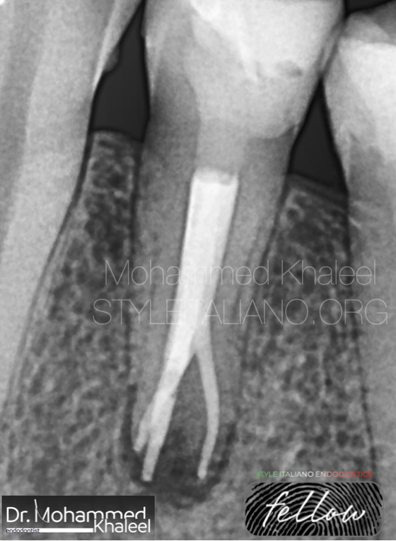
Fig. 7
Backfill the canal (middle and coronal)
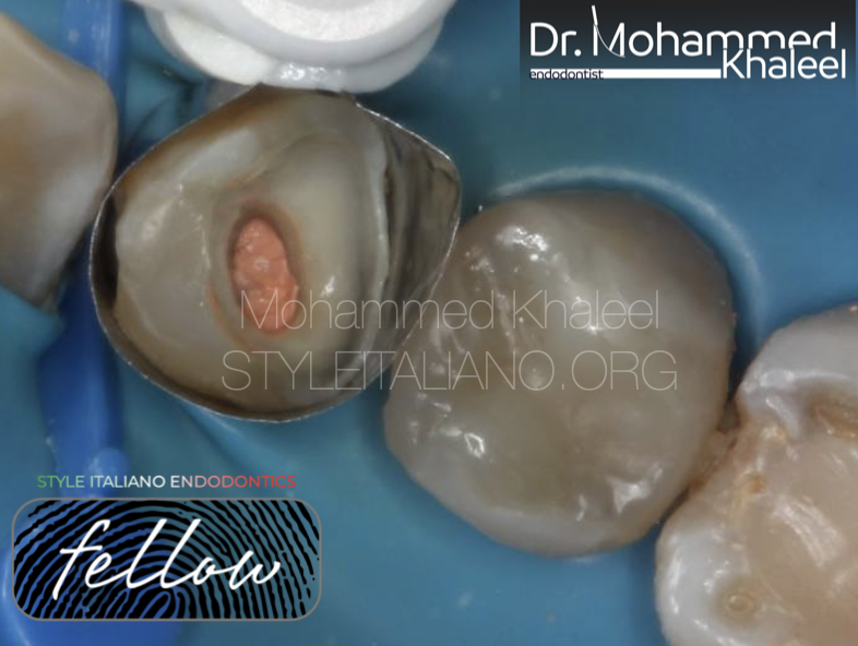
Fig. 8
Obturation
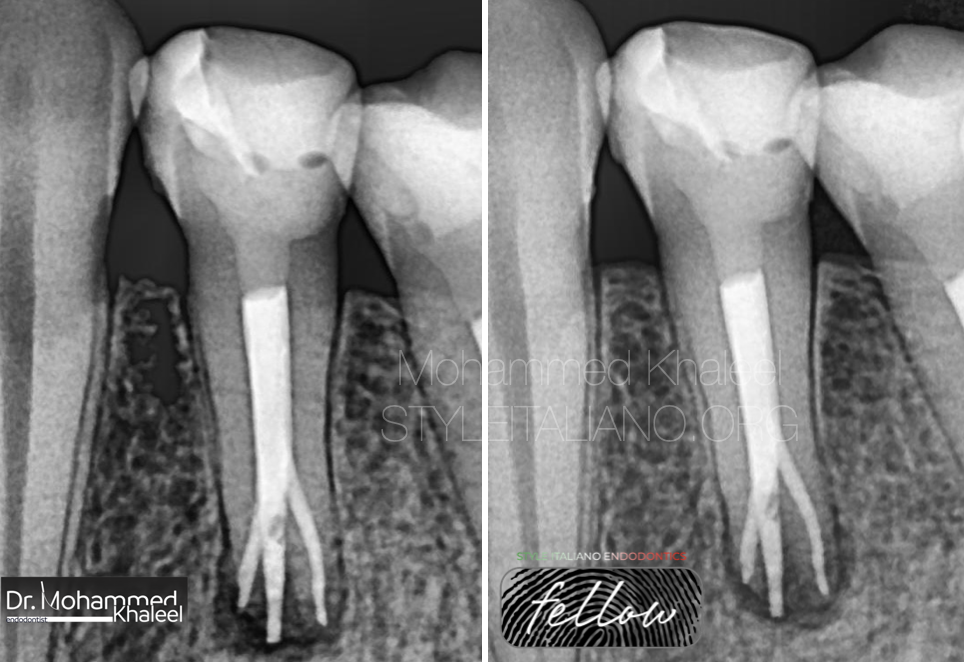
Fig. 9
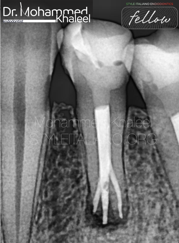
Post operative X-ray (left) and 1 year follow up (right)

Fig. 10
Graduated 2011 from Hawler medical university
Long term (5 modules ) Advance endodontic course 2018 .
Best case of the year 2021 from style italiano endodontics .
Fellow member of style italiano endodontics 2023 .
Conclusions
It is absolutely essential for an operator to form a mental picture of the pulp chamber in cross section & from the coronal aspect to the apical foramina.
Each canal may contain irregular & hidden configuration that should be taken into account during endodontic treatment.
Instruments must access these hidden regions and clean & shape them as maximally as possible to avoid or minimize treatment failure.
Bibliography
1-Zillich R, Dowson J. Root canal morphology of mandibular first and second premolars.Oral surg Oral Med Oral Patho.1973;36:738-44.
2-Ingle JI, Beveridge E,Glick D D,Weichman J. Ingle JI, Taintor JF, editors. The Washington study. Endodontics, Philadelphia.1994:1-53.Lea and Fabiger.
3-Vertucci FJ. Root canal morphology and its relationship to endodontic procedures.Endod Top.2005;10:3-29.