Management of a mandibular first premolar with four canals
04/05/2023
Mohamed Milad Ganawo
Warning: Undefined variable $post in /home/styleendo/htdocs/styleitaliano-endodontics.org/wp-content/plugins/oxygen/component-framework/components/classes/code-block.class.php(133) : eval()'d code on line 2
Warning: Attempt to read property "ID" on null in /home/styleendo/htdocs/styleitaliano-endodontics.org/wp-content/plugins/oxygen/component-framework/components/classes/code-block.class.php(133) : eval()'d code on line 2
Understanding of the root canal anatomy is crucial prerequisite for predictable successful long-term results. (1-5) Although basic applications of root canal therapy through biomechanical preparation of the entire root canal system to obturation and final restoration, it has been reported in the Literature that 42% of retreatment cases are due to missing canals. (2) Therefore, comprehensive preoperative evaluation of root canal anatomy significantly contributes to awareness to unusual root canal morphology.
Wide variations in root canal morphology of mandibular premolars have been described in the literature. Vertucci et al reported that the mandibular first premolar had only one root canal at the apex in 97.5% of the teeth studied, two canals in only 2.5% and the incidence of three roots canals were rare. However, to date, most studies report cases with 2-3 root canals while cases of four canals are extremely rare. (3-4)
The purpose of this case report to presents unusual case of mandibular first premolar with two fused roots and rare canal morphology , that started as one canal and divided into four canals in apical one third of the root. The obvious contribution to this case report was detection of this rare root canal morphology with different angled periapical radiographs and the use of operating microscope in treating such cases was essential tool for better vision and light brightness.
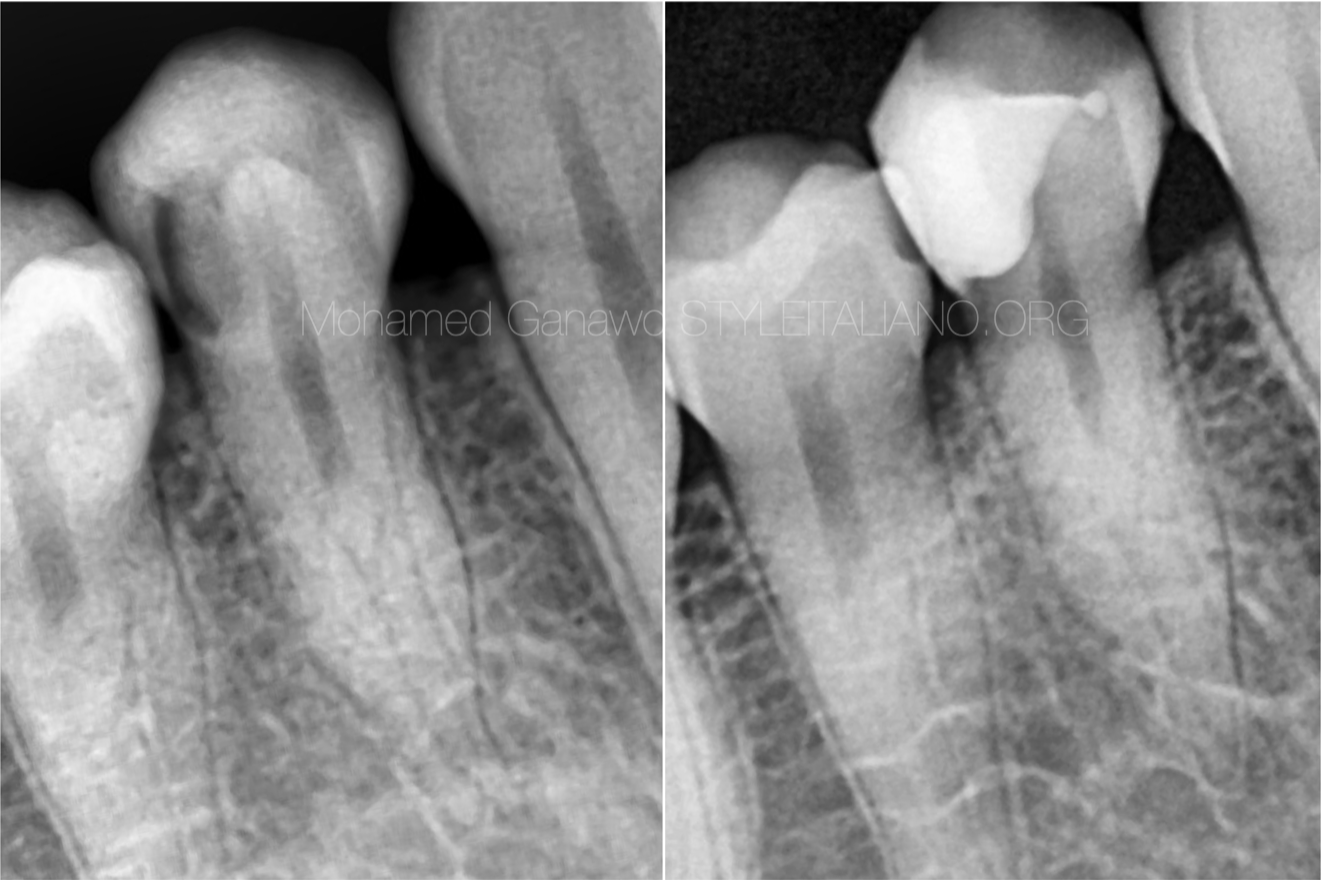
Fig. 1
Pre operative X-ray of mandibular right first premolar
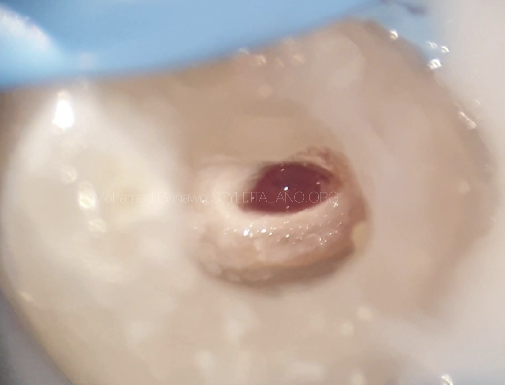
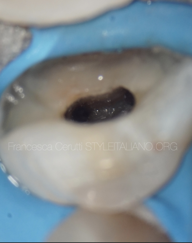
Fig. 2
Access opening of mandibular right first premolar
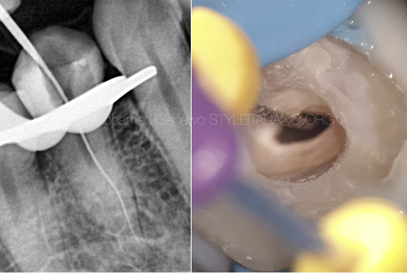
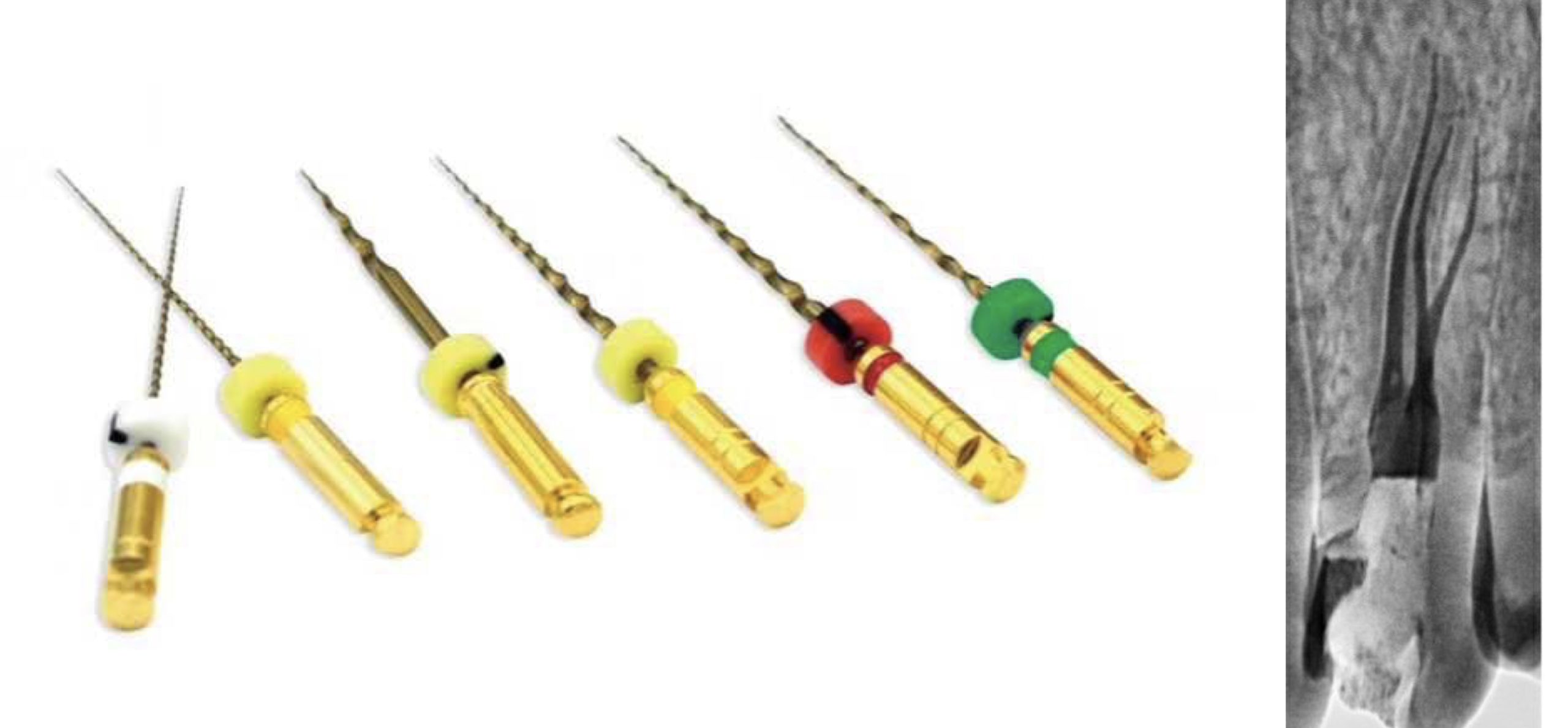
Fig. 3
Manual file goes into the first split
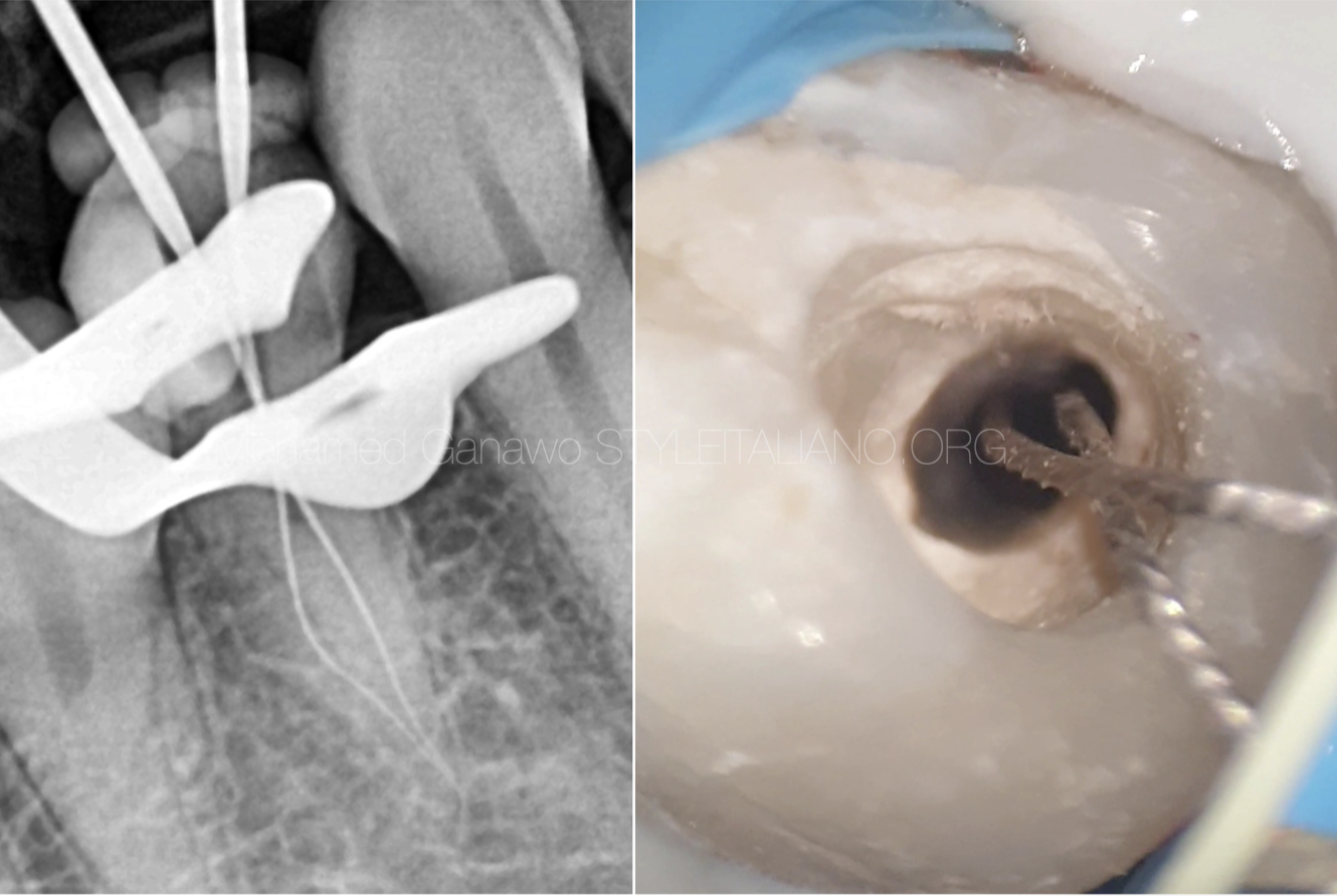
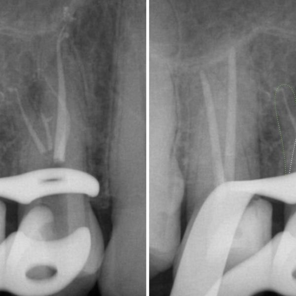
Fig. 4
this figure shows files in 2 splits radiographically and intra orally
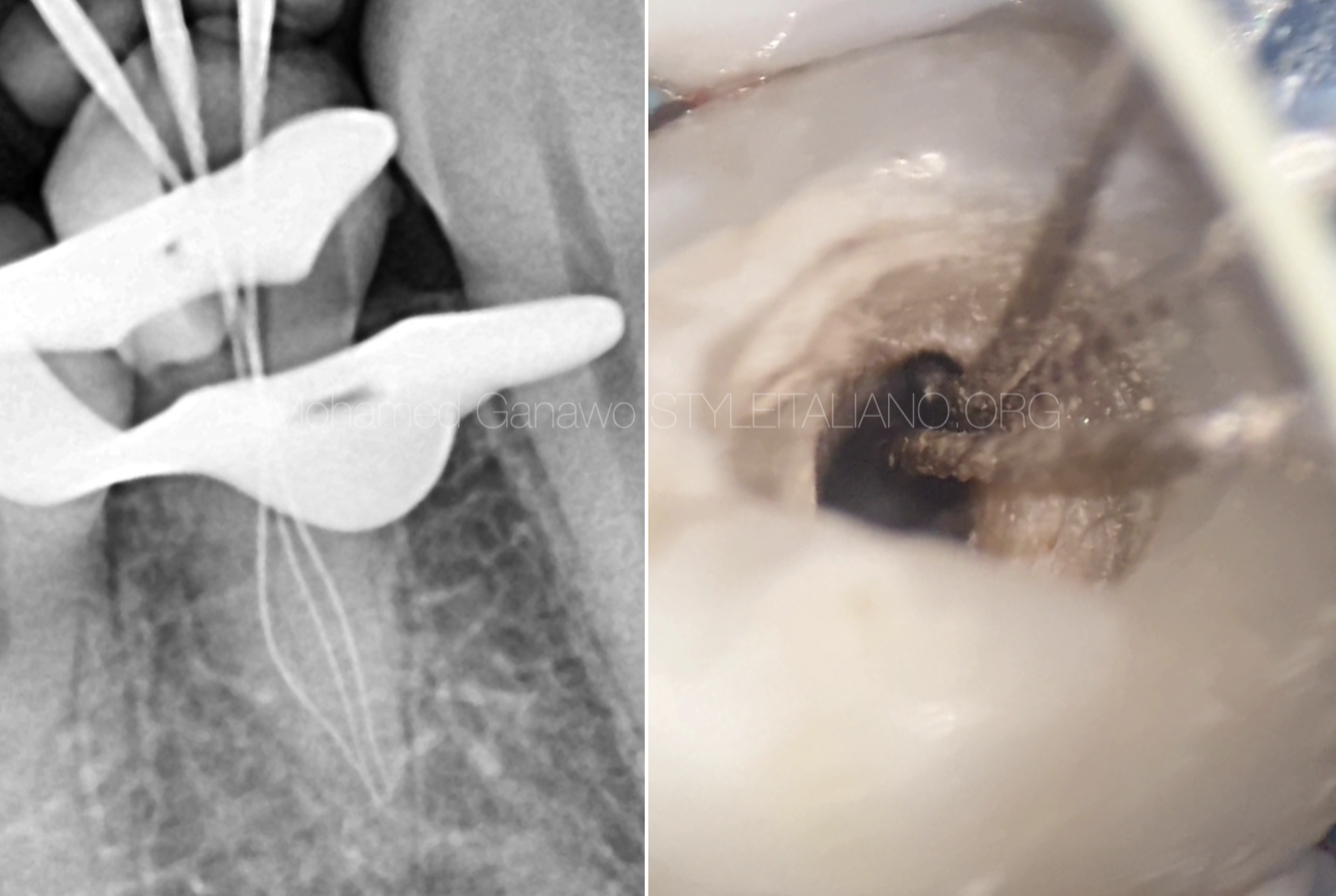
Fig. 5
this figure shows files in 3 splits radiographically and intra orally
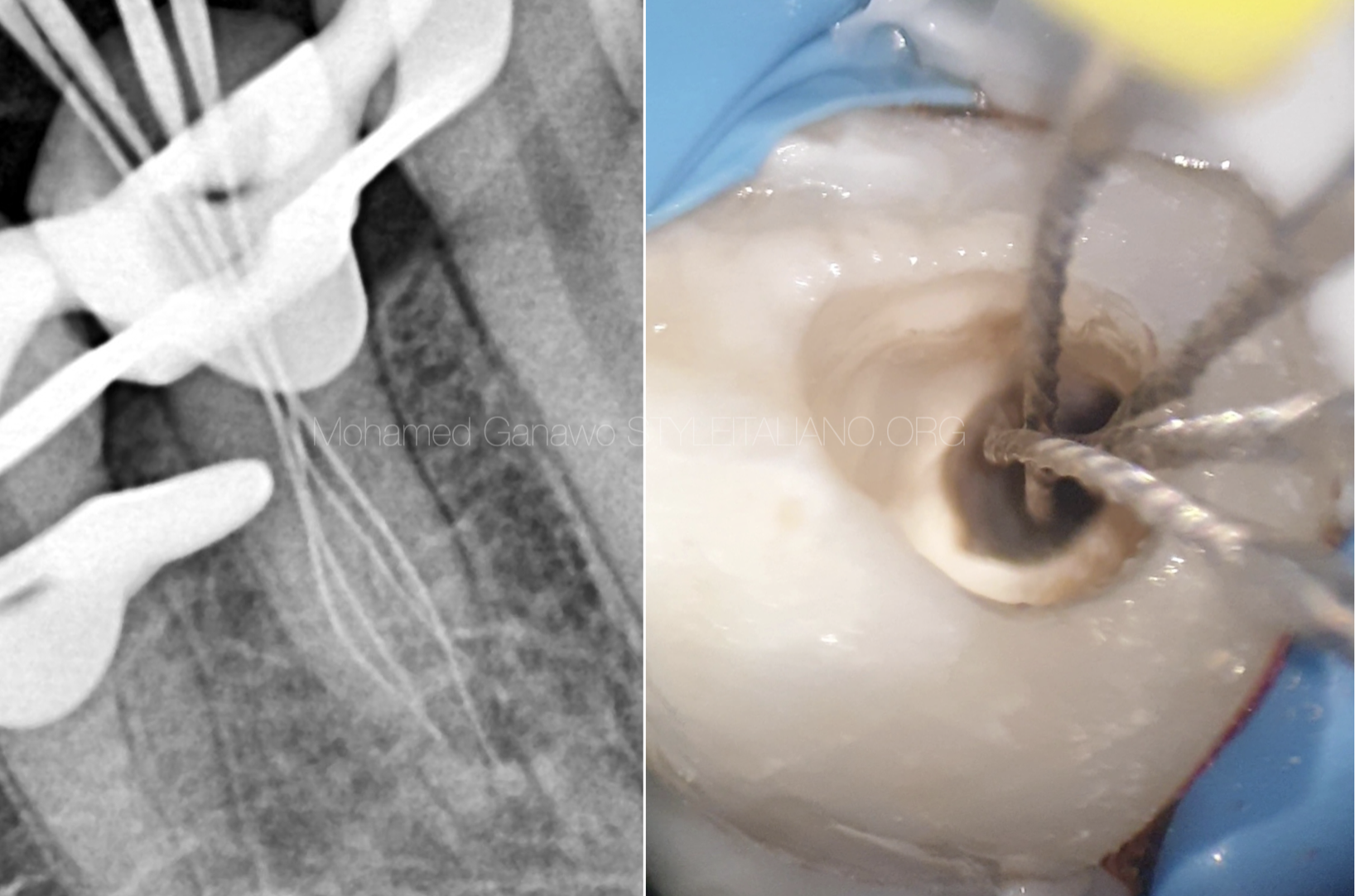
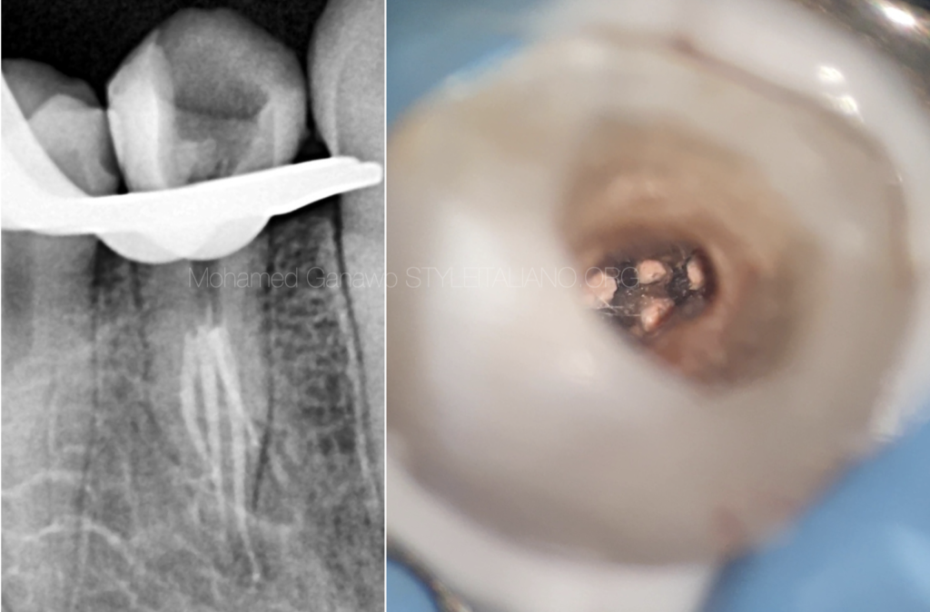
Fig. 6
this figure shows files in 4 splits radiographically and intra orally and work length determination
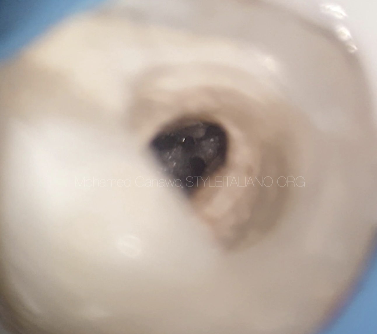
Fig. 7
microscopic shot shows 4 splits together
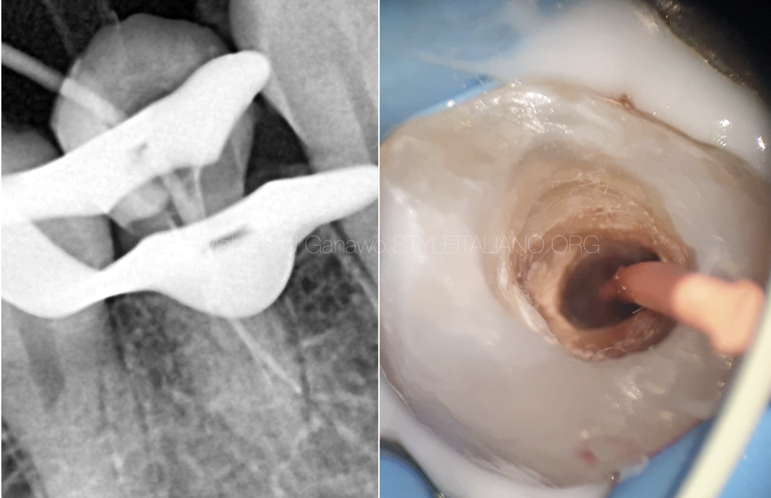
Fig. 8
Cone fit of first split
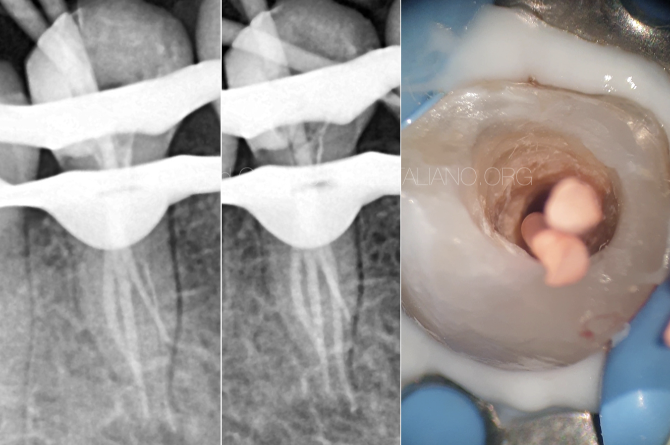
Fig. 9
obturation of 2 splits followed by insertion of 2 Gutta percha
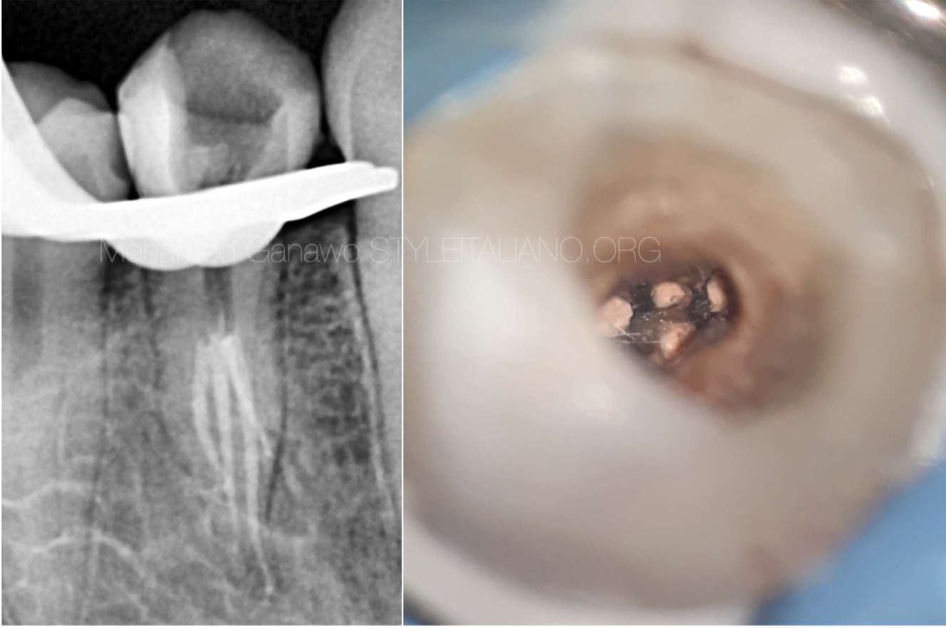
Fig. 10
microscopic shot shows obturation of 4 splits
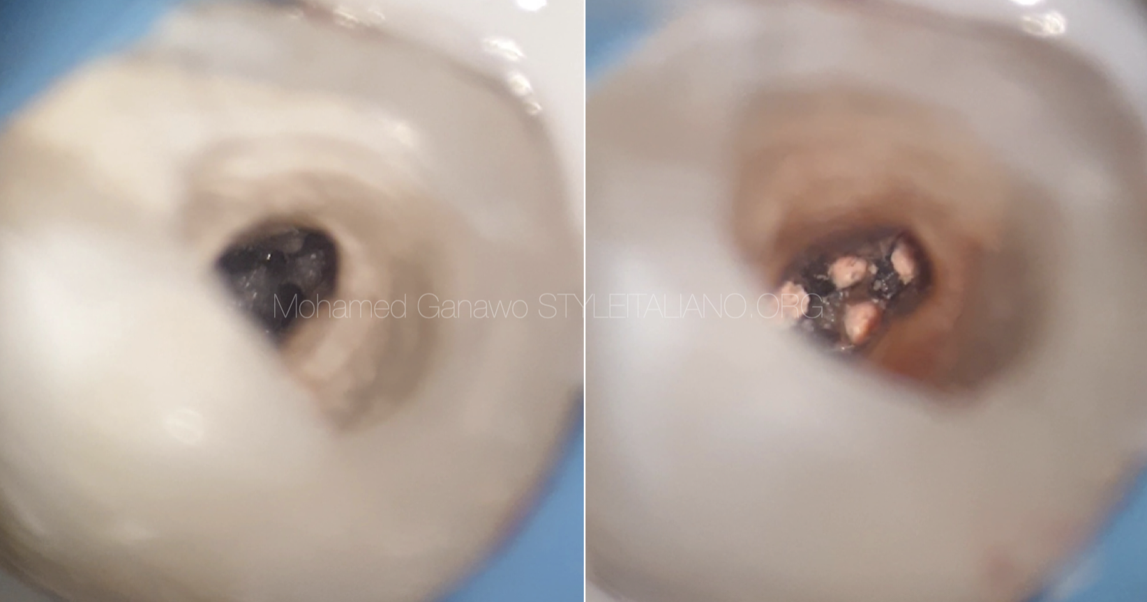
Fig. 11
microscopic shot shows 4 splits together before and after obturation
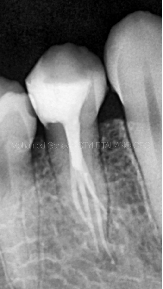
Fig. 12
Post operative radiograph
Full video of the case
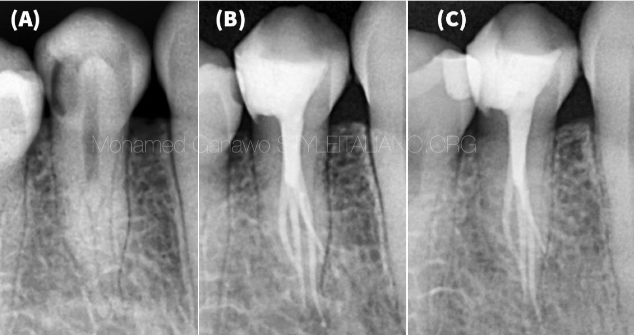
Fig. 13
A) pre-operative
B) post - operative
c) Follow up radiograph
Conclusions
In conclusion, this case report is unique case of mandibular first premolar with complex root canal morphology.
In this case report I showed the importance of understanding of root canal anatomy and its variation to provide the patients with long term successful results. Also, to emphasis on the importance of using of operating microscope that reflected positively to locate the extra canals especially in deep apical one third split.
Bibliography
Gurmeet et al. Endodontic Management of a Mandibular Second Premolar with Four Roots and Four Root Canals with the Aid of Spiral
Computed Tomography: A Case Report. 2008 by the American Association of Endodontists. doi: 10. 1016/j.joen.2007. 10.004
Hoen MM, Pink FE. Contemporary endodontic retreatments: An analysis based on clinical treatment findings. J Endod. 2002;28:834-6.
Vertucci FJ. Root canal anatomy of the human permanent teeth. Oral Surg Oral Med Oral Pathol. 1984;58:589-99
Ming Zhang et al. Mandibular first premolar with five root canals: a case report BMC Oral Health (2020) 20:253 https://doi.org/10.1186/$12903-020-01241-0
Carina Lea et al. American Association of Endodontists. 2014 http://dx.doi.org/10.1016/j.joen.2013.09.016


