Endodontic Management of Type II Dens Invaginatus in a Maxillary Lateral Incisor Using: A Case Report
01/10/2026
Warning: Undefined variable $post in /home/styleendo/htdocs/styleitaliano-endodontics.org/wp-content/plugins/oxygen/component-framework/components/classes/code-block.class.php(133) : eval()'d code on line 2
Warning: Attempt to read property "ID" on null in /home/styleendo/htdocs/styleitaliano-endodontics.org/wp-content/plugins/oxygen/component-framework/components/classes/code-block.class.php(133) : eval()'d code on line 2
Dens invaginatus is a developmental anomaly characterized by invagination of the enamel organ into the dental papilla before calcification, resulting in highly variable internal anatomy. The complexity of the root canal system may compromise diagnosis, debridement, irrigation and three-dimensional obturation. This case report describes the endodontic management of a Type II dens invaginatus in a 14-year-old male presenting with spontaneous pain, a sinus tract and periapical pathology associated with the maxillary right lateral incisor. The treatment was completed in a single session using microscopic visualization, conservative access, ultrasonic instrumentation, a OneFile system (COLTENE), activated irrigation and an apical plug of Bio-C Repair followed by heated gutta-percha and bioceramic sealer. Particular attention is given to the role of three-dimensional imaging. Although conventional periapical radiographs remain essential, cone-beam computed tomography (CBCT) can provide additional information about the extent and spatial relationships of the invagination and may improve treatment planning in anatomically complex cases. At one-year follow-up, the patient was asymptomatic and the periapical lesion had disappeared.
Dens invaginatus, also known as dens in dent, is a developmental dental anomaly resulting from invagination of the enamel organ into the dental papilla before calcification occurs [1]. It presents with a wide spectrum of morphological configurations and is most frequently observed in maxillary lateral incisors [2,3]. The reported prevalence varies considerably according to the population and diagnostic method used [2].
Oehlers classified dens invaginatus into three types according to the extent of the invagination [4]. Type I is limited to the crown; Type II extends beyond the cementoenamel junction into the root canal system but does not communicate with the periodontal ligament; and Type III extends toward the apical region and may communicate with the periodontal or periapical tissues. The altered anatomy, thin dentinal walls and possible communication pathways may facilitate bacterial contamination and make conventional endodontic treatment technically demanding [3,5].
Accurate three-dimensional assessment is particularly important when the morphology is complex. Conventional periapical radiographs provide essential information and should be obtained with different horizontal angulations; however, their two-dimensional nature may mask the true extent and spatial relationships of the invagination. Three-dimensional imaging, particularly CBCT, can provide additional information regarding the course and extension of the invagination, its relationship with the main canal, the thickness of surrounding dentine and the presence of periapical pathology. This information can be decisive for treatment planning in complex cases [6,7]. Pallavi Reddy et al. specifically reported the management of dens invaginatus diagnosed using spiral computed tomography, highlighting the limitations of conventional radiographs in fully characterizing complex anatomy [6].
The purpose of this case report is to describe the management of a Type II dens invaginatus with pulpal necrosis, chronic apical pathology and an incompletely formed apex, emphasizing the importance of magnification, conservative access, three-dimensional treatment planning, effective irrigation and three-dimensional obturation.
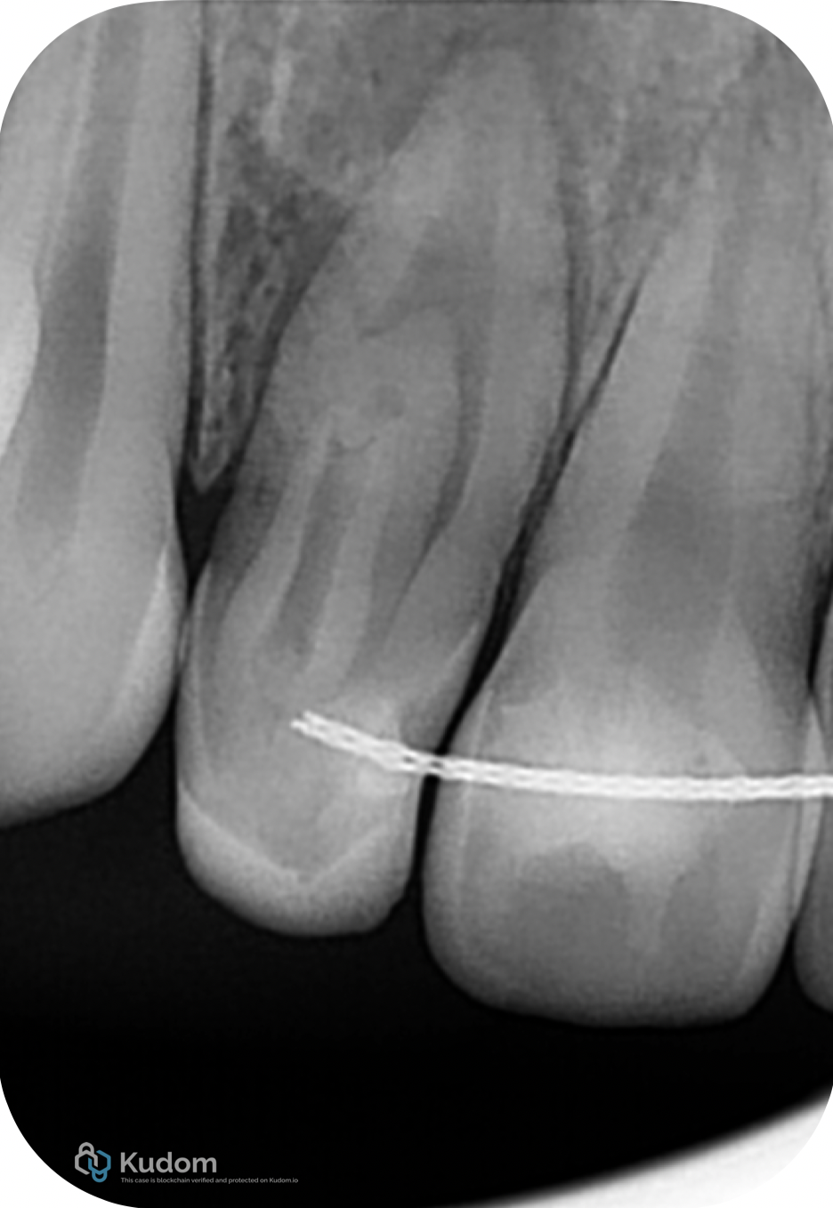
Fig. 1
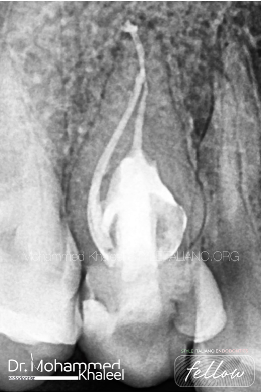
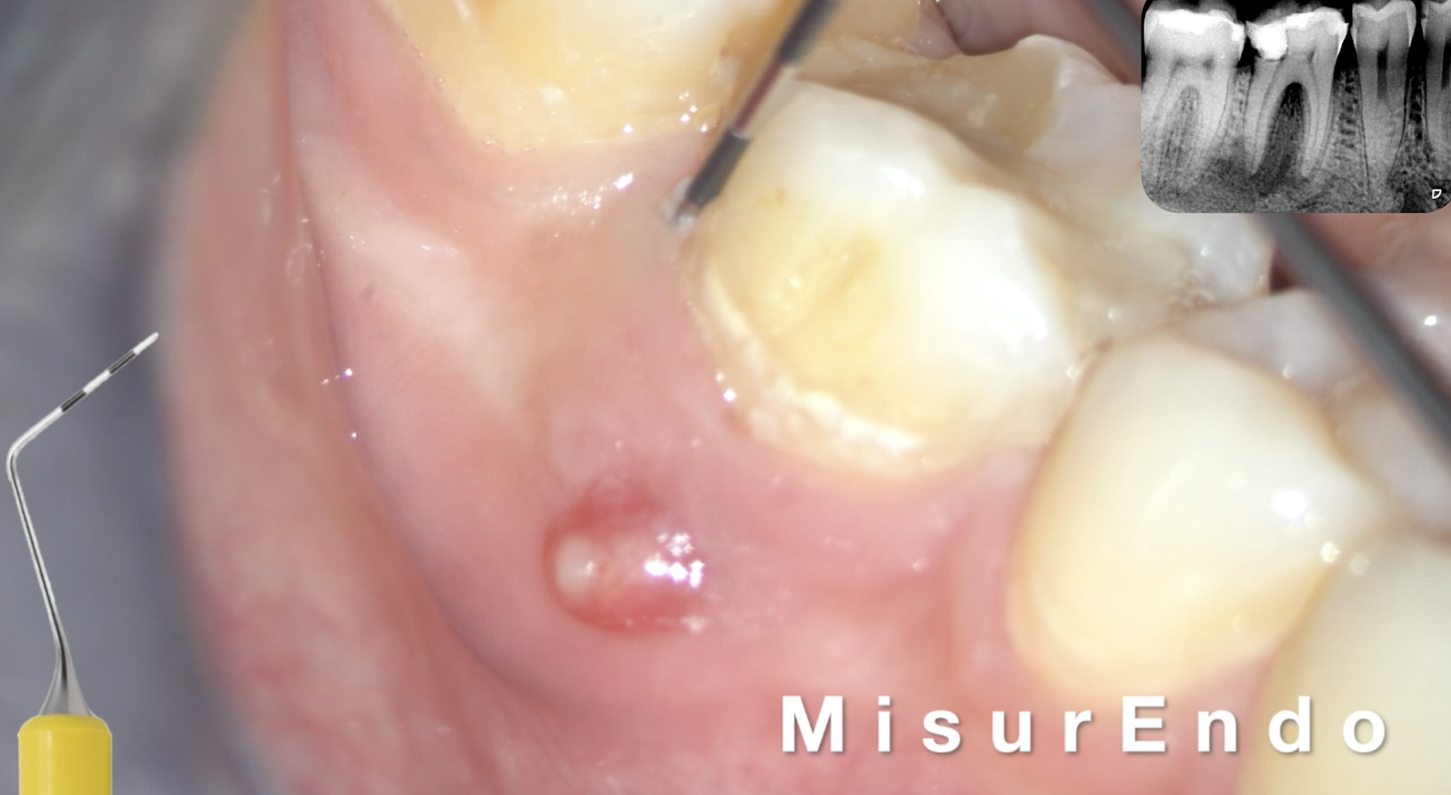
A 14-year-old male patient presented with spontaneous pain in the first quadrant. The affected tooth was the maxillary right lateral incisor (tooth 12). Clinical examination revealed discomfort on mucosal palpation and percussion, together with the presence of a sinus tract. Periapical radiographic examination showed a characteristic Type II dens invaginatus associated with a periapical lesion. The clinical diagnosis was pulp necrosis with chronic apical abscess.
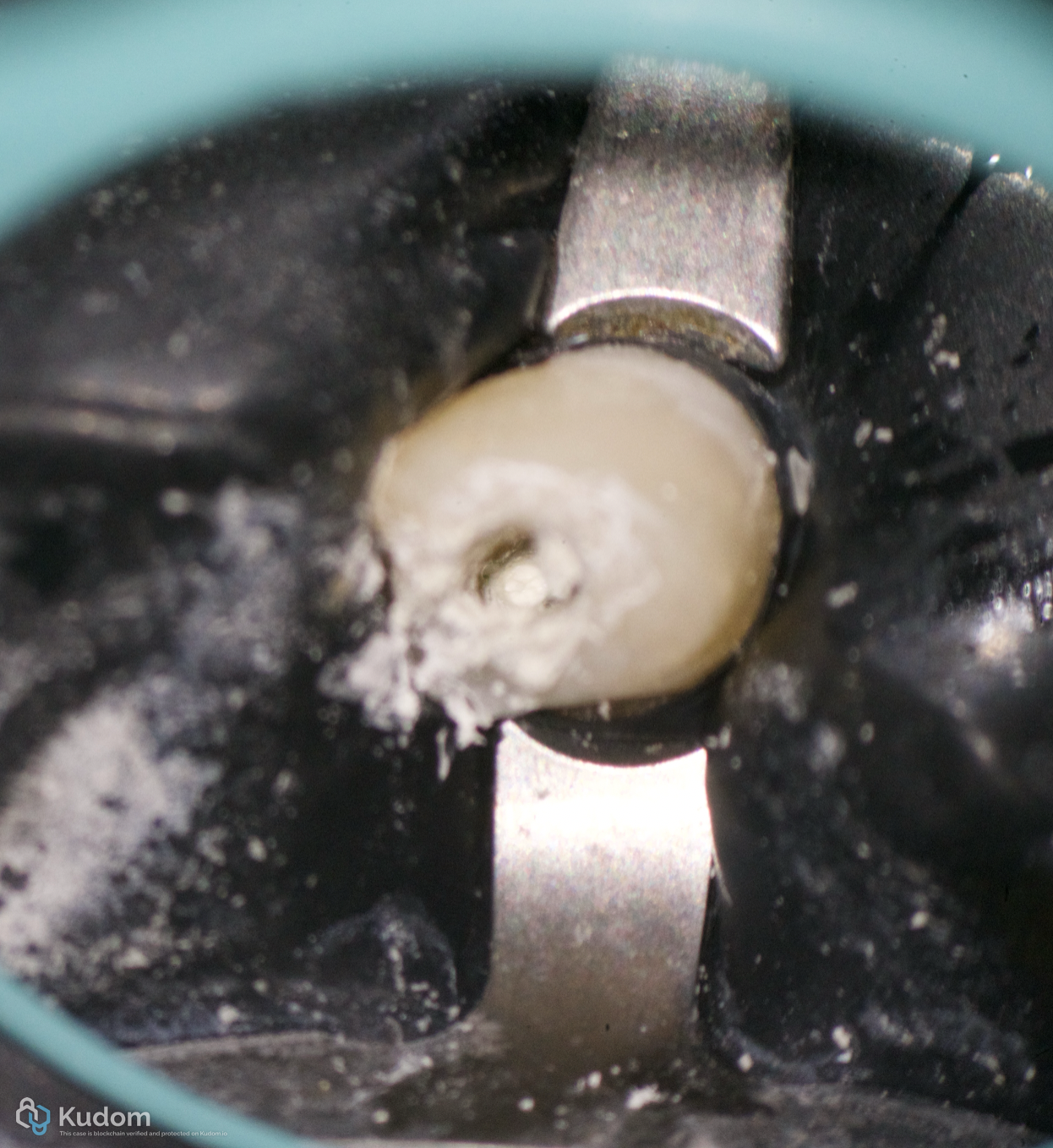
Fig. 2
Periapical radiographs were obtained using a paralleling technique. Different radiographic angulations are particularly useful in dens invaginatus because they may reveal the extent of the invagination and its relationship with the main root canal [5].
In complex cases of dens invaginatus, CBCT should be considered when the information obtained from conventional radiographs is insufficient for diagnosis or treatment planning. Its three-dimensional visualization can help determine the actual extension of the invagination, identify possible communications, assess the remaining dentine and visualize periapical anatomy without the superimposition inherent to two-dimensional radiography [6,7]. A systematic review and meta-analysis also found that the prevalence of dens invaginatus detected by CBCT was higher than that reported using conventional radiography, supporting its greater sensitivity for identifying this developmental anomaly [8]. In the present case, the treatment plan was based on the available clinical and periapical radiographic findings; the role of CBCT is particularly relevant as an additional planning tool when conventional imaging does not adequately define the anatomy.
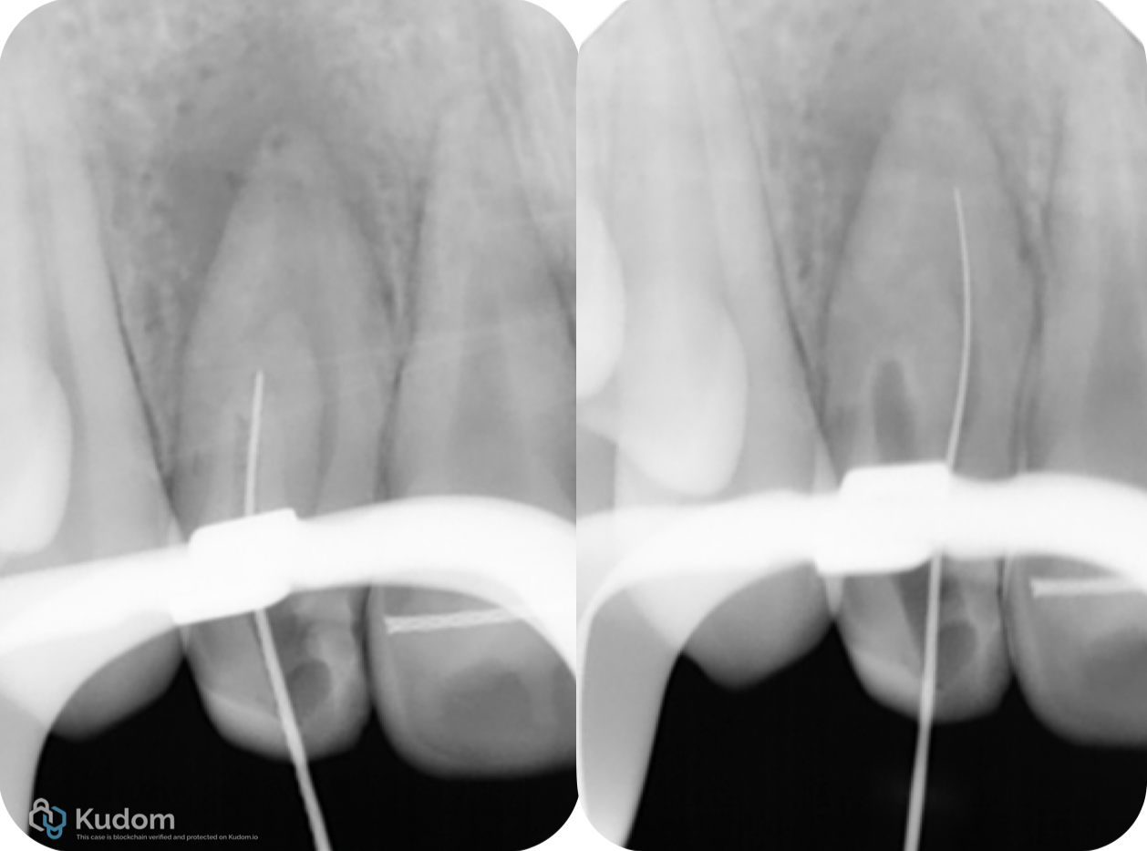
Fig. 3
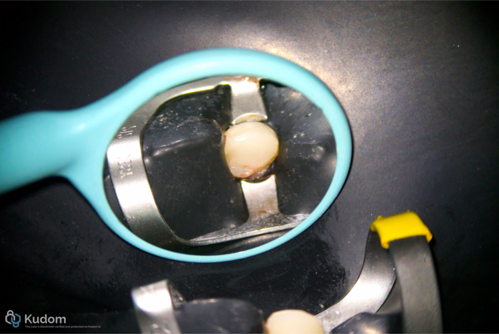
Local infiltrative anesthesia was administered using 4% articaine with 1:100,000 adrenaline (Inibsa). A periapical radiograph was obtained using the paralleling technique. The tooth was isolated with a rubber dam using a 212 clamp. A conservative access cavity was prepared with a round bur under microscopic visualization.
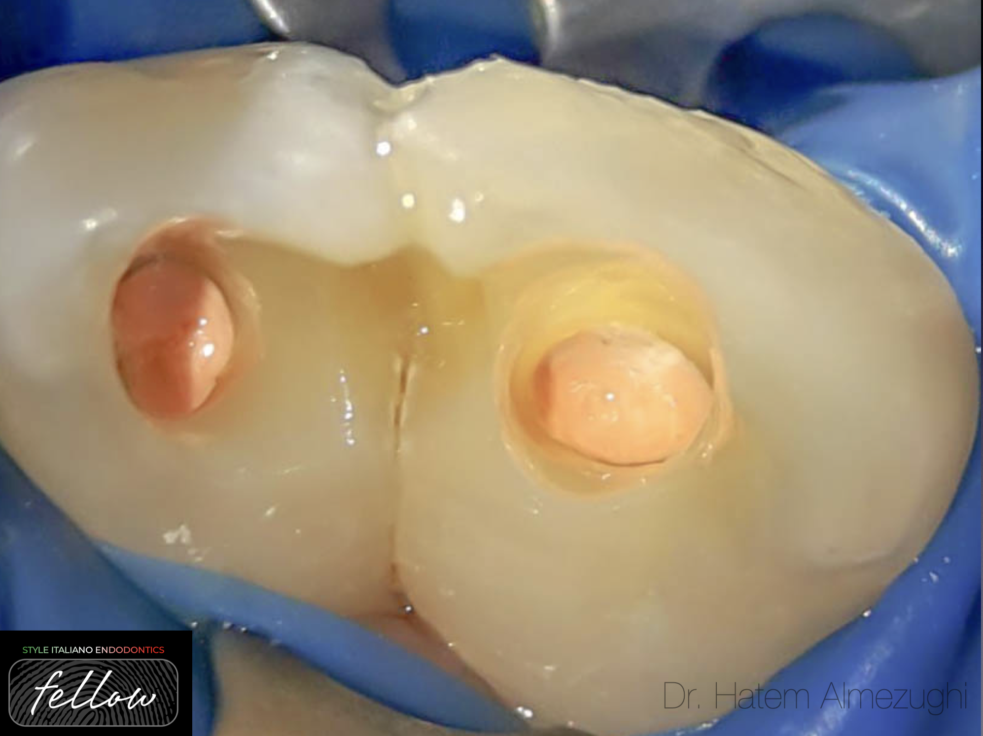
The treatment was completed in a single session. After access, the entrances of the two canals were identified. Initial scouting was performed with size 10 and 15 C-Pilot® files (VDW, Munich, Germany). Patency was confirmed and working length was established using an electronic apex locator (Root ZX®, Morita).
The canals were instrumented according to the manufacturer's protocol using OneFile (COLTENE, Switzerland). The use of magnification was essential for identifying and negotiating the complex internal anatomy while maintaining a conservative access and limiting unnecessary removal of tooth structure.
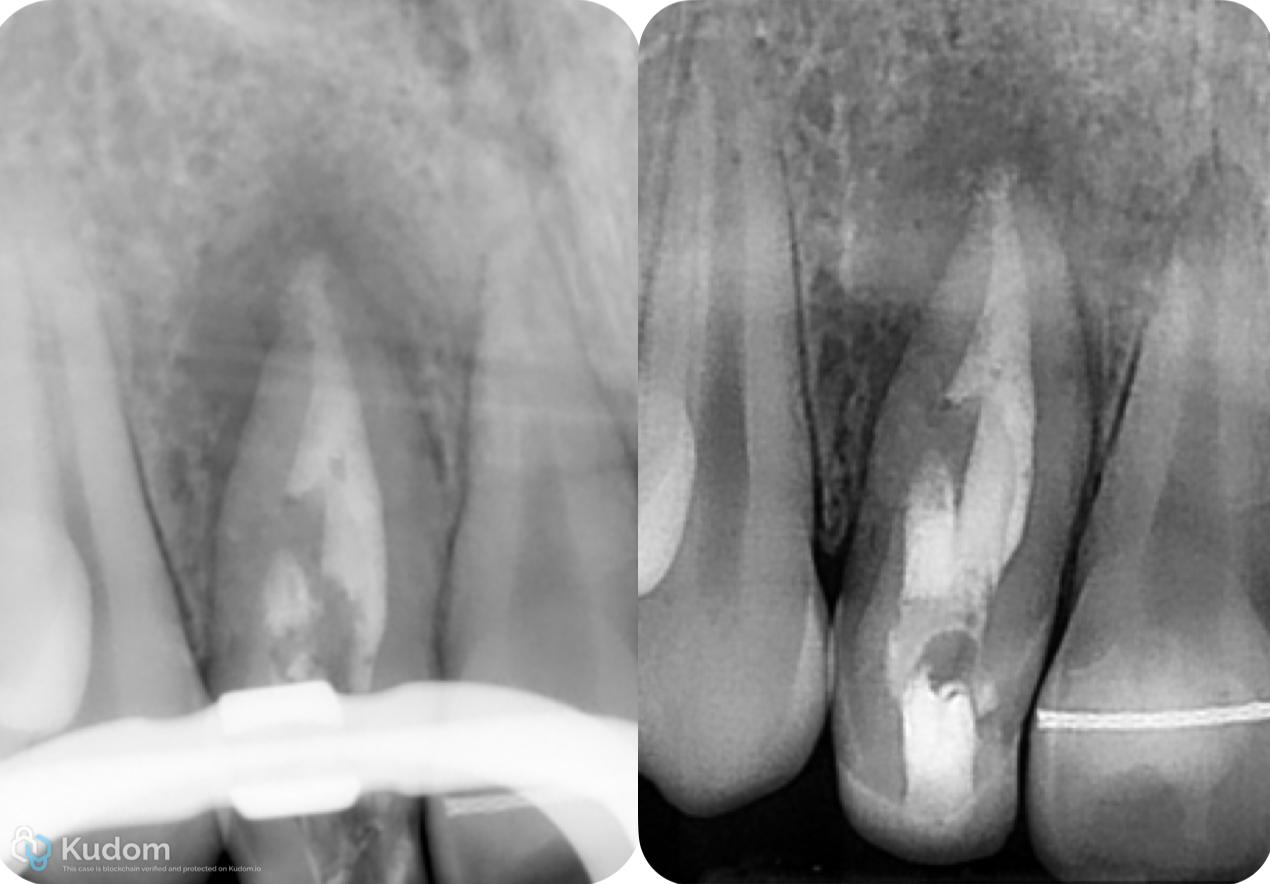
Fig. 4
Irrigation was performed with 6% sodium hypochlorite (NaOCl) using a 30G needle. Seventeen percent EDTA was subsequently used, with both solutions activated sonically using EDDY® (VDW, Munich, Germany). Saline was used between the two irrigants, and final irrigation was performed with saline.
An apical plug was placed using Bio-C Repair (Angelus, Brazil). The invaginated anatomy was then obturated, followed by backfilling of the middle and coronal thirds with heated gutta-percha and bioceramic sealer (Bio-C Sealer, Angelus, Brazil). The objective was to achieve a dense three-dimensional seal while respecting the complex morphology of the tooth.
Video of the procedure
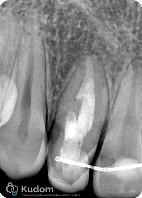
Fig. 5
1-Year Follow-Up
At the one-year follow-up examination, the patient was asymptomatic, with no clinical signs of infection and no recurrence of the sinus tract. Periapical radiographic examination demonstrated complete resolution of the previously observed periapical lesion, indicating a favorable healing response following the endodontic treatment.
Conclusions
The endodontic management of dens invaginatus requires a detailed understanding of the individual anatomy and a treatment strategy adapted to the morphology of the anomaly. In Type II cases with an open apex and periapical pathology, microscopic visualization, conservative access, ultrasonic assistance, effective irrigation and three-dimensional obturation are important components of treatment. CBCT should be considered when conventional radiographs do not provide sufficient anatomical information, as three-dimensional imaging can improve diagnosis and treatment planning in complex cases. In this case, the combination of a OneFile instrumentation approach, activated irrigation and bioceramic obturation allowed treatment to be completed in a single session, with resolution of the clinical signs and disappearance of the periapical lesion at one-year follow-up.
Bibliography
1. Shafer WG, Hine MK, Levy BM. A textbook of oral pathology. 4th ed. Philadelphia: W B Saunders Co; 1987.
2. Hovland EJ, Block RM. Nonrecognition and subsequent endodontic treatment of dens invaginatus. J Endod. 1977;3:360–362.
3. Hulsmann M. Dens invaginatus: etiology, classification, prevalence, diagnosis and treatment considerations. Int Endod J. 1997;30:79–90.
4. Oehlers FAC. Dens invaginatus (dilated composite odontome) I: variations of the invagination process and associated anterior crown forms. Oral Surg Oral Med Oral Pathol. 1957;10:1204–1218.
5. Yeh SC, Lin YT, Lu SY. Dens invaginatus in the maxillary lateral incisor. Oral Surg Oral Med Oral Pathol. 1999;87:628.
6. Pallavi Reddy Y, Kumaraguru Karpagavinayagam, Subbarao CV. Management of dens invaginatus diagnosed by spiral computed tomography: a case report. J Endod. 2008;34(9):1138–1142. doi:10.1016/j.joen.2008.06.001.
7. Zhu J, Wang X, Fang Y, Von den Hoff JW, Meng L. An update on the diagnosis and treatment of dens invaginatus. Aust Dent J. 2017;62 Suppl 1:43–55. doi:10.1111/adj.12513.
8. González Mancilla S, Montero Miralles P, Cabanillas-Balsera D, et al. Prevalence of dens invaginatus assessed by CBCT: systematic review and meta-analysis. Sci Rep. 2022;12:20379.