A case report
14/03/2024
Shadi Nagi
Warning: Undefined variable $post in /home/styleendo/htdocs/styleitaliano-endodontics.org/wp-content/plugins/oxygen/component-framework/components/classes/code-block.class.php(133) : eval()'d code on line 2
Warning: Attempt to read property "ID" on null in /home/styleendo/htdocs/styleitaliano-endodontics.org/wp-content/plugins/oxygen/component-framework/components/classes/code-block.class.php(133) : eval()'d code on line 2
This article / Case report is to illustrates a very rare anatomy of maxillary second molar using CBCT.
The endodontic treatment of teeth with unusual anatomy like dens invaginatus , talon cusp , dens evaginates , fusion , root dilaceration, etc, are really challenging to clinicians and endodontists.
At the same time These difficulties lead to endodontic treatment failure directly or indirectly if improperly managed. That’s why CBCT was requested and the images showed an extra root with a distinct canal space .
In the article we will discuss step by step the procedures of the management.
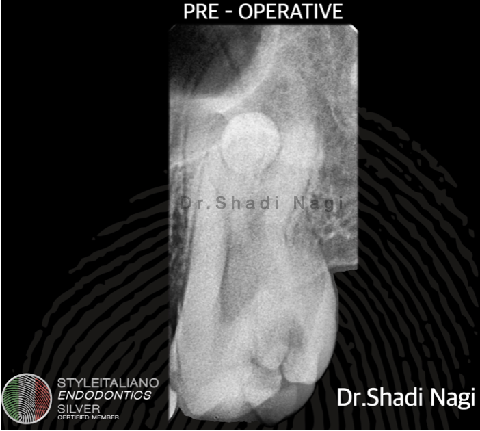
Fig. 1
A 17 years old patient referred to clinic with a severe pain related to tooth no 2.7
Pulp vitality tests gives a severe pain to cold compared with the control teeth.
The maxillary left second molar exhibited tenderness to percussion.
There was no sensitivity to palpation or abnormal pocket depth.
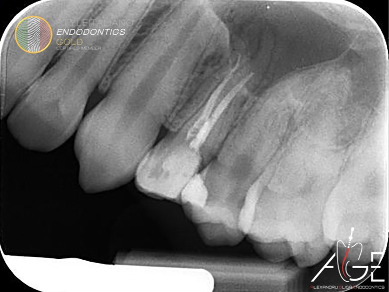
Notice from the pre operative radiograph that there is a well defined round anatomy , That’s why CBCT was requested for the proper assessment.
After clinical and radiographic examination the diagnosis was symptomatic irreversible pulpits.
Treatment plan RCT then follow up.
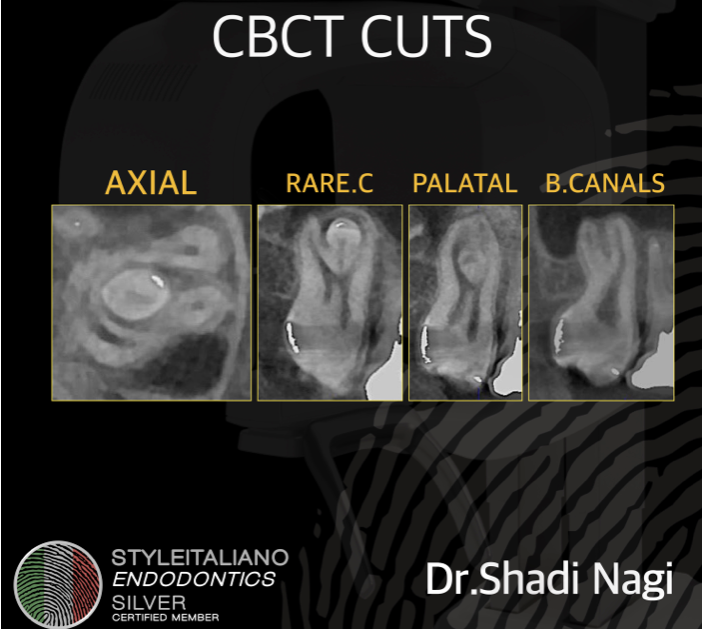
Fig. 2
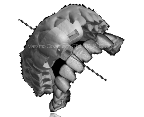
Because the limitations of the 2D radiography and to confirm the unusual anatomy CBCT was undertaken.
As we can see from the CBCT images , there is an extra round root with distinct canal space.
Two palatal canal and two buccal canals.
Surprisingly this tooth has Five canals with 5 portal of exit .
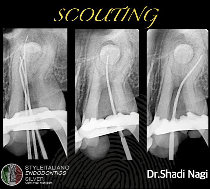
Fig. 3
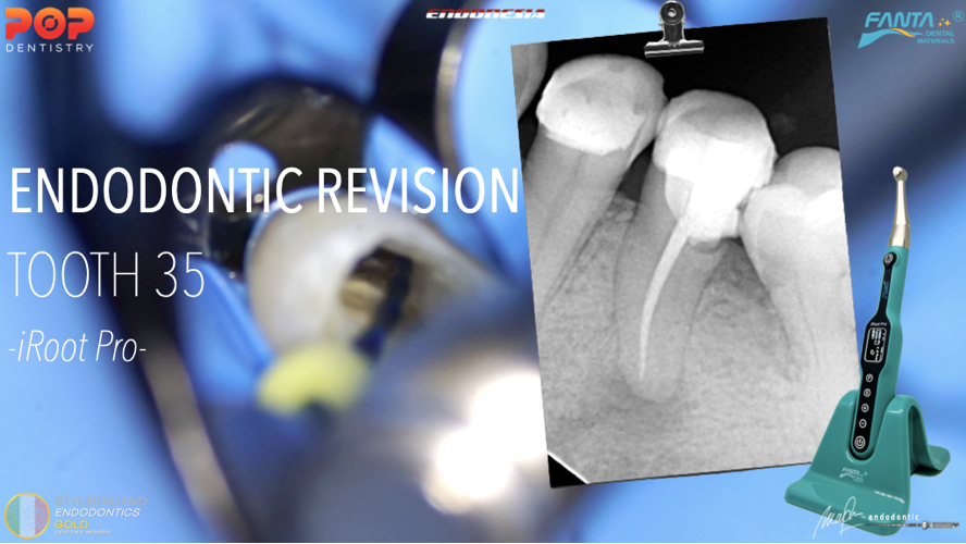
The Scouting radiograph.
‘NOTE’ The size of the MB canal’s foramen was more than ISO 80 so MTA apical plug was indicated with the use of collagen matrix to act as a barrier beyond the apex.
Here is the full case video.
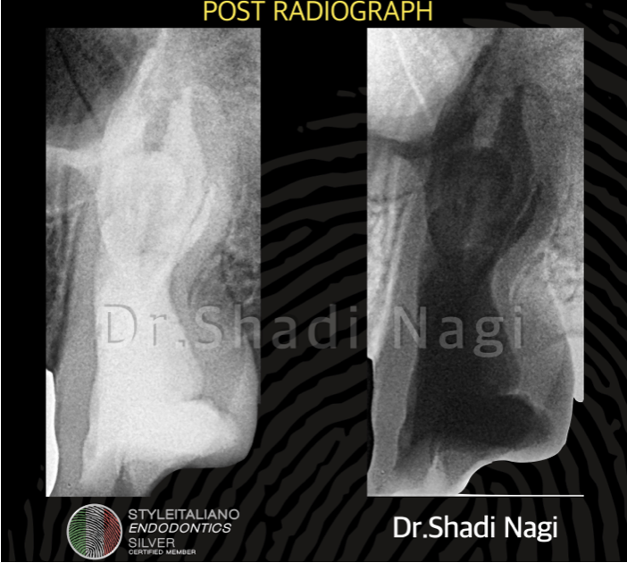
Fig. 4
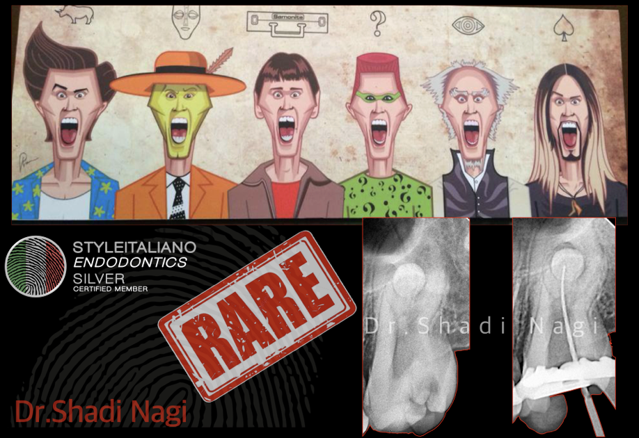
The post operative radiograph
The obturation technique was modified heat technique using bioceramic sealer and gutta percha for all canals EXCEPT the MB canal was MTA apical plug.
Conclusions
This case reports one of the very rare cases of maxillary second molars having unusual extra round root and it’s successfully management with the use of CBCT , floor dental map and magnification.
Bibliography
1- Hrudi Sundar Sahoo, R. Kurinji Amalavathy, D. Pavani, "A Case Report on Endodontic Management of a Rare Vertucci Type III Maxillary Canine”, https://doi.org/10.1155/2019/4154067
2- Fernando Branco Barletta; Fabiana Soares Grecca; Márcia Helena Wagner; Ronise Ferreira; Fernanda Ullmann Lopez. Endodontic treatment of a 36-mm long upper cuspid: clinical case report https://doi.org/10.1590/S1980-65232010000400017
3- Hela Zekri, Salima Bouaziz, Jihed Ben Ammar, Saida Sahtout, Hedia Ben Ghénaïa Jaouadi and Lotfi Bhouri. Nonsurgical endodontic treatment of an invaginatus maxillary lateral incisor DOI: 10.15761/DOCR.1000233


