Root Canal Retreatment of UL4 using the MG3 system from Perfect: A case report
19/10/2023
Alexandru Gliga
Warning: Undefined variable $post in /home/styleendo/htdocs/styleitaliano-endodontics.org/wp-content/plugins/oxygen/component-framework/components/classes/code-block.class.php(133) : eval()'d code on line 2
Warning: Attempt to read property "ID" on null in /home/styleendo/htdocs/styleitaliano-endodontics.org/wp-content/plugins/oxygen/component-framework/components/classes/code-block.class.php(133) : eval()'d code on line 2
Root canal retreatments have always been the elefant in the room. The preoperative X-ray may or may not predict future difficulties, thus the clinician has to stay constantly alert for any unplanned situations that have to be managed. Nonclinical curvatures, separated files, ledges, perforations and much more circumstances can be completely camouflaged inside a common periapical X-ray, sabotaging the clinician’s plan and increases the risks of iatrogenic errors. Without a CBCT investigation prior to the endodontic retreatment, any clinician should perceive that caution and safety approach, even thought it can be more time-consuming, enhances the chance of having a favourable outcome.
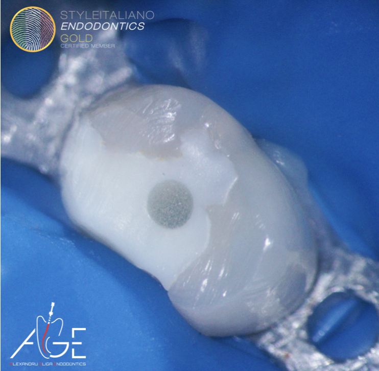
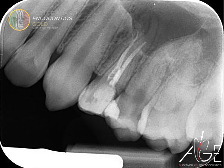
Fig. 1
The 24 years old patient was referred to our office by her general dental practitioner for endodontic retreatment. She complained about acute unprovoked pain in the upper left quadrant for the last 2 weeks.
The access cavity was driven by removing the coronal filling material and the infected dentine. Ultrasound was used to remove the fibre post.
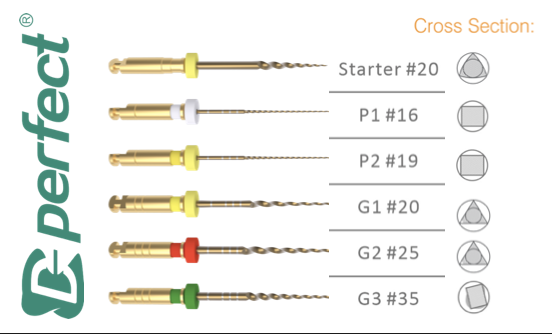
Fig. 2
The MG3 file system from Perfect was chosen for managing this case.
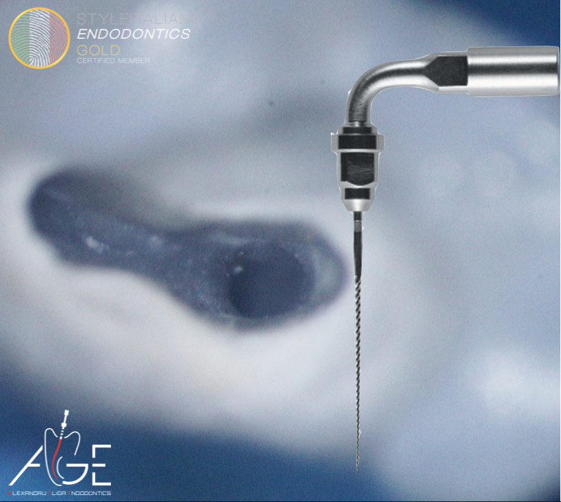
Fig. 3
The fibre post was removed from within the palatal canal with the use of 15 ISO U-file.
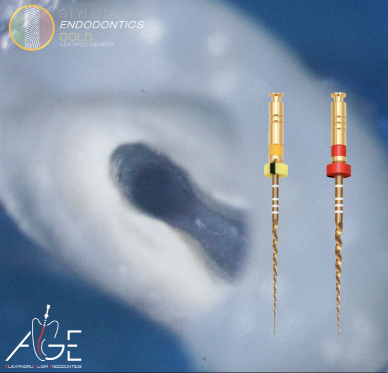
Fig. 4
Instrumentation sequence:
Perfect MG3: G1 and G2
Video of the instruments used
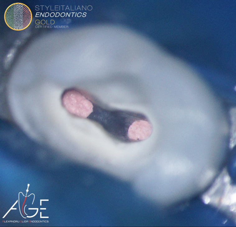
Fig. 5
Vertical warm condensation technique was performed to obturate the canals.
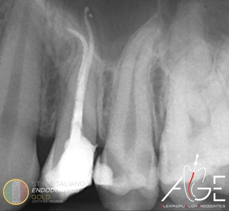
Fig. 6
Post operative X-ray
Conclusions
Before embarking into any endodontic treatment, clinicians should carefully consider if any investigation is required to improve the prediction of future clinical procedures.
Endodontic retreatments are like a Matryoshka doll (it opens up gradually) however, unlike the doll, retreatments very often hide inside the endodontic system many clinical unexpected surprises.
In conclusion, no matter the preoperative clinical investigations, clinicians should “hope for the best but prepare for the worse”.
Bibliography
Scarfe WC, Levin MD, Gane D, Farman AG. Use of cone beam computed tomography in endodontics. Int J Dent. 2009;2009:634567. doi: 10.1155/2009/634567. Epub 2010 Mar 31. PMID: 20379362; PMCID: PMC2850139.
Patel S, Durack C, Abella F, Shemesh H, Roig M, Lemberg K. Cone beam computed tomography in Endodontics - a review. Int Endod J. 2015;48(1):3-15. doi:10.1111/iej.12270
Bertani, P., Gagliani, M. & Gorni, F. 2020, Retreatments. Solutions for Periapical Diseases of Endodontic Origin, Edra.