Retreatment episodes, series 1
08/04/2021
Ibrahim El Naggar
Warning: Undefined variable $post in /var/www/vhosts/styleitaliano-endodontics.org/endodontics.styleitaliano.org/wp-content/plugins/oxygen/component-framework/components/classes/code-block.class.php(133) : eval()'d code on line 2
Warning: Attempt to read property "ID" on null in /var/www/vhosts/styleitaliano-endodontics.org/endodontics.styleitaliano.org/wp-content/plugins/oxygen/component-framework/components/classes/code-block.class.php(133) : eval()'d code on line 2
Root canal retreatment seems to be one of the biggest challenges in the Endodontic therapy, in the retreatment episodes we will review different scenarios with different cases.
In Endodontics we are dealing with a lot of challenges such as anatomy, mishaps, instrument separation, etc. but one of the biggest challenges is to deal safely with cases of retreatment that may present unusual anatomies or missed canals.
First of all, in case of retreatment, the clinician must have a strong idea about the causes that led to the failure of the initial treatment: it is impossible to know the exact cause in every case but, in order to reduce the risk of failure, assessment of the most likely reason of failure must be done. If we identify this reason, we can easily avoid it during the Retreatment, hence increasing the success of the therapy.
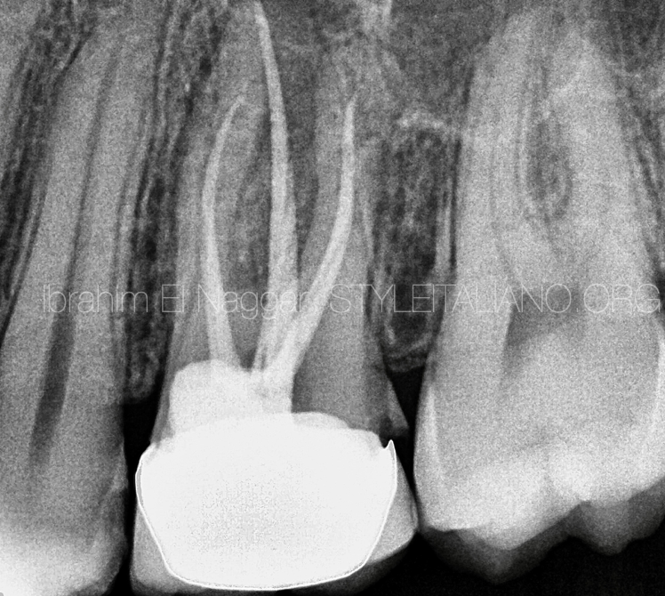
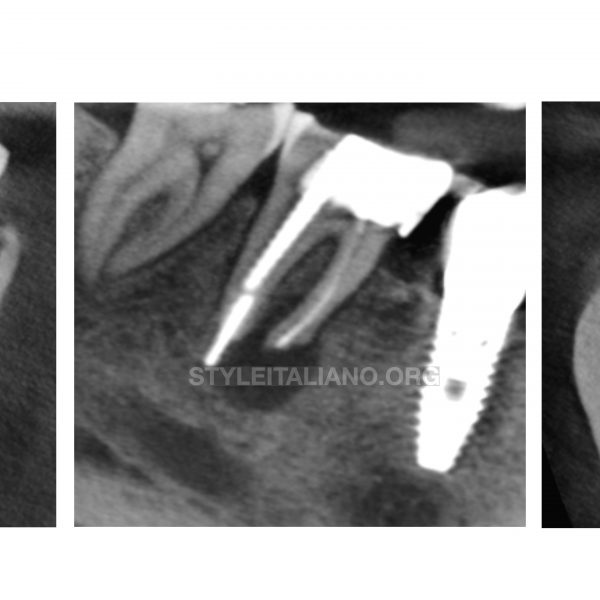
Fig. 1
The Pre-OP Radiograph shows :
Open margin Crown
Severe unnecessary cutting toward the Mesial.
Short Filling on DB canal.
Shadow of missing MB2
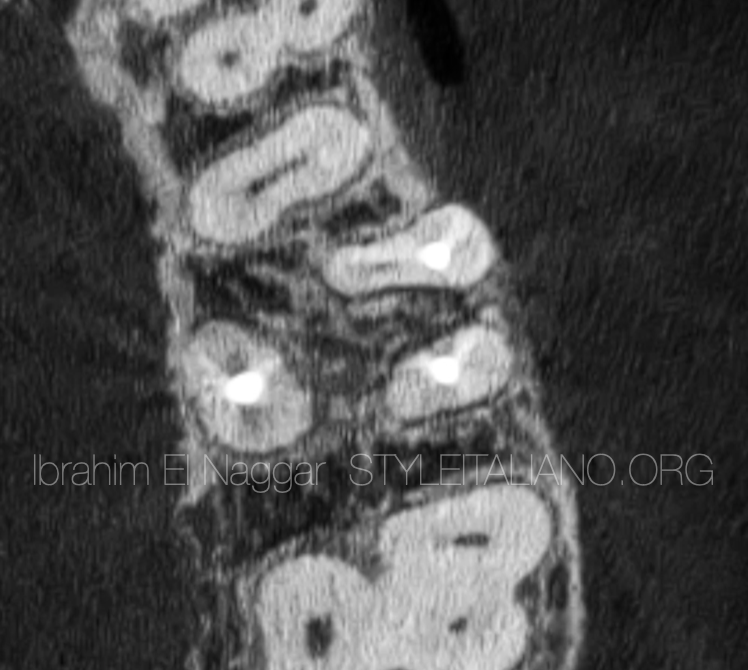
Fig. 2
Axial cut of CBCT scanning shows a clearly missed MB2 canal
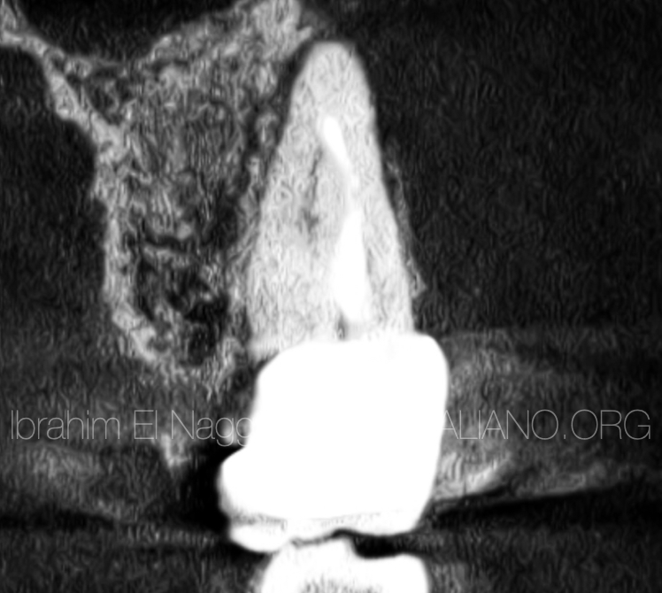
Fig. 3
The coronal cut shows the MB system: MB1 & MB2 are confluent.
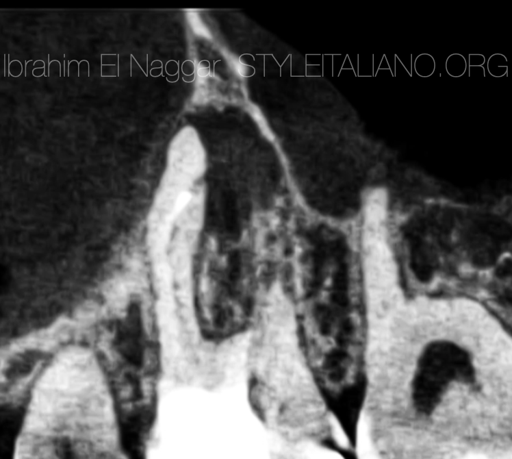
Fig. 4
The sagittal cut shows the presence of apical periodontitis around the MB system with the foramen being in a lateral position.
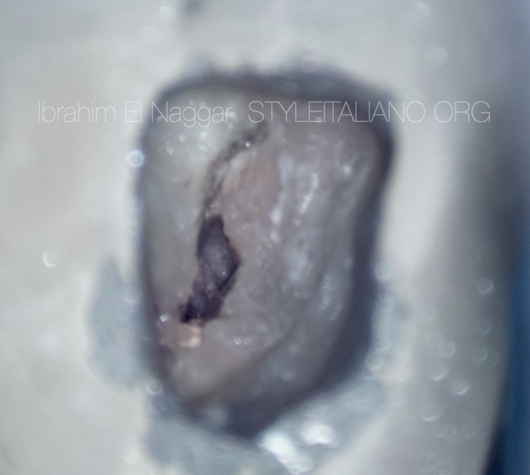
Fig. 5
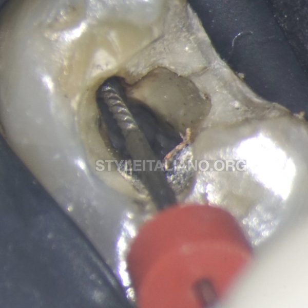
Access Opening through the crown.
The use of ultrasonic tips helps to remove obstacles and redefine the access cavity.
The removal of the filling materials done with a rotary file in the presence of irrigant solution.
MB2 Detection using Sharp DG16 Probe.
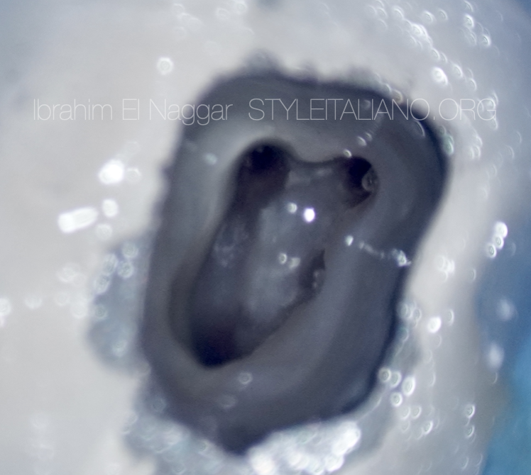
Fig. 6
After gutta-percha removal and access refinement
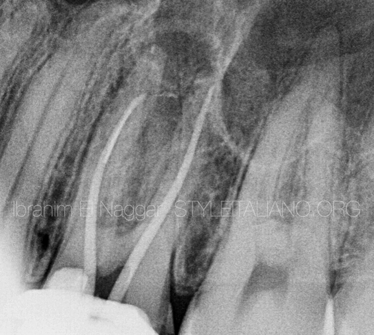
Fig. 7
Master Cone Fitting for DB & MB1
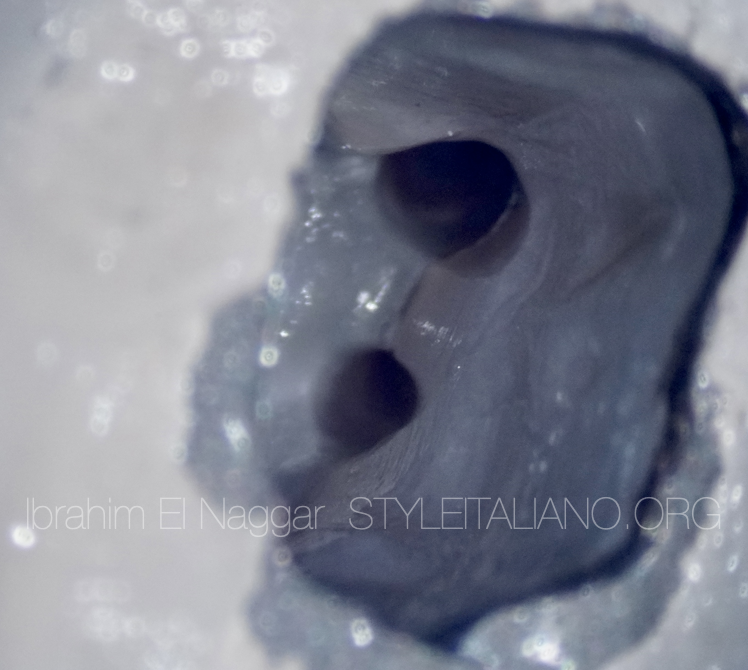
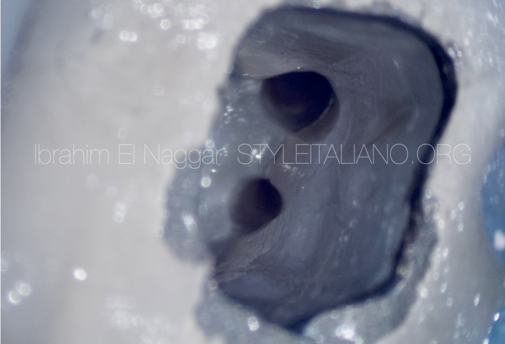
Fig. 8
MB1 and MB2 after shaping and dryness.
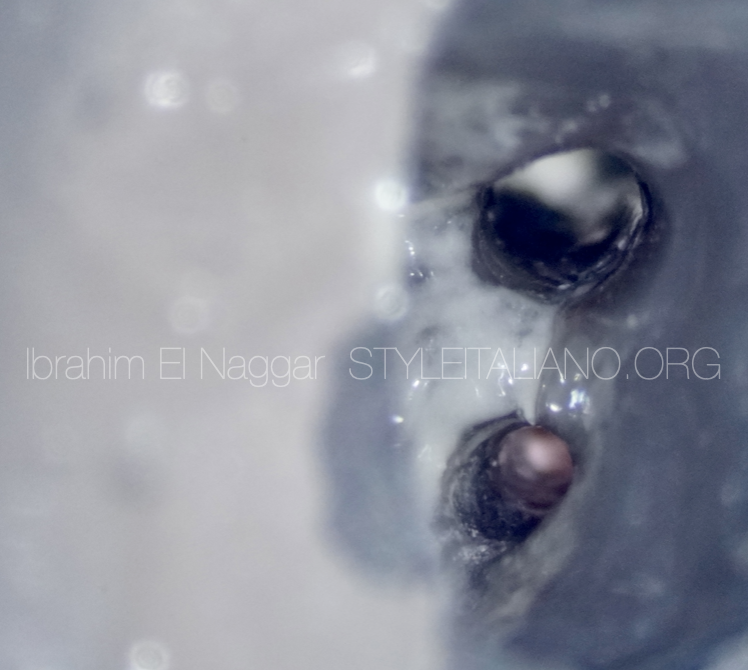
Fig. 9
Confluence shown with the down-packing
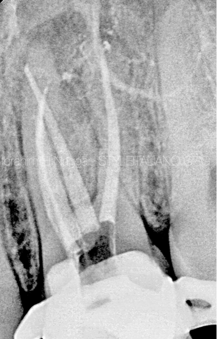
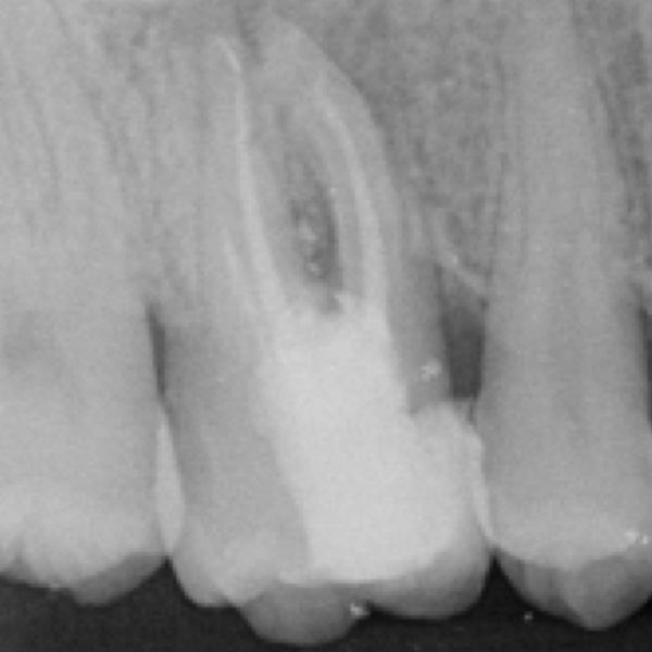
Fig. 10
Post-op radiograph with DB Delta in the apical third & MB2 filled.
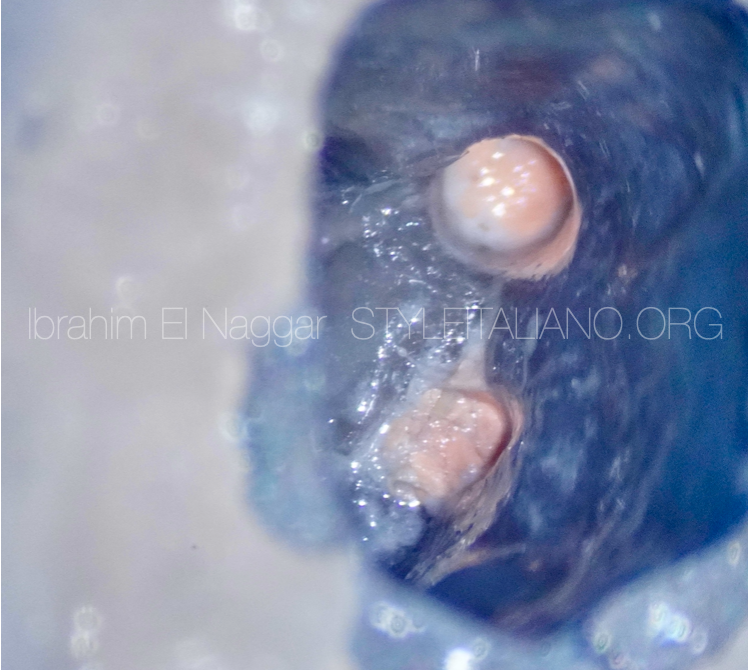
Fig. 11
Post-OP Obturation
Conclusions
It is very important to assess the cause of the treatment failure before starting a new treatment and also be aware with every step in the treatment, including the armamentarium for a better treatment outcome and for apical periodontitis prevention.
Bibliography
Baruwa AO, Martins JNR, Meirinhos J, Pereira B, Gouveia J, Quaresma SA, Monroe A, Ginjeira A. The Influence of Missed Canals on the Prevalence of Periapical Lesions in Endodontically Treated Teeth: A Cross-sectional Study. J Endod. 2020 Jan;46(1):34-39.e1. doi: 10.1016/j.joen.2019.10.007. Epub 2019 Nov 14. Erratum in: J Endod. 2020 Jun;46(6):881. PMID: 31733814.