MOLARIZED PREMOLAR Management of three rooted premolar
08/04/2022
The Community
Warning: Undefined variable $post in /var/www/vhosts/styleitaliano-endodontics.org/endodontics.styleitaliano.org/wp-content/plugins/oxygen/component-framework/components/classes/code-block.class.php(133) : eval()'d code on line 2
Warning: Attempt to read property "ID" on null in /var/www/vhosts/styleitaliano-endodontics.org/endodontics.styleitaliano.org/wp-content/plugins/oxygen/component-framework/components/classes/code-block.class.php(133) : eval()'d code on line 2
Maxillary first premolars are commonly known by having two canals in one or two separate roots (60-65%), meanwhile Maxillary second premolars are known by having one canal in one root (38-48%).
Three separate roots in Maxillary first premolar has shown very rare incidence (2.5-5%), even rarer are reported to Maxillary second premolars
This article describes the diagnosis and clinical management of four clinical cases of three rooted maxillary premolars.
Only one case is primary treated and the other three cases were previously treated, this reminds us that successful root canal treatment relies on correct access cavity preparation with location of all existing canals, sufficient cleaning, adequate shaping, and complete 3D obturation plays an important role in the success of initial treatment procedures.
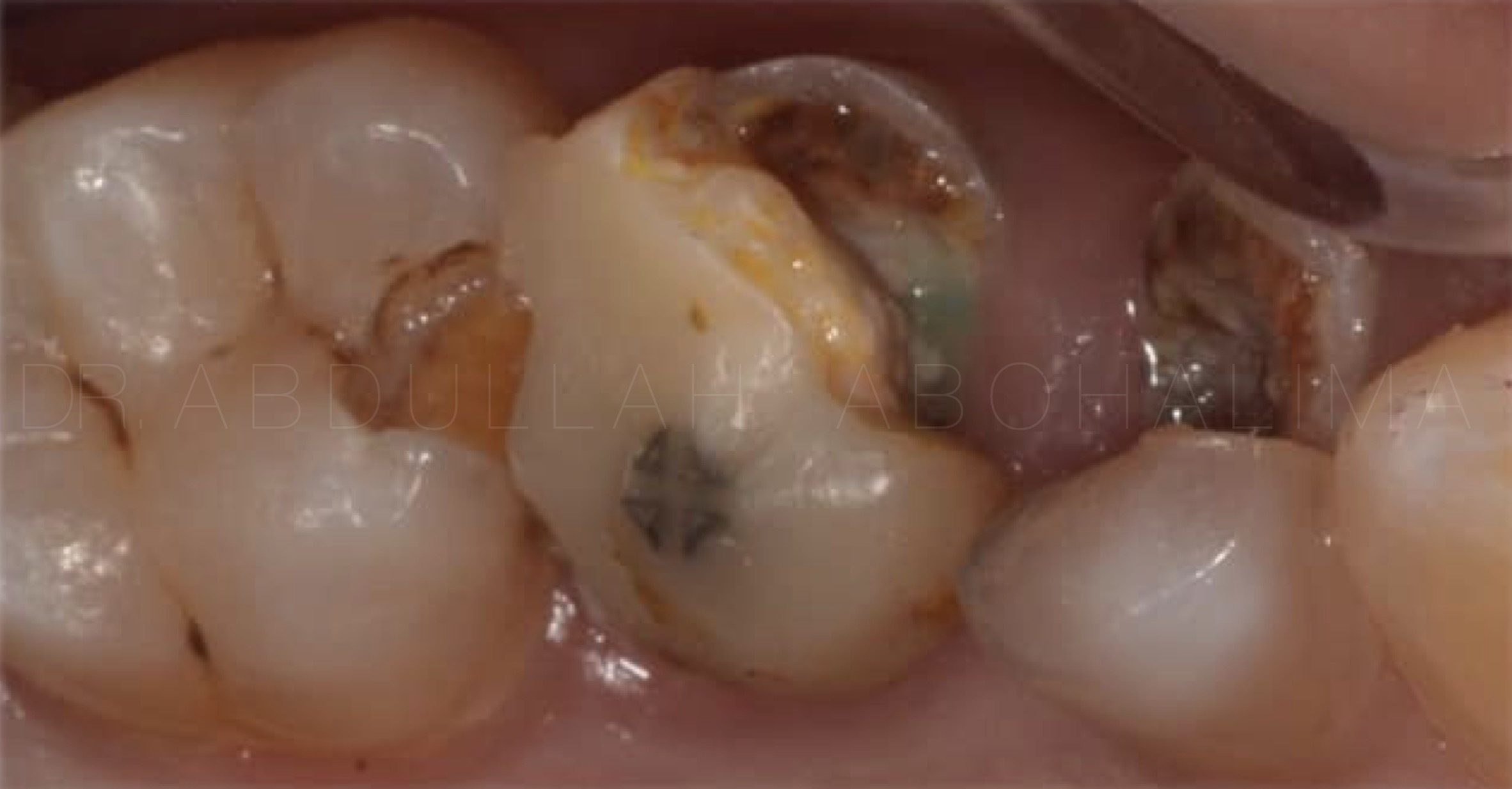
Fig. 1
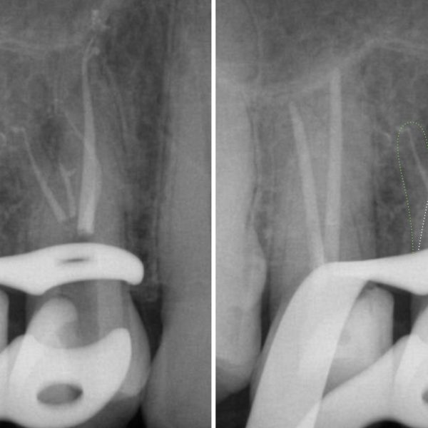
This case shows Symptomatic Apical Periodontitis (SAP) associated with a failed primary root canal treatment.
Initial situation of maxillary second premolar with a large carious lesion, poor filling and metal post presented with spontaneous pain, tender to percussion
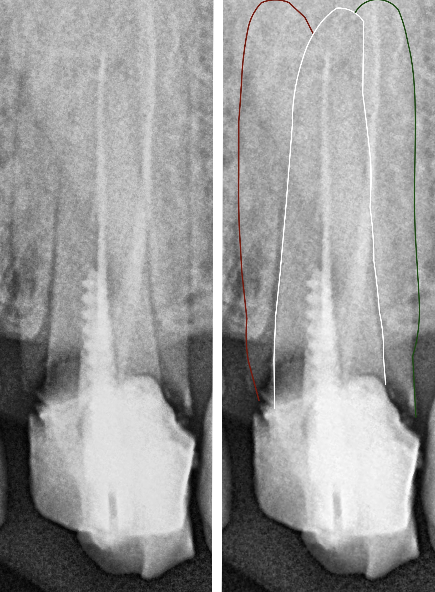
Fig. 2
Pre-op X-Ray showed previously insufficient root canal treatment for buccal canal, disto-palatal canal and complete neglect of mesio-palatal canal (MP)
Radiographic assessment should be done carefully.
Asymmetrical root to guttapercha thickness indicates a missed canal in the remaining empty space (mesial side of the case )
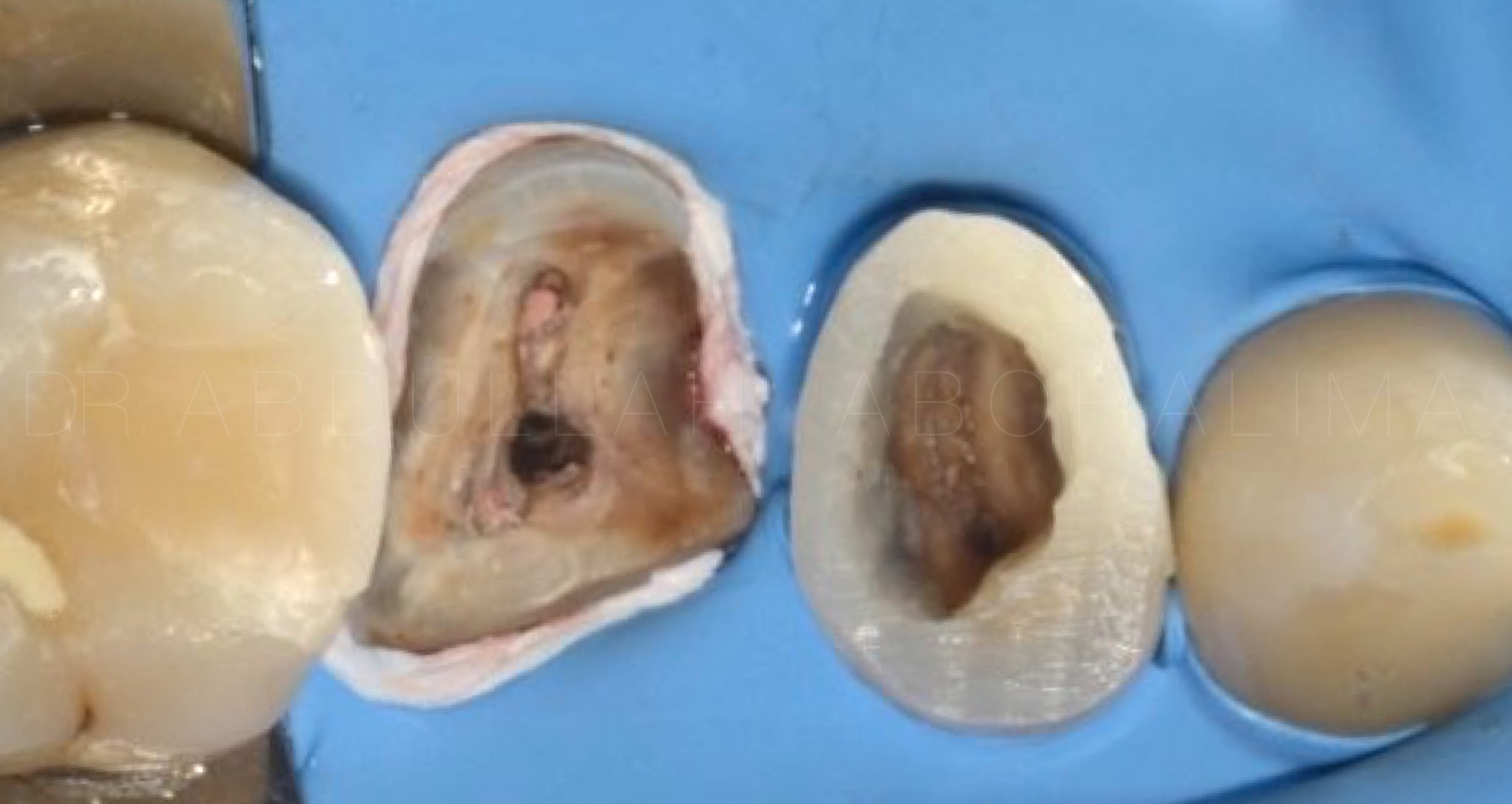
Fig. 3
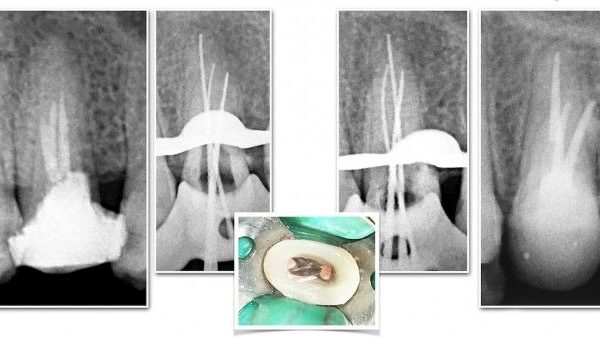
Multiple Isolation, metal post removal and missed canal located
During the Retreatment you may notice a sealer / guttapercha spot at the orifice level of the missed canal which is very helpful in the location and diagnosis of such cases .
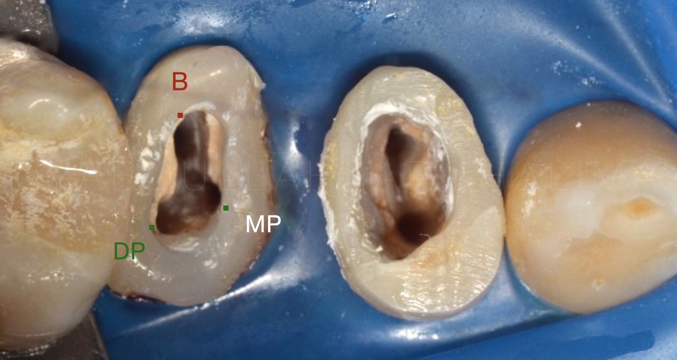
Fig. 4
Build up.
pay attention to the fact that maxillary first premolar having 3 roots is a rare incidence of 5 to 6%, while maxillary second preomalrs have less incidence of 0.3 to 2% in lab studies
Even rarer are maxillary second premolars with 1 buccal and 2 palatal canals which what we have here

Fig. 5
Master cone fit
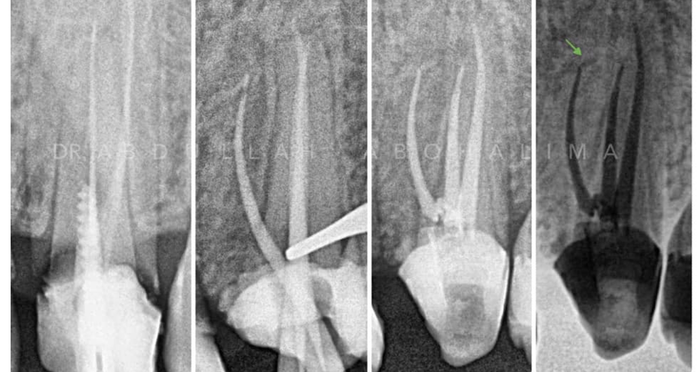
Fig. 6
Full case management
Fig. 7
In this case of maxillary first premolar: 2 separate canals present in the buccal root with 1 palatal canal
Access cavity modifications is required for stress free entry to complex anatomy. Higher magnification and illumination in addition to ultrasonics can be useful for access cavity preparation and recognizing and locate additional canals.
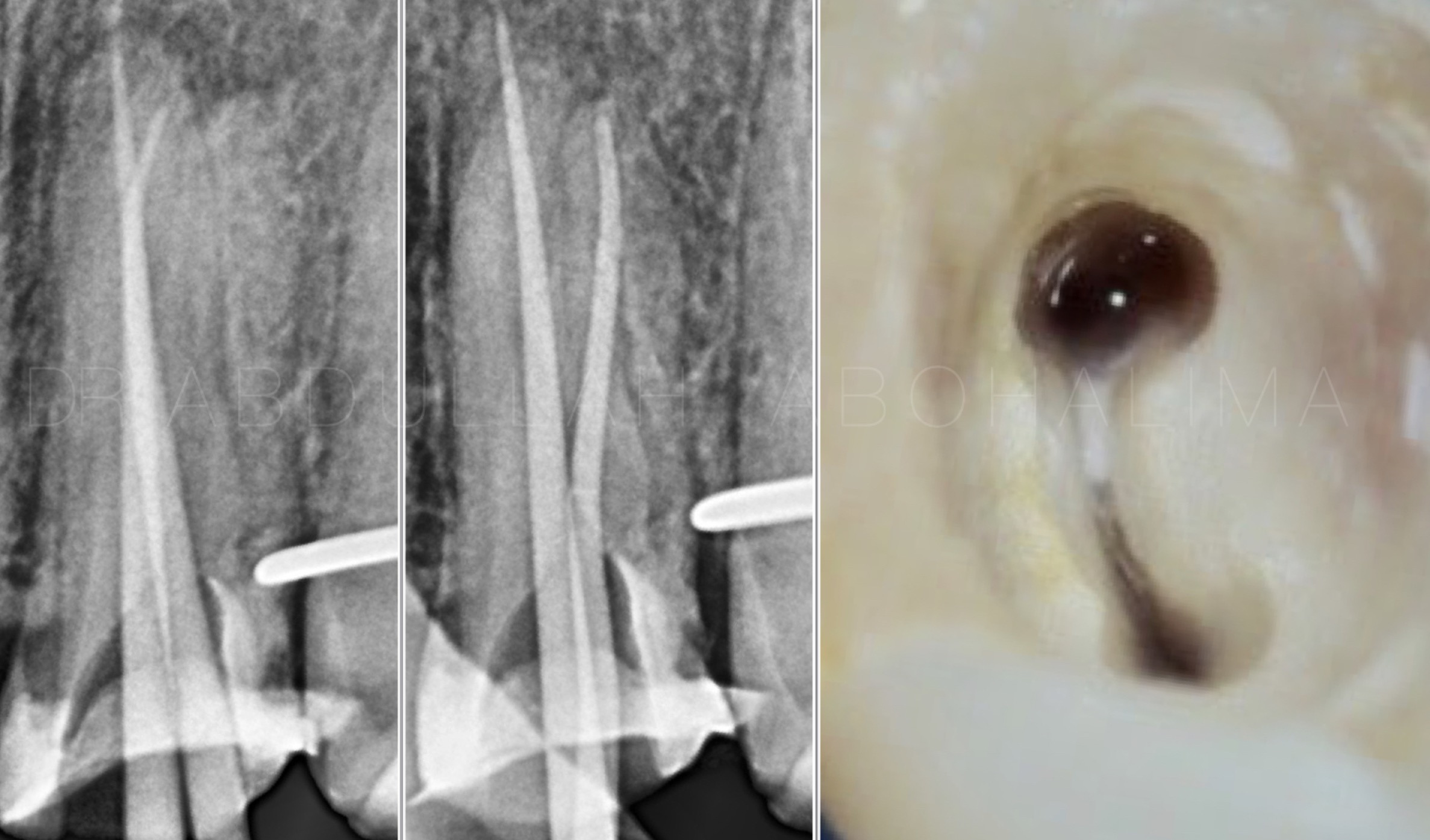
Fig. 8
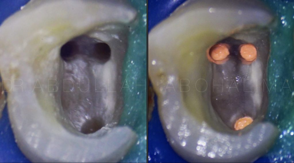
During master cones fit and obturation for buccal canals each cone has been inserted separately due to the closeness of the canals, so proper insertion should have been done in this way
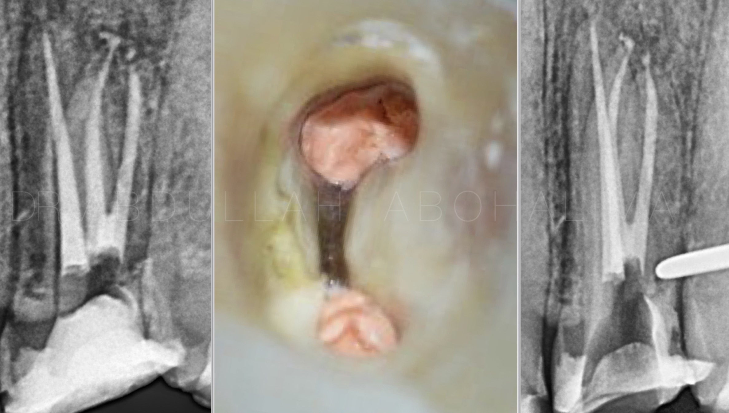
Fig. 9
Obturation,
Clean chamber is very important for post-endo restoration.
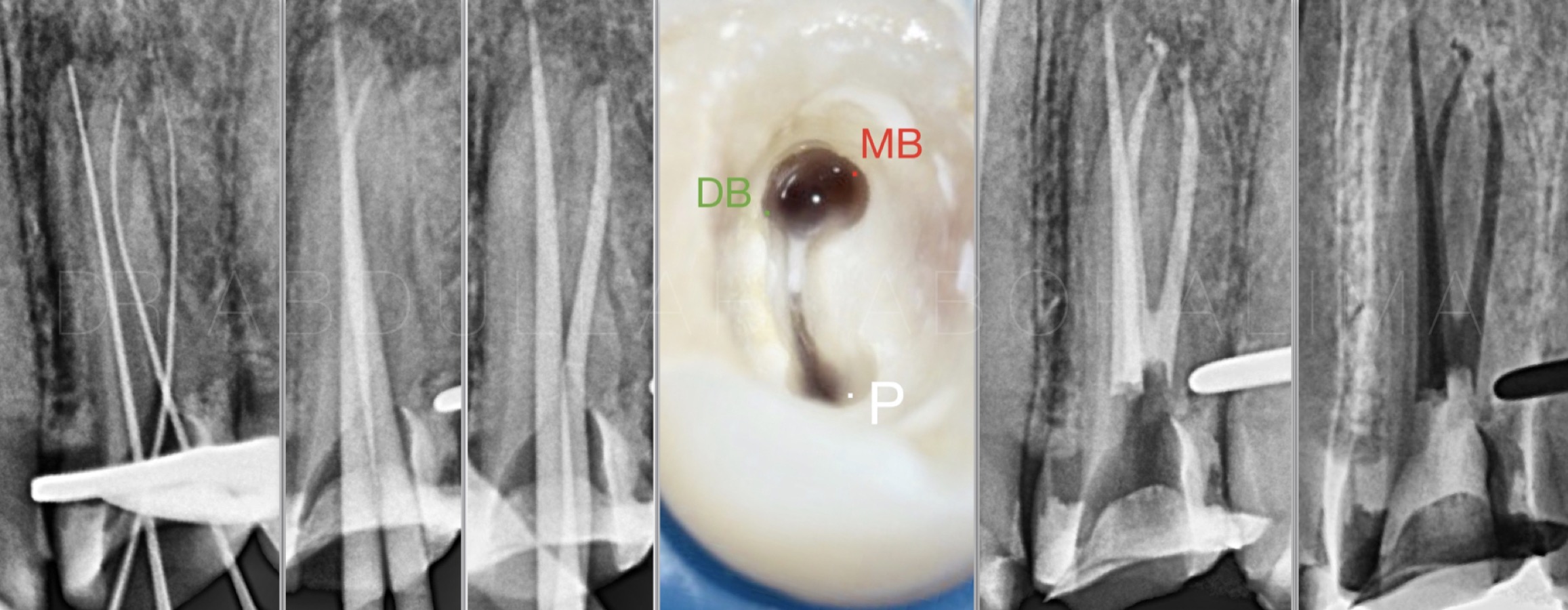
Fig. 10
In premolars if the mesio-distal width of the mid-root is equal to or greater than the mesio-distal width of the crown, three root canals will be most likely present.
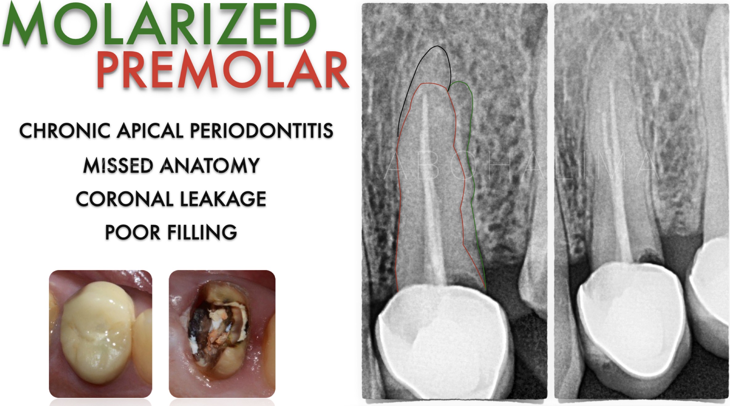
Fig. 11
In this case of retreatment of the maxillary first premolar. It was evident the presence of two buccal canals and one palatal canal with neglect of Disto-buccal canal
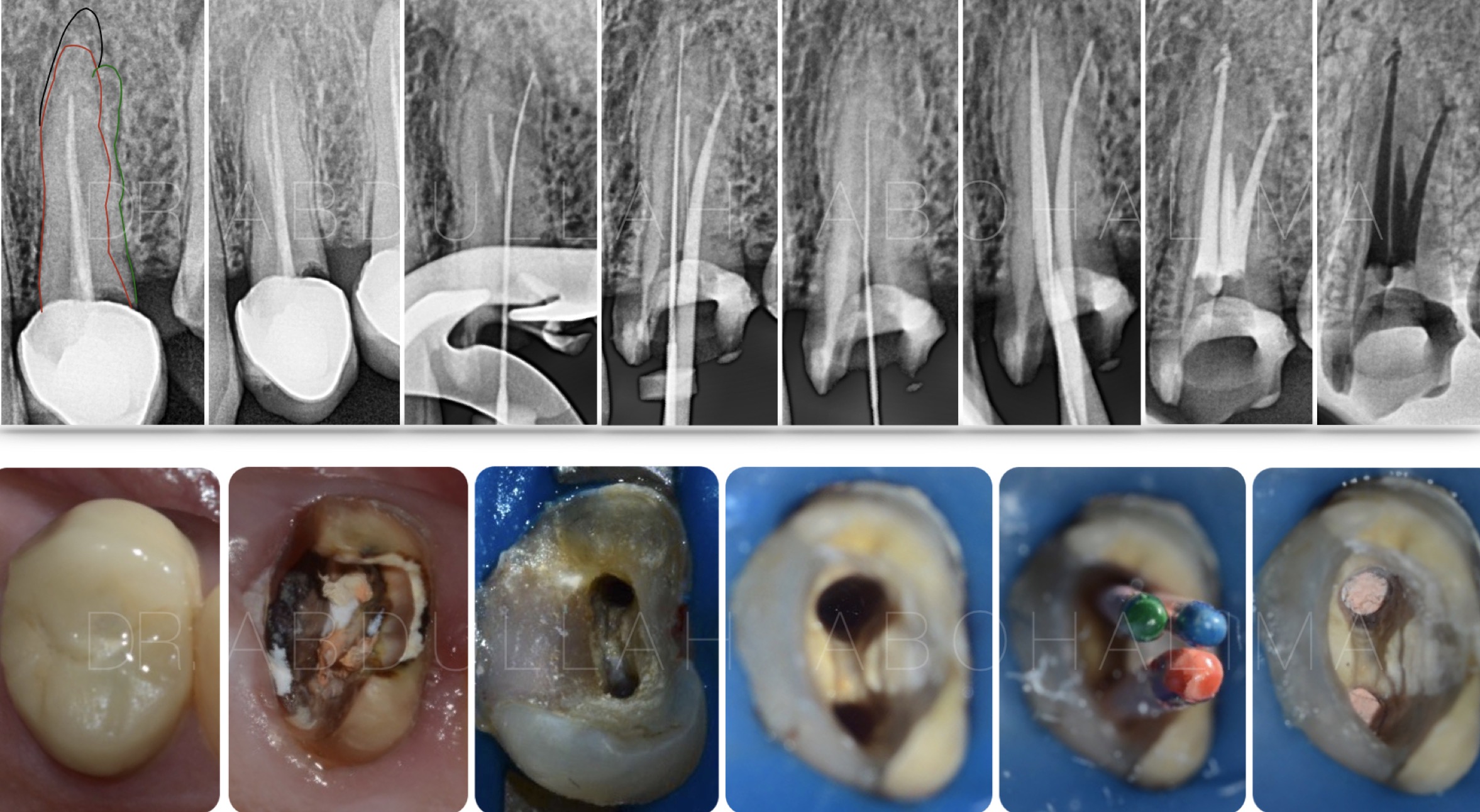
Fig. 12
Full case with locating of the missed canal and removal of the main cause of the failure
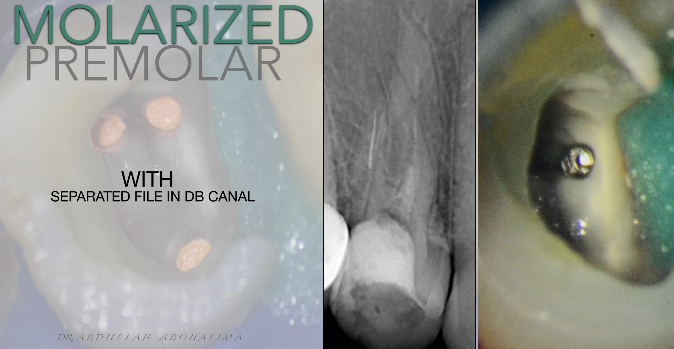
Fig. 13
Analyzing the data from preoperative radiograph was showing three rooted maxillary first premolar with separated file in disto-buccal canal
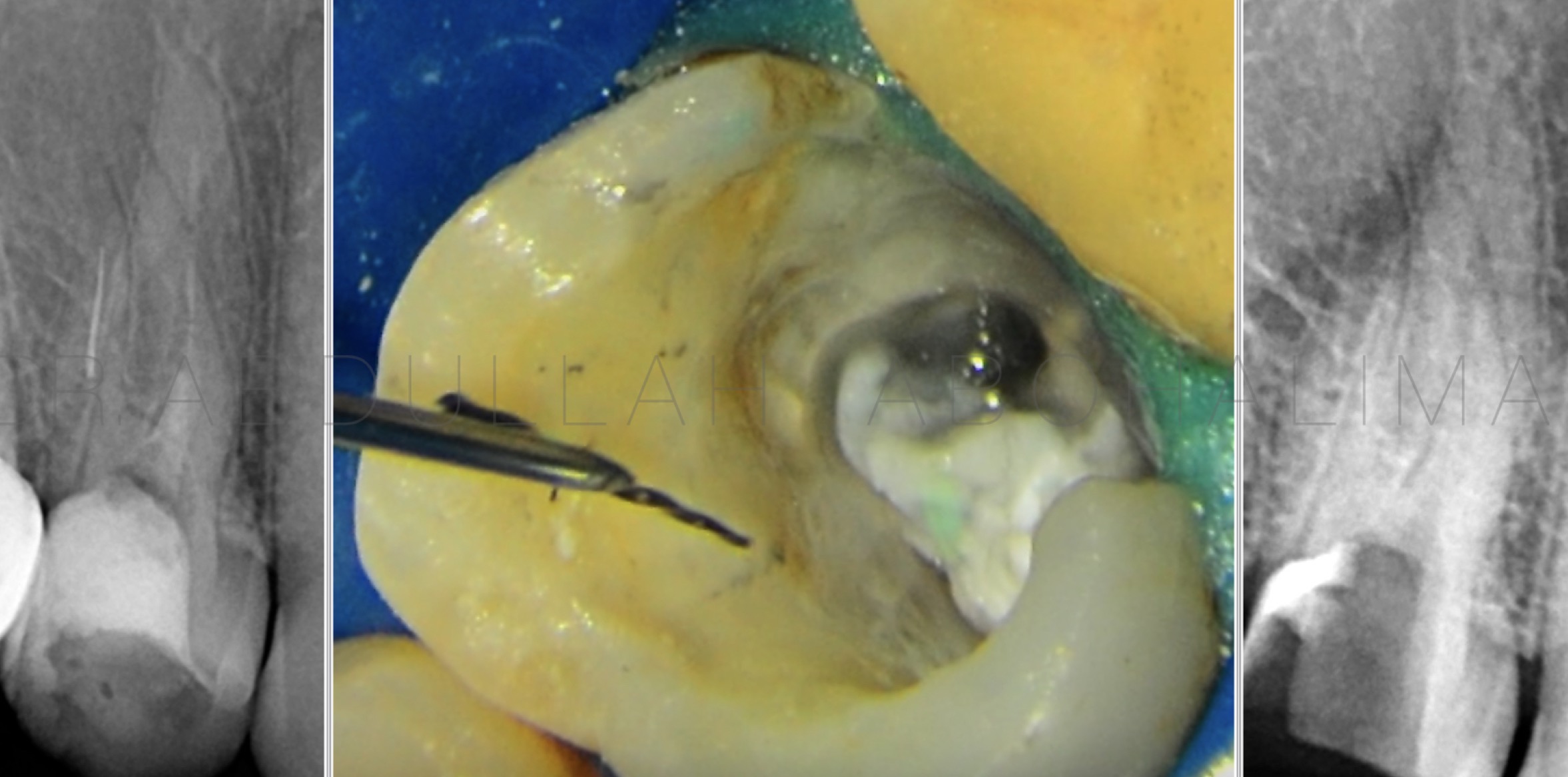
Fig. 14
Separated file retrieval using ultrasonic after deep marginal elevation, multiple isolation and coronal flaring
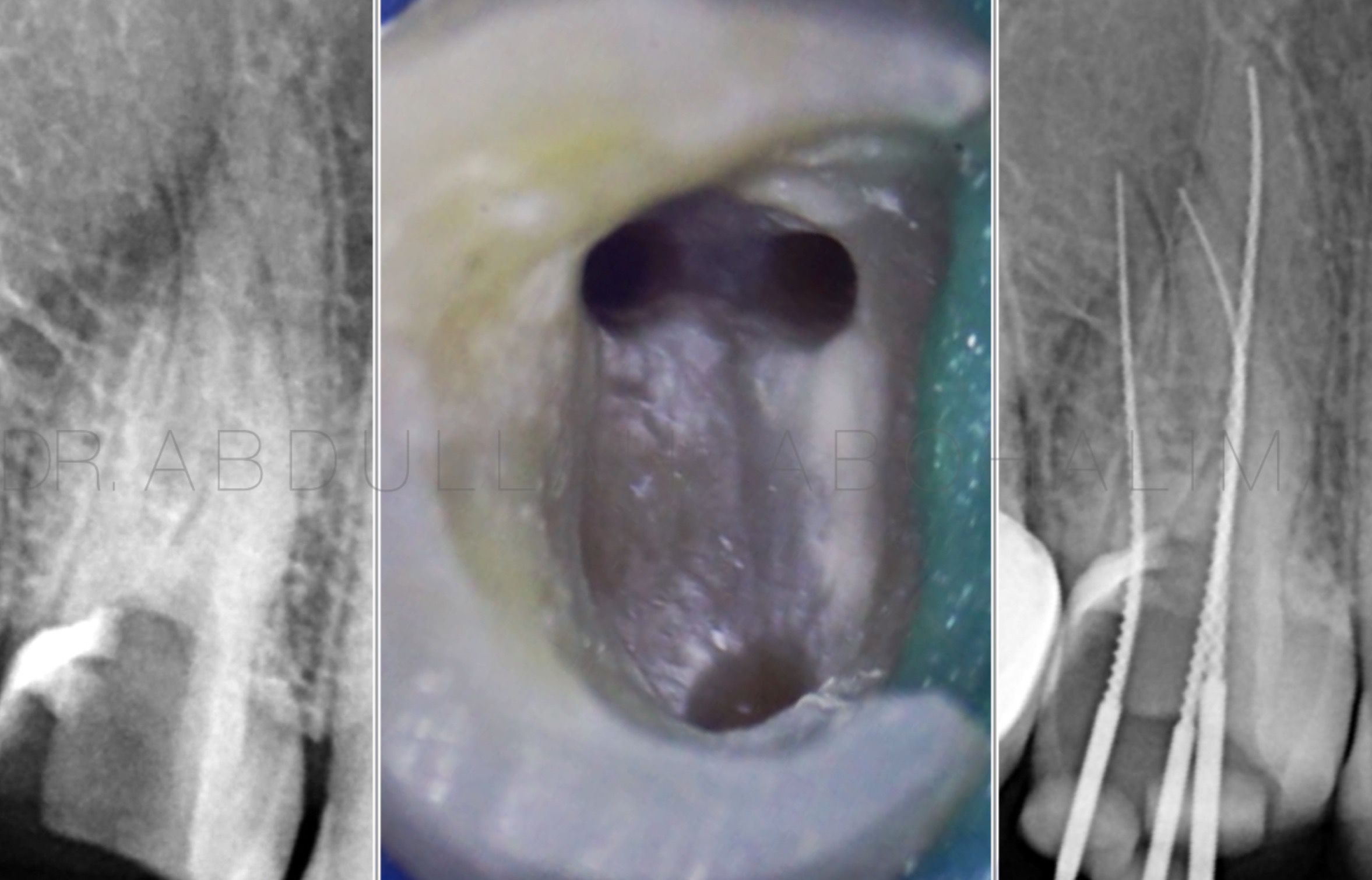
Fig. 15
Working length determination
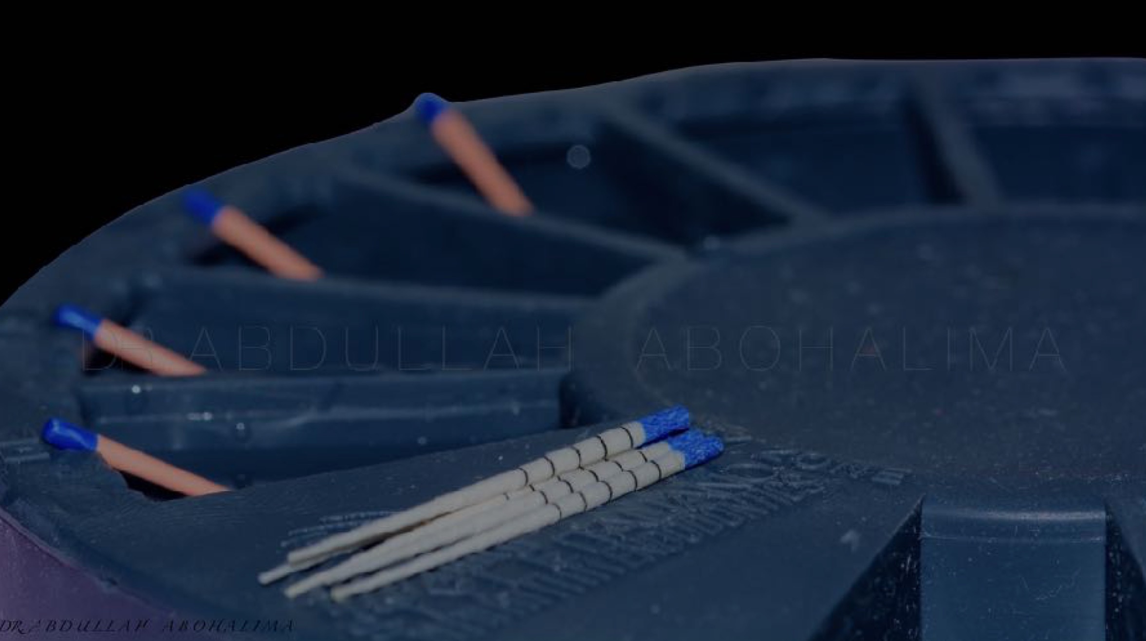
Fig. 16
Plano from Styleitaliano Endodontics to organize the work
Disinfection of gutta percha before the obturation 1 minute soaked in full concentrated sodium hypochlorite 5.25%
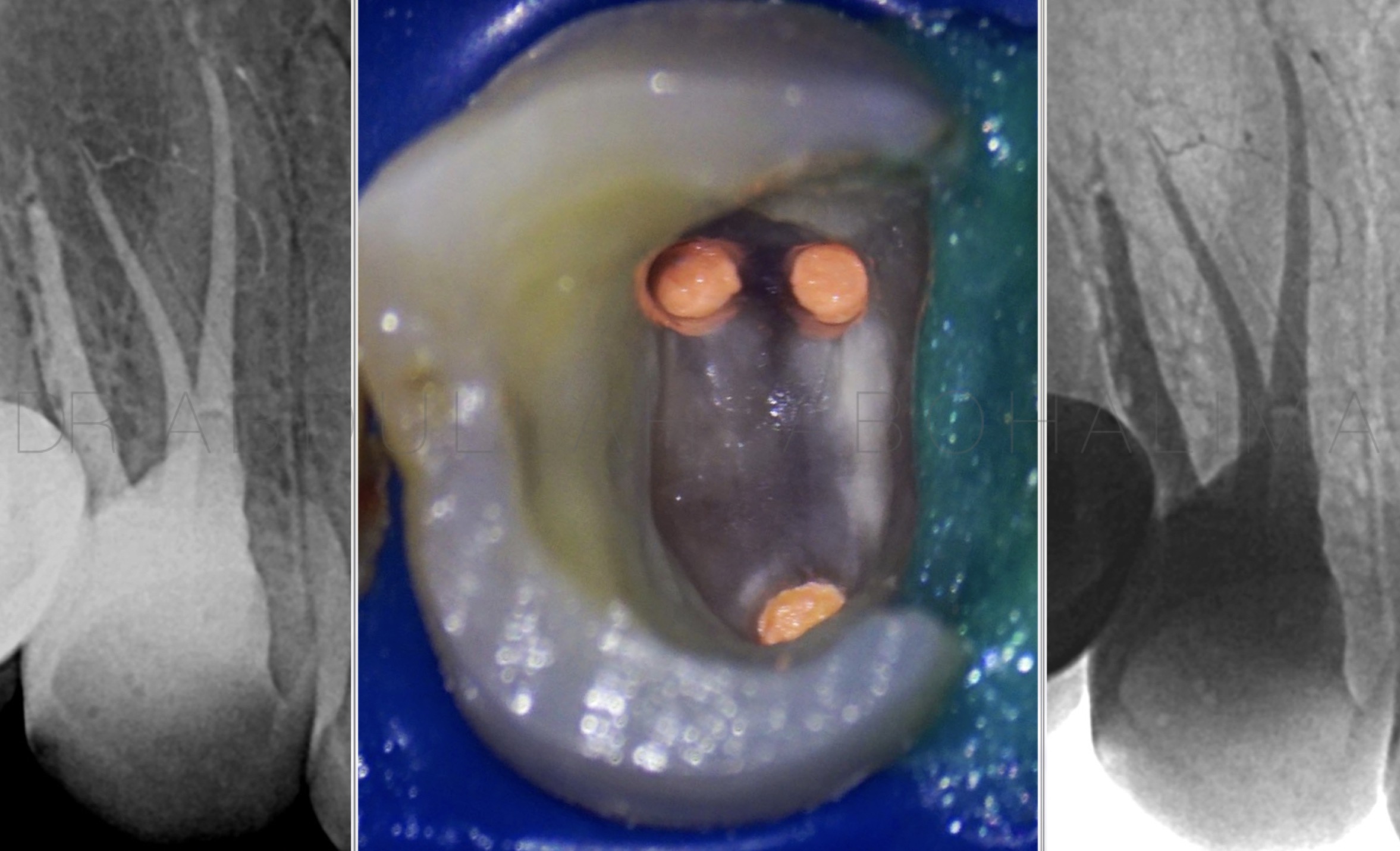
Fig. 17
Obturation with clean pulp chamber
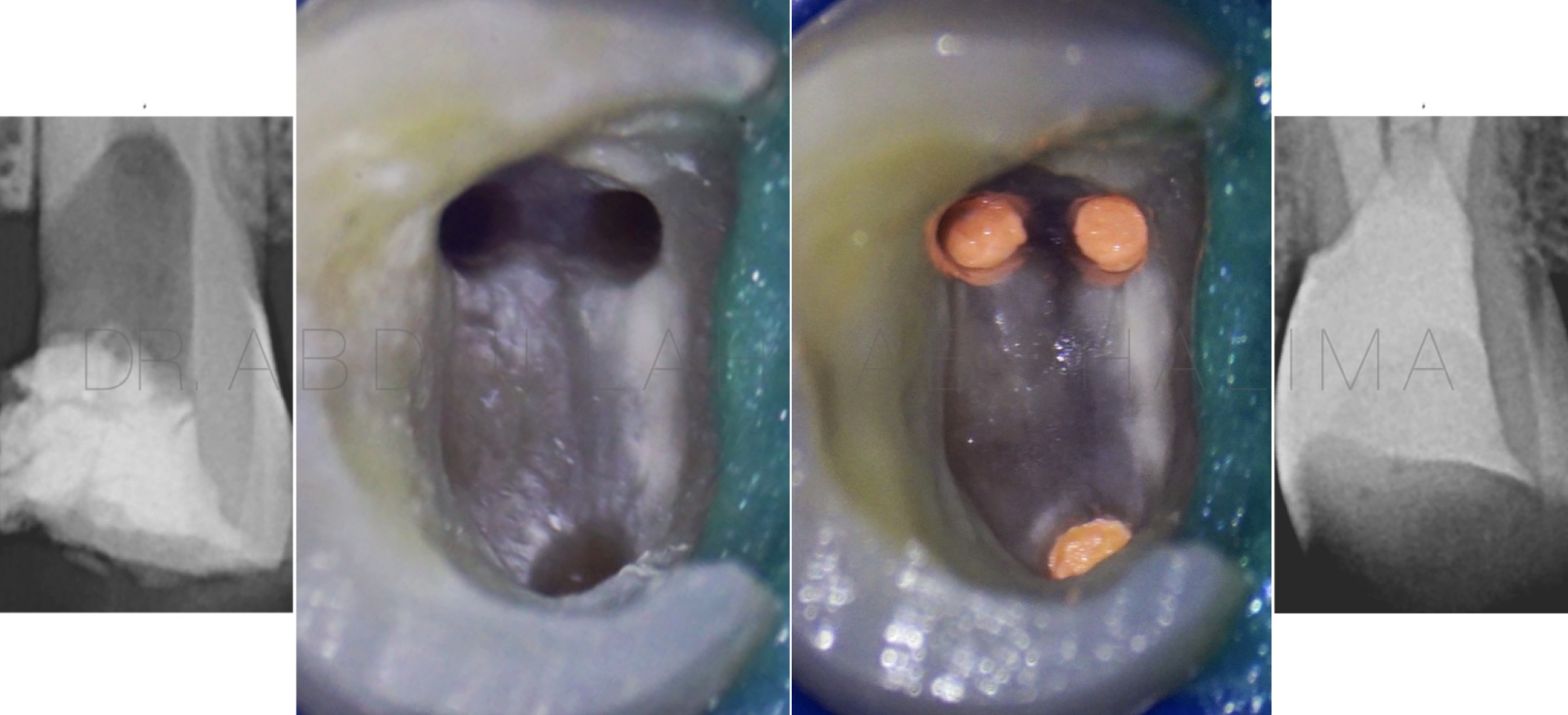
Fig. 18
Post-endo and deep marginal elevation
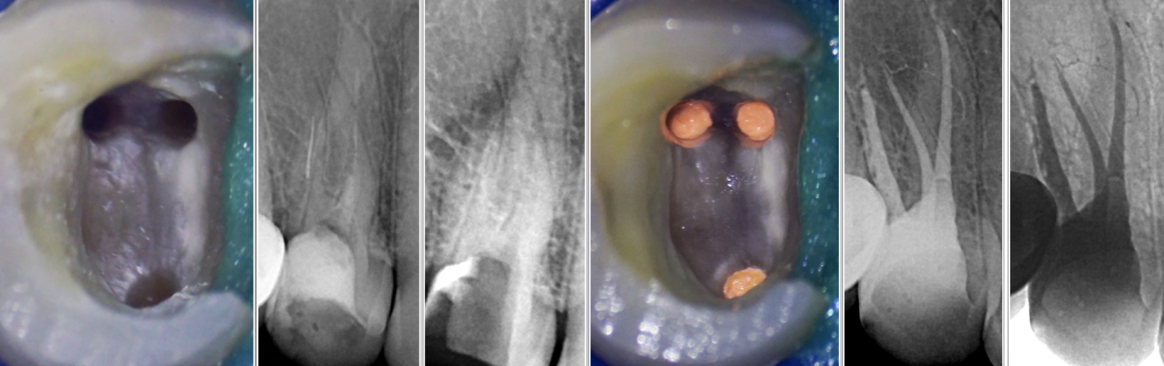
Fig. 19
Full case management
Full video of the procedure

Fig. 20
The author:
Dr Abdullah Abouhalima
-Endodontist at microscopia dental clinic
-Limited practice to microendodontics & Restorative
-Speaker and clinical practice coordinator in different courses done by Dr Hytham Abdelaziz educational center in alexandria
-won the award of best case of the year 2021 contest by SIE group “3rd place”
Conclusions
Maxillary premolars show many morphological variations in their pulpal anatomy and failure may be associated with the presence of infection due to incomplete elimination of microorganisms of the missed canals, so The key to successful outcome and predictable prognosis is to dedicate plenty of time in careful analysis of angulated radiographs with good knowledge of the anatomical variation of the canal system also Clinical examination with proper magnification to enhance the visualization of the pulp chamber and canal orifices is always needed to obtain optimum results .
Bibliography
1-Vertucci FJ. Root canal anatomy of the human permanent teeth. Oral Surg Oral Med Oral Pathol Oral Radiol Endod 1984: 58: 589–599.
2-Sert S, Bayirli GS. Evaluation of the root canal configurations of the mandibular and maxillary permanent teeth by gender in the Turkish population. J. Endod 2005: 30: 391–399.
3- F, Seelig A, Gillis R. Root canal morphology of the human maxillary second premolar. Oral Surg. 1974;38:456–464.
4- Sieraski SM, Taylor GT, Kohn RA. Identification and endodontic management of three-canalled maxillary premolars. J Endod. 1985;15:29–32.
5- Coutinho Filho T, La Cerda RS, Gurgel Filho ED, de Deus GA, Magalhães KM. The influence of the surgical operating microscope in locating the mesiolingual canal orifice: a laboratory analysis. Braz Oral Res. 2006;20:59–6