Mandibular Premolars: Teeth not to underestimate
12/07/2016
Mohamad Zaafrany
Warning: Undefined variable $post in /var/www/vhosts/styleitaliano-endodontics.org/endodontics.styleitaliano.org/wp-content/plugins/oxygen/component-framework/components/classes/code-block.class.php(133) : eval()'d code on line 2
Warning: Attempt to read property "ID" on null in /var/www/vhosts/styleitaliano-endodontics.org/endodontics.styleitaliano.org/wp-content/plugins/oxygen/component-framework/components/classes/code-block.class.php(133) : eval()'d code on line 2
The success of endodontic treatment depends on removing microorganisms from root canal system, three dimensionally sealing and the placement of a good coronal seal to prevent communications between oral cavity and the periradicular tissues (1-4).
The operator should be able to assess the endodontic case and know about tooth anatomy and the suspected anatomical variations one may face during treatment.
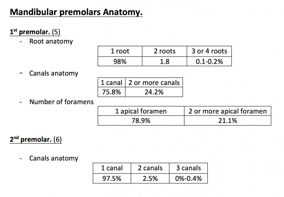
Fig. 1
These number and percentages show that these situations or these anatomies already exist but the clinician should be aware of any abnormalities or any deviation from these situations. Few studies report ethnic difference regarding canal numbers in mandibular premolars. Trope et al. found ethnic differences in their study of mandibular premolars: 2 or more canals present is 32.8% of patients while its 13.7% in Caucasian (7). Amos also reported Bifurcated canals 21% in African American and 16% in Caucasian (8). Caliskan et al. found 2 or more canals in around 36% in Turkish people (9). Zaatar et al. found 2 or more canals in 40% in Kuwaiti patients (10).
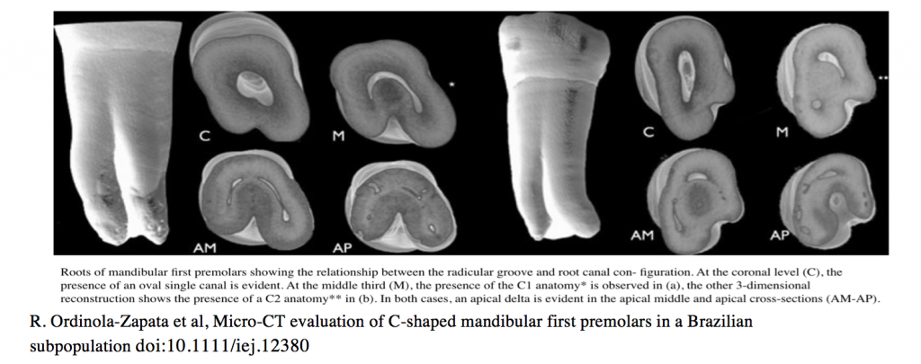
Fig. 2
C- shaped anatomy; Micro CT evaluation of mandibular first premolar in Brazilian subpopulation showed that C-shaped canal configuration of the root canal system was found in 67% of extracted mandibular premolars with radicular groove, C-shaped cross sections were more prevalent in the middle third with the presence of apical delta as a common feature in the apical third (11). In 3D morphological analysis of roots in mandibular first premolar in Chinese population It was found that the incidence of C-shaped anatomy was high when roots exhibit a radicular groove on its outer surfaces compared to roots without radicular groove(12). The presence of radicular groove on the outer root surface from the proximal aspect play an important role on having anatomical variations as sometimes we have one or two developmental grooves which exhibit 2 or more roots or root canals which is more common in mandibular first premolar as dentin deposition and calcification by age will show many anatomical variations and more challenges during root canal treatment in such cases (11). We can understand that the mandibular premolars can exhibit complex canal system morphology which if not considered during root canal treatment might lead to poor prognosis and subsequently failure of the treatment. Management: knowledge about root morphology and the nature of root canal systems and suspected deviation as well as rare conditions.
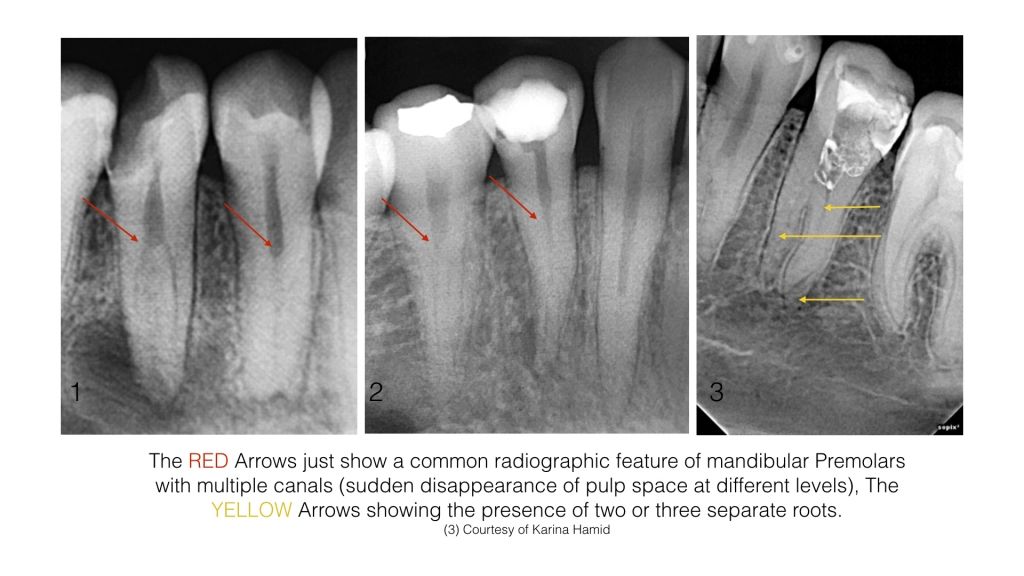
Fig. 3
Assessment to preoperative radiographs: preoperative radiograph is of great significance in determination of root canal configuration, canal numbers and curvatures. At least 2 radiograph should be taken preoperatively. One parallel and the other one with horizontally angulated cone as it will reveal more details about the anatomy were dealing with. One of the common radiographic feature in mandibular premolars with 2 or more canals is the absence of continuity of pulp space at different levels (cervically, mid root and apically). Tracing of periodontal ligament space around the tooth could diagnose multiple roots, bifurcated roots or aberrant anatomy. Tracing of sinus tracts if present could reveal presence of more than one root. Presence of radiolucent lesion un centered with the radiographic apex might be indication of presence of multiple roots of multiple POEs. Radiographic appearance of neighboring or contralateral teeth might be an indication of suspected aberrant root canal anatomy. CBCT is also a great tool for preoperative assessment (13).
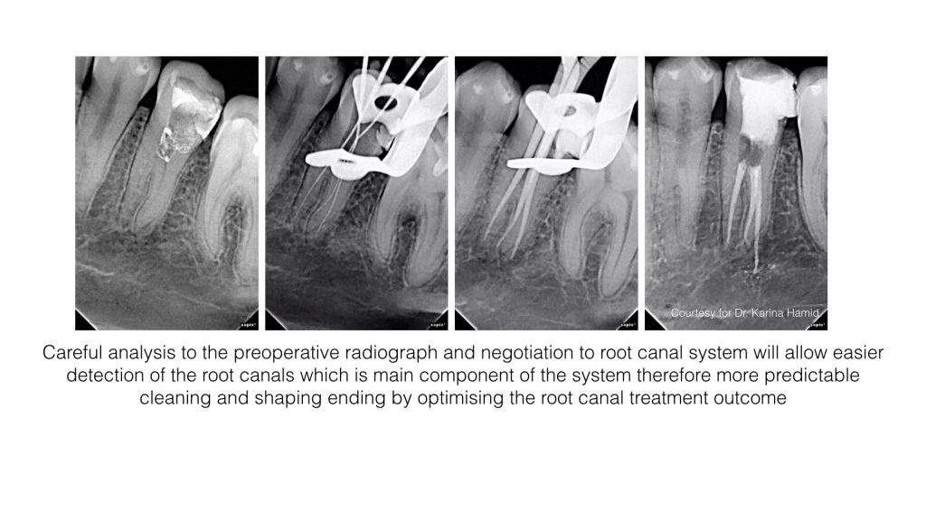
Fig. 4
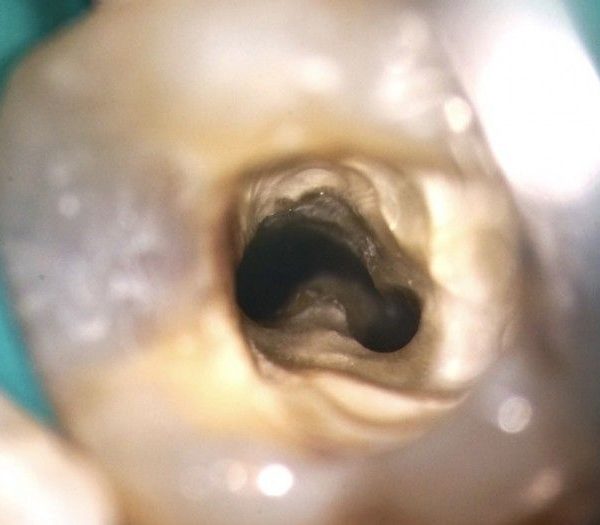
Access opening
Access opening of mandibular premolars usually is oval shape extended buccolingually with orifices located under B,L cusps.
Floor anatomy: visualization of floor anatomy and map will help in detection of extra canal orifices if exist. Either the use of long shafted bur mounted on low speed hand piece or ultrasonics could be used to detect hidden or extra canal orifices with less damaging to tooth structure (14). The use of Dental Operating Microscope with good illumination will significantly improve the access cavity preparation and allow easier detection of canal orifices (15).
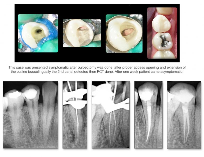
Fig. 5
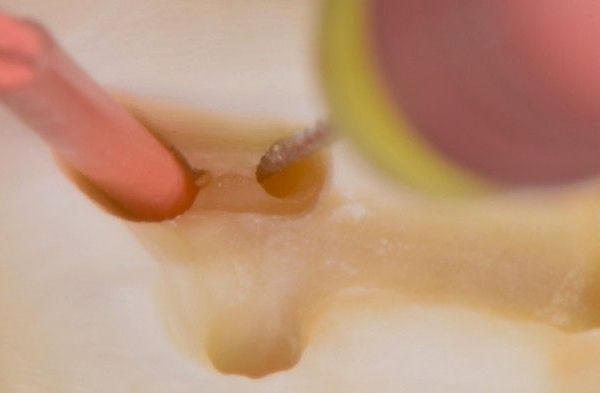
During Canals negotiation
The Use of EDTA during negotiation will allow easier movement of the files along canal walls (16). The operator should be aware of any stoppage for the negotiating files during the gentle movement on the canal walls which might be an indication of presence of another canal or split. Careful moving of the precurved files along the canal walls in multiple direction without any estimation the extra canal should be here or there will allow easier detection of the entrance if canal located in unsuspected place. (Precurving also should be tried at smooth and sharp angles).
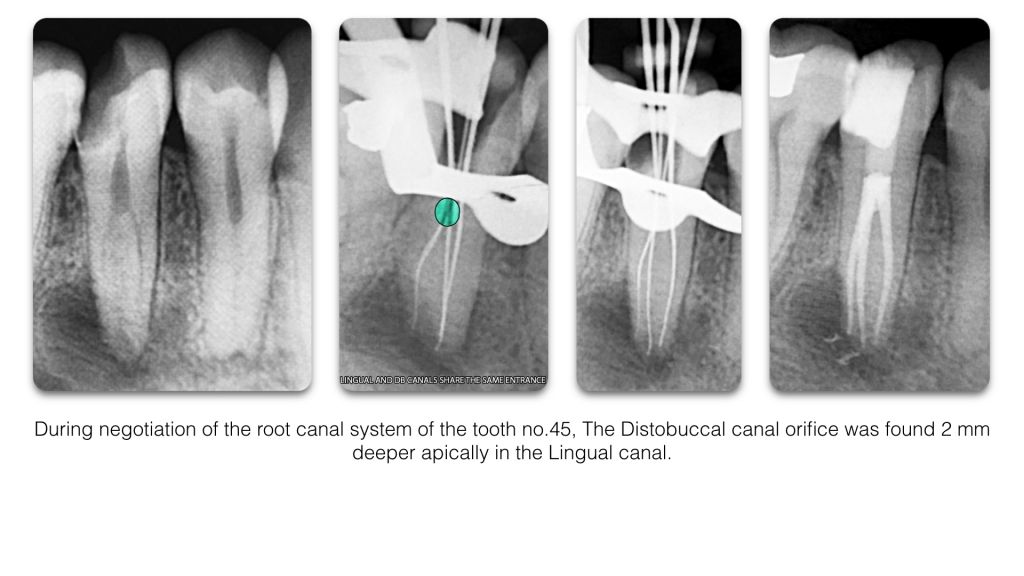
Fig. 6
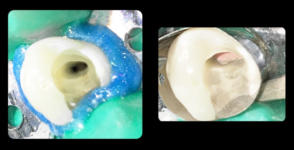
During Bio Mechanical Preparation
RCT is a dynamic process, Extra canals could be detected during any phase of treatment, The Operator should be aware of any slippage of rotary or reciprocating files in different direction during this process. Controlled Memory files is a greet tool for managing canal splits and deep divisions as its prependable so it can be used to mechanically clean and shape these canals without the need of sacrificing more coronal dentin (17).
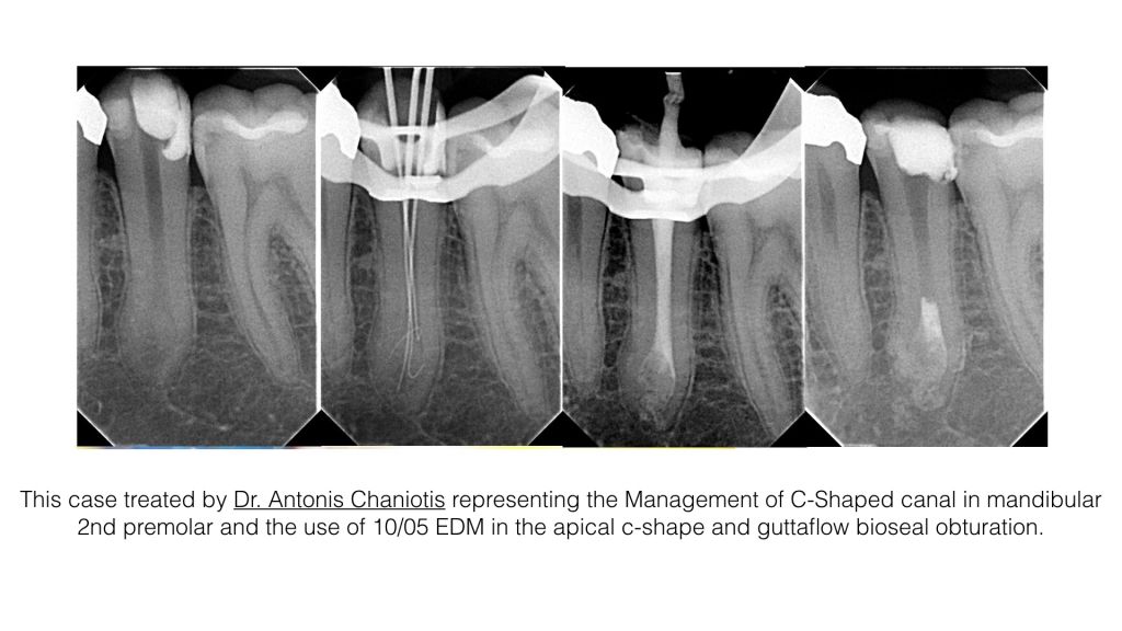
Fig. 7
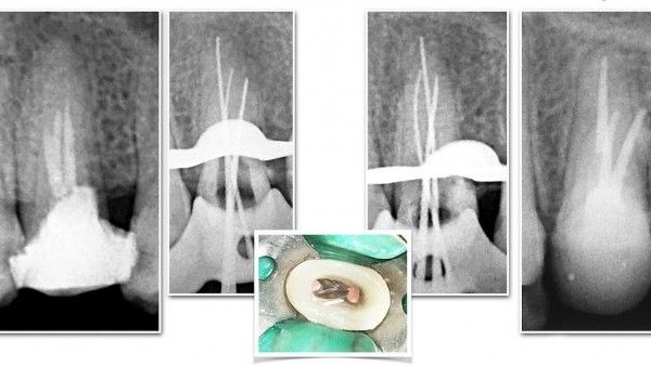
After Obturation
Careful reading to Radiographic image at that time is so important for inspection of seepage of obturation material in different direction could indicate the presence of extra canals (the operator should be able to differentiate between lateral or accessory canals and missed main canal).
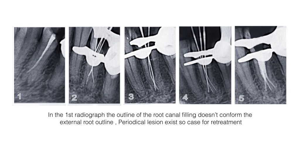
Fig. 8
Placement of root canal filling
If the root canal filling outline doesnt coincide with or conform the external root geometry or the periodontal ligament space, this might be an indication of missed anatomy.nIn these situations, Retreatment might be considered.
Conclusions
Mandibular premolars can exhibit complex and aberrant root canal anatomy which is of a great challenge to diagnose and to manage during root canal treatment.
Bibliography
- Orstavik D. Time-course and risk analyses of the development and healing of chronic apical periodontitis in man. IEJ 1996: 29: 150155.
- Friedman S. Considerations and concepts of case selection in the management of post-treatment endodontic disease (treatment failure). Endod Topics 2002: 1: 5478.
- Kirkevang LL, Horsted-Bindslev P. Technical aspects of treatment in relation to treatment outcome. Endod Topics 2002: 2: 89102.
- Vertucci FJ. Root canal morphology and its relationship to endodontic procedures. Endod Topics 2005: 10: 329.
- Cleghorne BM, Christie WH, Dong CC. The root and root canal morphology of the human mandibular first premolar: a literature review. J Endod 2007: 33: 509 516.
- Zillich R, Dowson J. Root canal morphology of mandibular first and second premolars. Oral Surg Oral Med Oral Pathol 1973: 36: 738744.
- Trope M, Elfenbein L, Tronstad L. Mandibular premolars with more than one root canal in different race groups. J Endod 1986;12:3435.
- Amos ER. Incidence of bifurcated root canals in mandibular bicuspids. J Am Dent Assoc 1955;50:70 1.
- Caliskan MK, Pehlivan Y, Sepetcioglu F, Turkun M, Tuncer SS. Root canal morphology of human permanent teeth in a Turkish population. J Endod 1995;21:200 4.
- Zaatar EI, al-Kandari AM, Alhomaidah S, al-Yasin IM. Frequency of endodontic treatment in Kuwait: radiographic evaluation of 846 endodontically treated teeth. J Endod 1997;23:453 6.
- R. Ordinola-Zapata et al. Micro-CT evaluation of C-shaped mandibular first premolars in a Brazilian subpopulation doi:10.1111/iej.12380
- B. Fan et al. Three-dimensional morphological analysis of C-shaped canals in mandibular first premolars in a Chinese population iej doi:10.1111/j.1365-2591.2012.02070.x
- William C. Scarfe et al, Use of Cone Beam Computed Tomography in Endodontics,International Journal of Dentistry Volume 2009, Article ID 634567, 20 pages doi:10.1155/2009/634567
- Arnaldo Castellucci, Endodontics text book, Access Cavity and Endodontic Anatomy P.244.
- John S. Mamoun ;A rationale for the use of high-powered magnification or microscopes in general dentistry; General Dentistry, January/February 2009 ;Pg. 18-26.
- Putzer P, Hoy L, Gunay H. Highly concentrated EDTA gel improves cleaning efficiency of root canal preparation in vitro. Clin Oral Investig 2008; 12:319-24.
- Journal of Natural Science, Biology and Medicine, DOI: 10.4130/0976-9668.160032.