Management of the complex anatomy of a lower first premolar
03/11/2020
Mohamed Milad Ganawo
Warning: Undefined variable $post in /var/www/vhosts/styleitaliano-endodontics.org/endodontics.styleitaliano.org/wp-content/plugins/oxygen/component-framework/components/classes/code-block.class.php(133) : eval()'d code on line 2
Warning: Attempt to read property "ID" on null in /var/www/vhosts/styleitaliano-endodontics.org/endodontics.styleitaliano.org/wp-content/plugins/oxygen/component-framework/components/classes/code-block.class.php(133) : eval()'d code on line 2
An accurate diagnosis of the morphology of the root canal system is crucial in order to obtain a successful root canal treatment.
The prevalence of three canals with three separate foramina in mandibular premolars is very rare like in the case that will be shown in this report: one canal leaves the pulp chamber, and then divides into three separate canals with three separate apical foramina - additional type (Vertucci )Type (1‐3).
This clinical case will describe the retreatment of a lower first premolar with three canals

Fig. 1
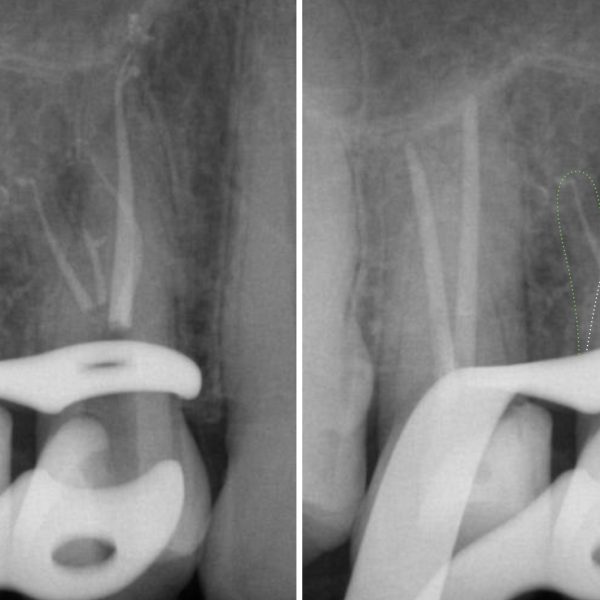
A 35 years old female was referred for retreatment of tooth 44, which was symptomatic. The initial x-ray revealed a periapical lesion around root in the presence of a poor endodontic therapy, as shown in figure .
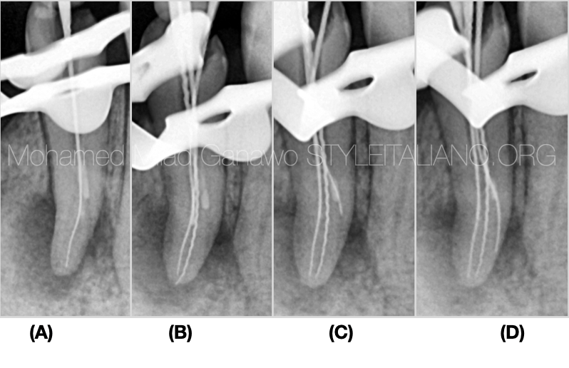
Fig. 2
A- insertion of a #10 K-file in the main buccal canal and glide path to full length to achieve patency
B- insertion of another manual file in the second buccal canal, confirming another buccal split
C- an attempt to negotiate the lingual split to gain glide path while 2 files in buccals .
D- reaching full length in lingual split .
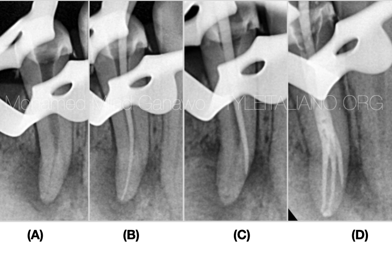
Fig. 3
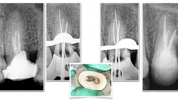
A- x-ray shows entire removal of old gutta percha
B- cone fit in the buccal canal
C- cone fit in the lingual split
D- cone fit in all canals
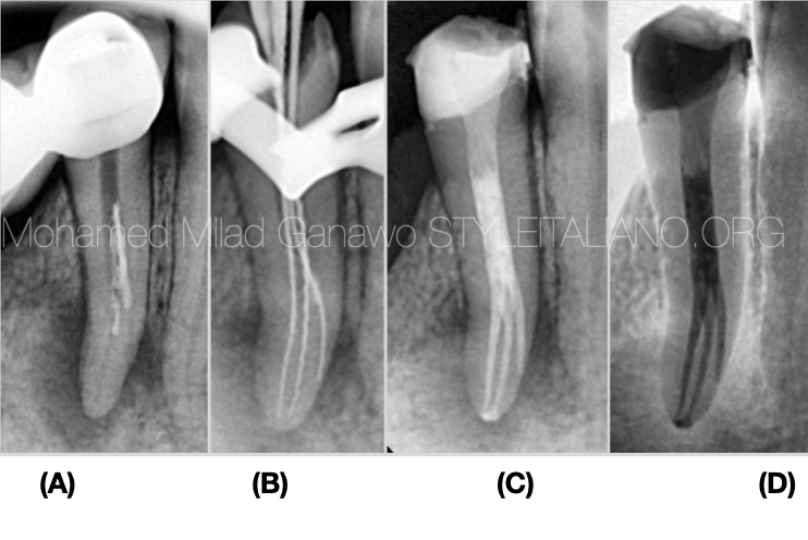
Fig. 4
Summary of the steps of the retreatment, starting from the radiographic interpretation and ending up with obturation.
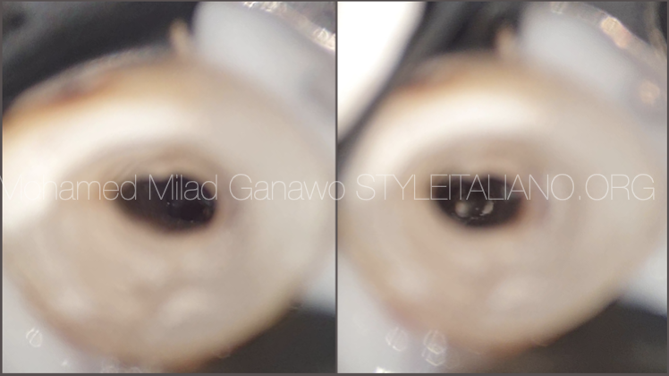
Fig. 5
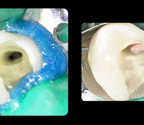
Intra oral Microscopic photos show conservative access before and after obturation .
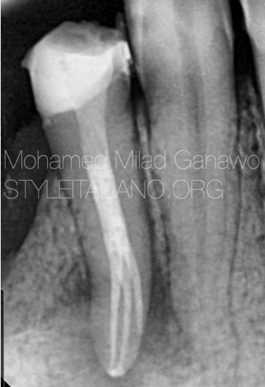
Fig. 6
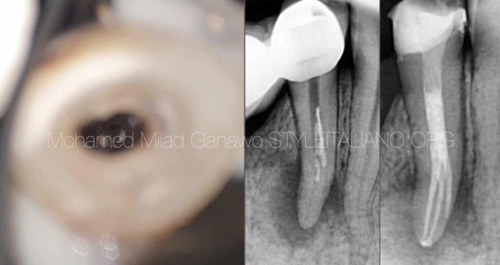
The final x-ray shows the obturation of three canals followed by good coronal seal. The tooth is planned to be crowned soon.
Conclusions
Mandibular premolars can present a complex root canal morphology, which if not considered during the diagnosis and treatment, may lead to post-treatment complications or failure.
Use of an operating microscope or loop can promote the visualization of the pulp chamber and extra canal orifices.
Therefore, the understanding of abnormal anatomy and variations is necessary to guarantee predictable result and successful treatment.
Bibliography
Vertucci FJ. Root canal morphology of mandibular premolars. J Am Dent Assoc
1978;97:47-50.
Cleghorn BM, Christie WH, Dong CC. The root and root canal morphology of the human mandibular first premolar: a literature review. J Endod. 2007 May;33(5):509-16
J. Kottoor, D. Albuquerque, N. Velmurugan, and J. Kuruvilla, “Root anatomy and root canal configuration of human permanent mandibular premolars: a systematic review,” Anatomy Research International, vol. 2013, Article ID 254250, 14 pages, 2013.
Vertucci FJ. Root canal anatomy of the human permanent teeth. Oral Surg Oral Med Oral Pathol 1984;58:589 –99.
Rhodes JS. A case of unusual anatomy: a mandibular second premolar with four canals. Int Endod J 2001;34:645– 8. 15. D
England MC Jr, Hartwell GR, Lance JR. Detection and treatment of multiple canals in mandibular premolars. J Endod 1991;17:174 – 8.
Nallapati S. Three canal mandibular first and second premolars: a treatment approach. J Endod. 2005 Jun;31(6):474-6.