Unusual anatomy of a maxillary first molar with two palatal canals
18/11/2024
Mohamed Milad Ganawo
Warning: Undefined variable $post in /home/styleendo/htdocs/styleitaliano-endodontics.org/wp-content/plugins/oxygen/component-framework/components/classes/code-block.class.php(133) : eval()'d code on line 2
Warning: Attempt to read property "ID" on null in /home/styleendo/htdocs/styleitaliano-endodontics.org/wp-content/plugins/oxygen/component-framework/components/classes/code-block.class.php(133) : eval()'d code on line 2
A thorough understanding of root and root canal anatomy is imperative for successful root canal treatment. The chemo-mechanical preparation of root canal systems aims to diminish intra-canal bacterial populations, facilitating periapical tissue healing (1-3). However, variability in internal anatomy and overlooked canals are common factors contributing to endodontic treatment failures, particularly in maxillary molars, which often exhibit three roots with diverse canal configurations (4). Among maxillary molars, the mesiobuccal root presents the greatest anatomical variability, with over half of cases featuring two or more canals. Conversely, the palatal root typically demonstrates minimal variability, with a single canal and apical foramen in 99% and a single apical foramen in 98.8% of cases (4,5). Despite the overall low prevalence of anatomical variations in the palatal canal of maxillary molars (<2%), it can reach up to 33% in maxillary first molars and up to 14% in maxillary second molars in certain ethnic groups (1). Neglecting missed canals or incomplete obturations can present significant challenges in endodontic treatment and lead to subsequent failure (5,6).
Detecting anatomical variations beyond the standard 1-1 configuration can be challenging on two-dimensional periapical radiographs. Therefore, precise interpretation of radiographs is crucial for treatment planning, with advanced imaging modalities like CBCT offering valuable insights into complex cases. Nevertheless, experienced endodontic clinicians may overlook a bifurcated palatal canal if they are not aware of anatomical variations. Therefore, utilizing proper magnification and meticulous access cavity preparation is essential for performing successful endodontic treatment.

Fig. 1
This is a pre operative radiograph of tooth 16 shows a composite fill placed 6 months ago for 30 years old patient.
The patient approached the clinic with severe pain related to the tooth
Always radiographic interpretation is crucial as we can notice the apical part of palatal root is flat rounded which may indicate complex apical anatomy
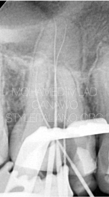
Fig. 2
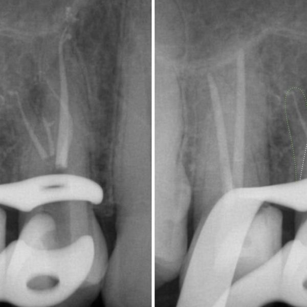
This figure shows working length confirming K files following the right path especially palatally.
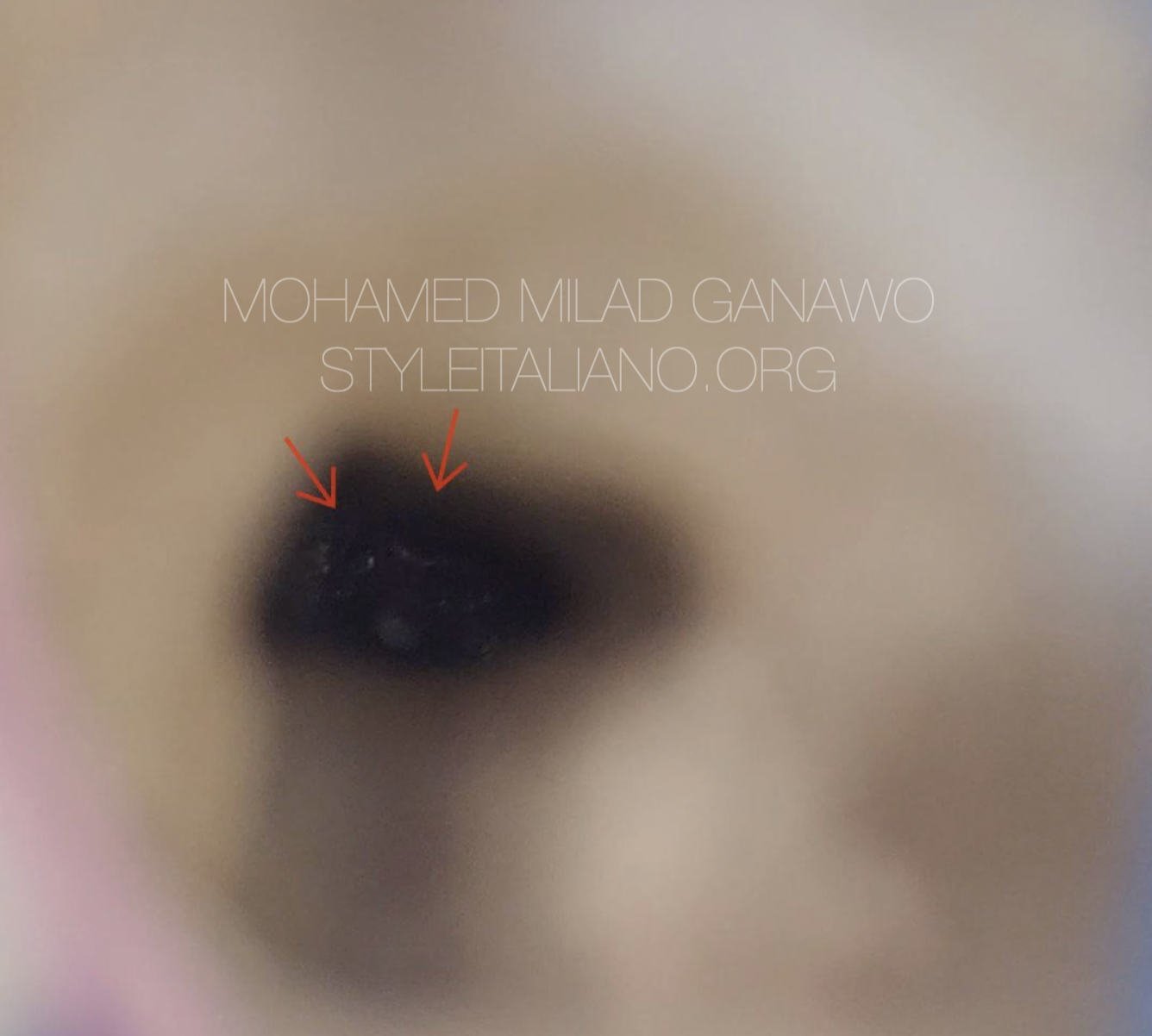
Fig. 3
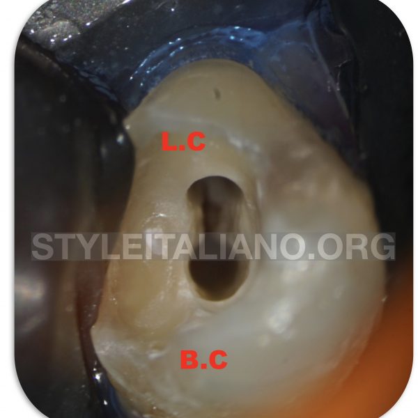
This zoomed in microscopic photo shows the orifices of two PALATAL splits.

Fig. 4
This figure shows cone fitting where two gutta percha placed in palatal canal due to presence of two canals palatally.
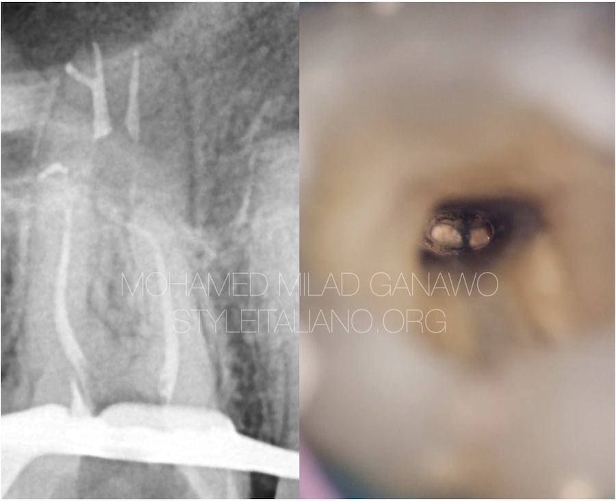
Fig. 5
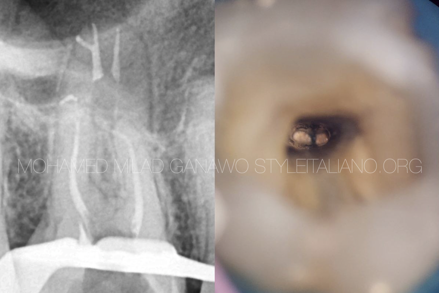
This figure shows stages of down packing in palatal, I could manage to pack deeper apically, about 3mm away from the apex by aid of microscope of course and managed to take a clear microscopic shot as shown in the photo.
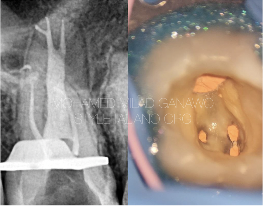
Fig. 6
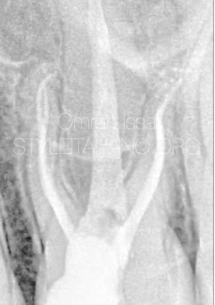
This figure shows both radiograph and intra oral photo of final obturation
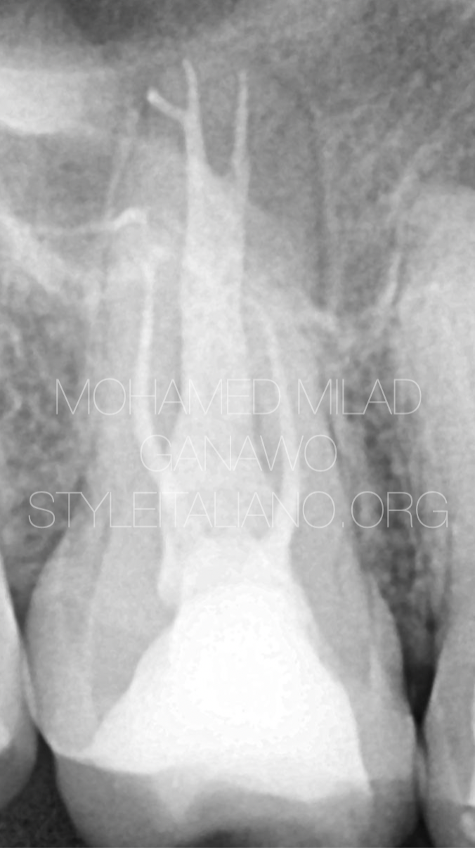
Fig. 7
post operative x.ray

Fig. 8
This figure summarises the step from cone fitting till obturation followed by permanent core placement
Video of the procedure
Conclusions
This article describes a non-surgical treatment of a maxillary first molar with are palatal canal configuration, highlighting the importance of addressing anatomical complexities for optimal treatment outcomes.
In conclusion, understanding the diverse anatomical variations within root canal systems is paramount for successful endodontic therapy. Meticulous examination and thoughtful treatment planning are imperative, especially in cases of rare anatomical anomalies like maxillary molars.
Bibliography
Nosrat A, Verma P, Hicks ML, Schneider SC, Behnia A, Azim AA. Variations of Palatal Canal Morphology in Maxillary Molars: A Case Series and Literature Review. J Endod. 2017 Nov;43(11):1888-1896. doi: 10.1016/j.joen.2017.04.006. Epub 2017 Jun 30. PMID: 28673493.
Ng YL, Mann V, Rahbaran S, et al. Outcome of primary root canal treatment: systematic review of the literature–Part 2. Influence of clinical factors. Int Endod J 2008; 41:6–31.
Azim AA, Griggs JA, Huang GT. The Tennessee study: factors affecting treatment outcome and healing time following nonsurgical root canal treatment. Int Endod J 2016;49:6–16.
B. M. Cleghorn, W. H. Christie, and C. C. Dong, “Root and root canal morphology of the human permanent maxillary first molar: a literature review,” Journal of Endodontics, vol. 32, no. 9, pp. 813–821, 2006.
S. Holderrieth and C. R. Gernhardt, “Maxillary molars with morphologic variations of the palatal root canals: a report of four cases,” Journal of Endodontics, vol. 35, no. 7, pp. 1060– 1065, 2009.


