Ultrasonics in Endodontics: Part 1
10/06/2016
Enrico Cassai
Warning: Undefined variable $post in /home/styleendo/htdocs/styleitaliano-endodontics.org/wp-content/plugins/oxygen/component-framework/components/classes/code-block.class.php(133) : eval()'d code on line 2
Warning: Attempt to read property "ID" on null in /home/styleendo/htdocs/styleitaliano-endodontics.org/wp-content/plugins/oxygen/component-framework/components/classes/code-block.class.php(133) : eval()'d code on line 2
Around the end of 1950 the ultrasonic technology has had a wide spread not only in the field of hygiene and periodontology, but also in endodontics.
Nowadays the operators thoroughly know the benefits of the use of ultrasound in the endodontic field, where a minimally invasive approach is always needed, together with magnification, in order to correctly manage tissues and have a better visibility.
The purpose of this article is to illustrate the history and progress that the ultrasonic technology and related techniques have had over the years, to classify the various ultrasonic tips available on the market and to evaluate the different clinical applications.
When ultrasound or ultrasonic instrumentation was born, it was primarily intended for the realization of cavity preparations with the use of an abrasive slurry. Despite receiving favorable opinions, the technique has never been widely applied as it had to compete with a much quicker and effective technique: the high-speed handpiece.
Only in 1955 Zinner introduced ultrasound technology in periodontology suggesting its use for removal of debris from the tooth surface. By 1960 Johnson and Wilson improved the ultrasound technique as long as it became an established tool in the periodontal field in the removal of tartar and plaque.
The first to introduce the concept of ultrasound in endodontics was Richman, around 1957.
In 1970 the ultrasonic technology found a field of application in treatment of TMJ dysfunction and in measurement of translatory movements of the condyle during motion.
In 1976 Martin published his first paper on increasing the efficacy of bactericidal root canal irrigation if associated with ultrasonic technique. In the same year Bertrand et al published an article about what is presumed to be the first use of a modified ultrasonic tip for a retropreparation during an apicoectomy.
In 1980, Martin et al. discovered an increase in the cutting capacity of a k-files, activated by ultrasound and emphasized its potential for use in root canal preparation before filling. In 1984-85 Martin and Cunningham coined the term `endosonic´ to define the synergistic action of instrumentation and disinfection the root canal system by ultrasonics.
Around 1990s, after the introduction of the first ultrasonic tips by Gary Carr, the focus shifted to the use and possible consequences of ultrasonic root-end preparations during apicoectomy.
The introduction of the piezoelectric device and numerous drawings of ultrasonic tips after 1990 has allowed clinicians to remove dentin or other dental materials in a very controlled and precise manner, using tips that are often approximately the same size as a root canal or smaller.
In the same time were introduced in the market tips designed to deliver vibrational energy in a focused manner, without the intention to cut tooth structure.
DEVICES FOR ULTRASOUND PRODUCTION
Ultrasound is sound energy with a frequency above the range of human hearing, which is 20 kHz. The frequencies originally employed in ultrasonic units ranged between 25 and 40 kHz; later on, low-frequency ultrasonic handpieces operating from 1 to 8 kHz were developed, to produce lower shear stresses, in order to reduce risk for alteration to the tooth surface.
There are two basic methods for ultrasound production:
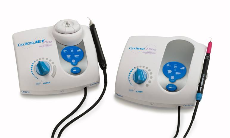
Fig. 1
MAGNETOSTRICTION
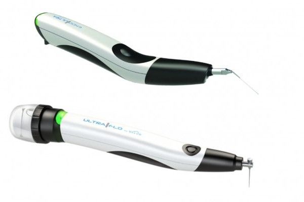
Magnetostriction converts electromagnetic energy into mechanical energy. A stack of magnetostrictive metal strips in a handpiece is subjected to a standing and alternating magnetic field, as a result of which vibrations are produced. A magnetostrictive device creates more of an elliptical motion, which is not ideal for either surgical or nonsurgical endodontic use and have also the disadvantage that the stack generates heat, thus requiring adequate cooling.
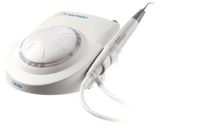
Fig. 2
PIEZOELETRIC
Based on the piezoelectric principle, which uses a crystal that changes dimension when an electrical charge is applied. Deformation of this crystal is converted into mechanical oscillation without heat production.
Piezoelectric units have some advantages with respect to earlier magnetostrictive units as they offer more cycles per second, 40 versus 24 kHz. The tips of these units work in a linear, back-and-forth, piston-like motion, which is ideal for endodontic applications.
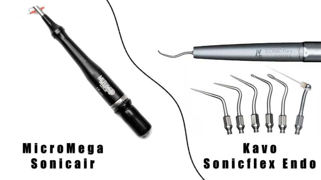
Fig. 3
Besides ultrasonic devices, also sonic instruments are used in endodontics, with a frequency from 1500 to 6000 hz (Micro-Mega Sonic Air, KaVo SonicFlex Endo) for detection and preparation of the canal orifices, for removal of soft materials and for canal preparation with continuous irrigation.
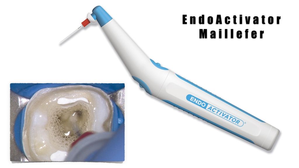
Fig. 4
Another sonic device (EndoActivator) is used to activate intra-canal irrigants during endodontic treatment.
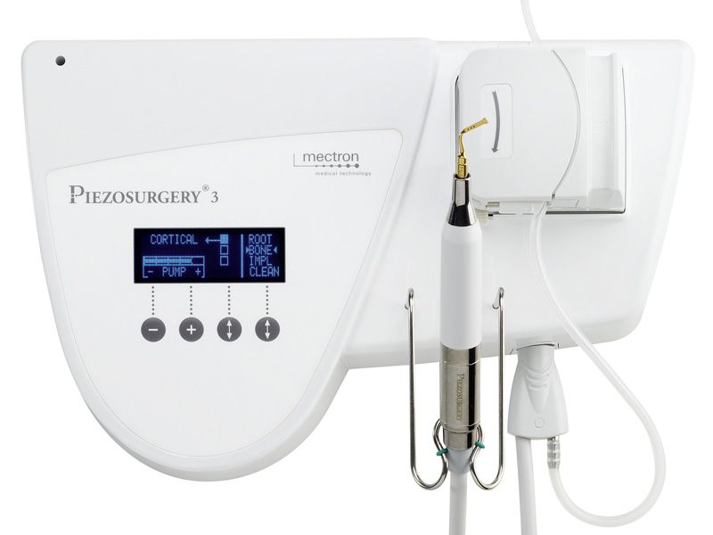
Fig. 5
Piezosurgery devices have been developed for bone surgery, and have found applications in endodontic surgery, for osteotomy, root-end resection and retropreparation. A recent literature review (Abella JOE 2014) found no published data on the effect of piezosurgery on the outcomes of endodontic surgery; no study has evaluated the effect of piezosurgery on root-end resection, and only one has investigated root-end morphology after retrograde cavity preparation using piezosurgery. From anecdotical reports, it looks that piezosurgery results in less bleeding, less swelling and less postoperative pain.
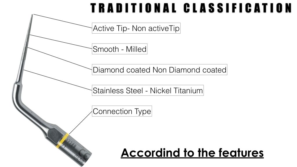
Fig. 6
STANDARD CLASSIFICATION FEATURES
To this day, a variety of ultrasonic tips have been introduced on the market, both for orthograde and retrograde RCT.
All of these can normally be used on different piezoelectric ultrasonic devices, but one must check that the thread pattern on the unit and the tip is compatible (E-thread and an S-thread are currently used). The tips are also manufactured from a range of metal alloys, such as stainless-steel and titanium alloys, and can be coated with an abrasive such as diamond or zirconium nitride in order to increase the cutting efficiency of the tip.
Many of the tips incorporate a built-in water port so that debris can be washed away and cooling can take place if desired.
As a result of the variety of tips available, there is an appropriate tip design for virtually every step of endodontic treatment, from access to obturation, each to be used in the recommended power setting range.
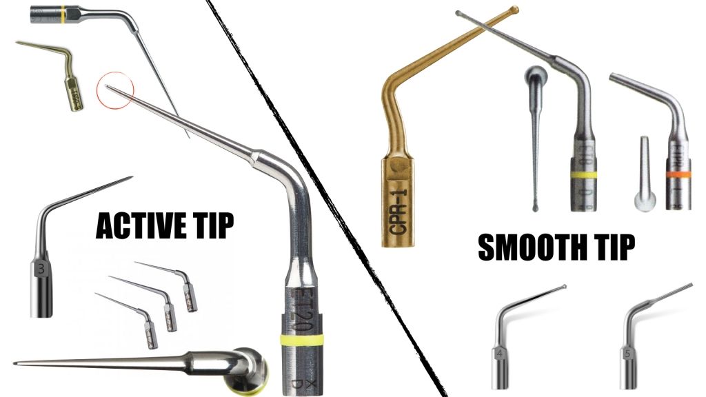
Fig. 7
ACTIVE TIP / SMOOTH TIP
The active tip makes an extremely effective tool when used for fiber post and obstacles present in pulp chamber removal and in all the circumstances in which there is a good view and a low risk of creating iatrogenic injury.
The smooth tip is useful in the cases in which cutting action is not necessary on the tip but is exerted by the body of the instrument. This is useful in pulp stone and intracanal obstructions (such as post) removal.
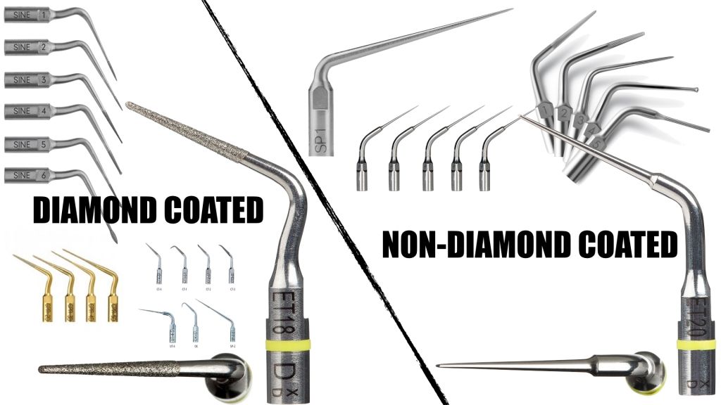
Fig. 8
DIAMOND COATED / NON DIAMOND COATED
The diamond coating of an ultrasonic tip makes it much more effective and abrasive.
This kind of tip, especially if used without irrigation, tend to get soiled with dentinal debris thus loosing cutting efficacy. Furthermore, the diamond part is likely to, overtime, get consumed and to detach from its site.
Surface coatings on ultrasonic tips are intended to increase efficiency and durability; diamond-coated tips have been shown to require less time than stainless-steel tips or zirconium nitride tips to cut similar preparations.
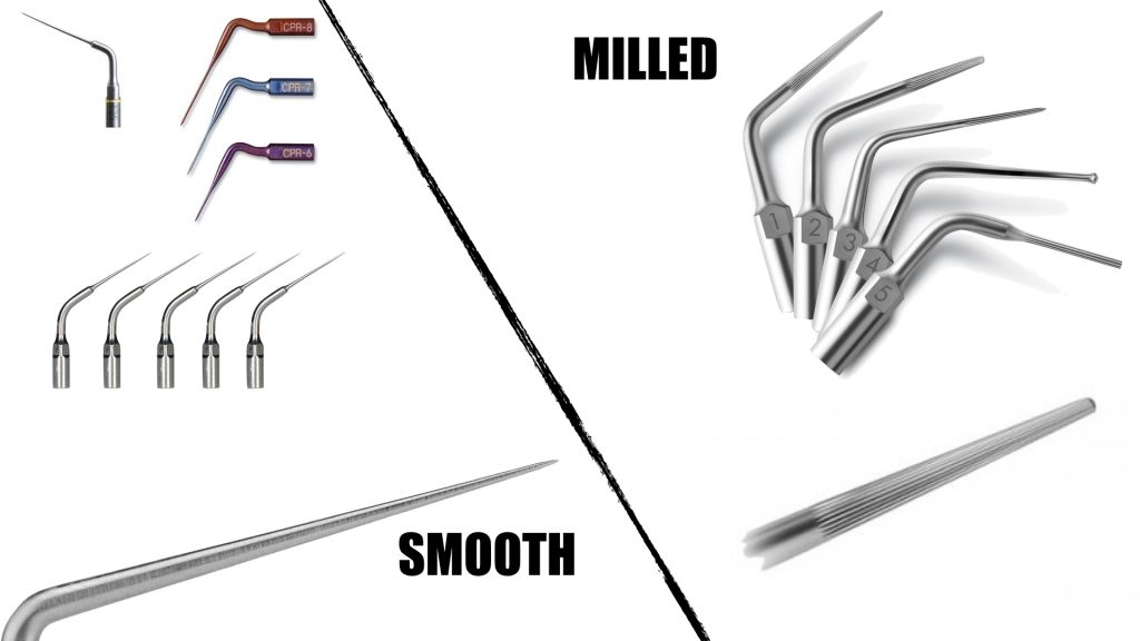
Fig. 9
SMOOTH / MILLED
Among the uncoated diamond tips we can find those with a smooth surface and those milled surface.
The tips with milled surface have a higher lateral cutting ability and longer lasting even compared to the coated diamond tips.
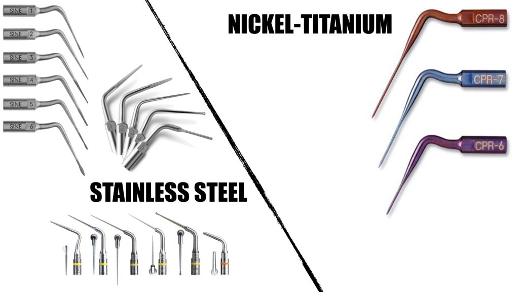
Fig. 10
STAINLESS STEEL / NICKEL TITANIUM
The ultrasonic tips Nickel-Titanium are much more fragile than stainless-steel, are used to work at a low intensity within the channel. They should be activated when in contact with the canal walls otherwise they tend to fracture.
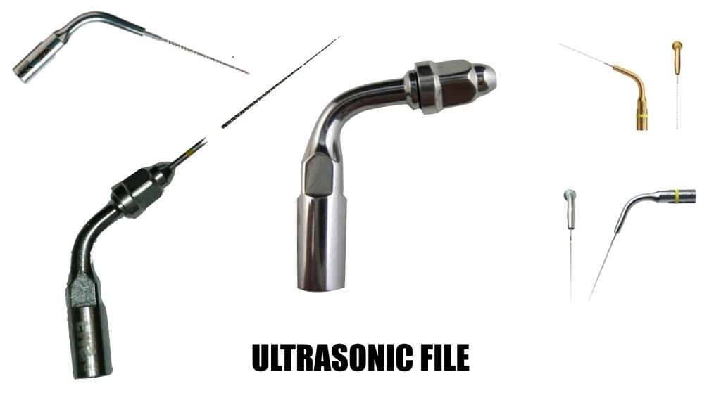
Fig. 11
ULTRASONIC FILES
The endodontic instruments as K-files mounted on Endochuck or as independent files inserts may be used for:
- Activation of irrigants to increase its effectiveness
- Removal of obstacles intracanal, especially positioned in the medium-apical third canal (like broken files)
- Allow the achieving and positioning of MTA in the third apical canal
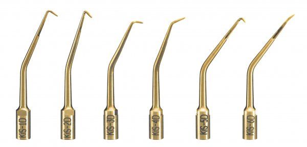
Fig. 12
SURGICAL ENDODONTICS TIPS
Ultrasonic technique has changed completely the endodontic surgery, opening the way towards endodontic microsurgery, along with magnification and coaxial illumination. Specially designed ultrasonic tips are used for root-end preparation, allowing an accurate, centered and deep preparation of the canal.
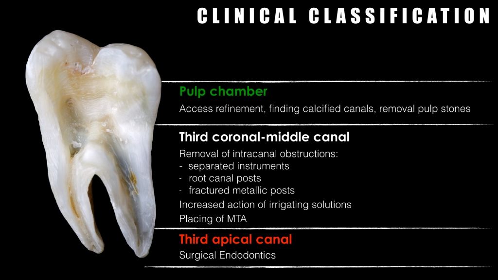
Fig. 13
As for the root canal instruments, also for the use of the ultrasonic instruments, it is appropriate to make a clinical classification according to the tooth section in which they must operate.
Therefore the choice of each tip type (active/non active, smooth/milled), the intensity of use, the use with or without water will be in close relationship with the area in which it will have to work.
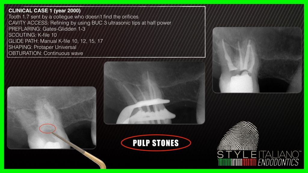
Fig. 14
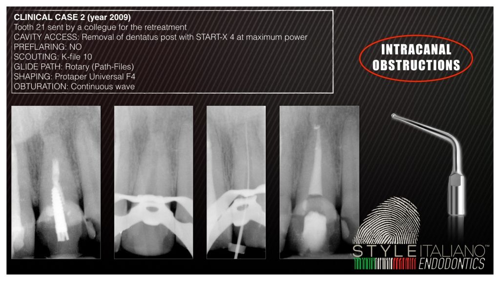
Fig. 15
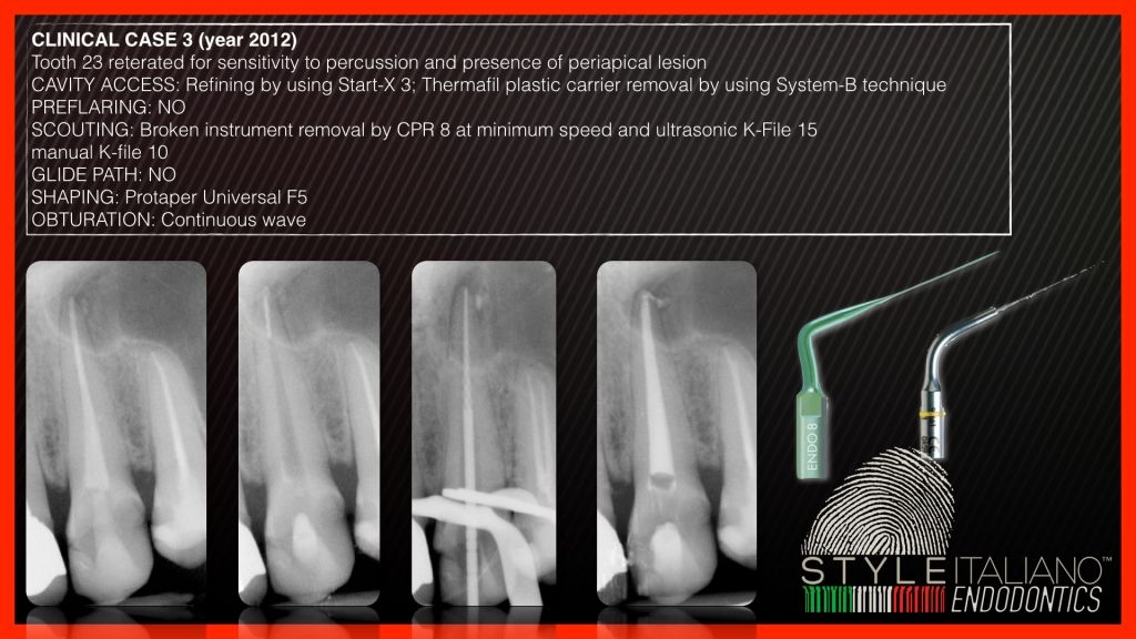
Fig. 16
Conclusions
It is interesting to consider that only 20 years ago, the rationale for the use of ultrasonics in endodontics was considered controversial, as it is now an integral tool during most tasks and challenges of root canal therapy.
In the next article, Ultrasonics in Endodontics Part 2, we will look at the different tips available on the market in relation to the site application and the clinical purpose.
Bibliography
- Catina MC. Ultrasonic energy:a possible dental application.Preliminary report of an ultrasonic cutting method. Ann Dent 1953.
- Postle HH. Ultrasonic cavity preparation. J Prosthet Dent 1958.
- Balamuth L. The application of ultrasonic energy in the dental field. In: Brown B, Gordon D, eds. Ultrasonic techniques in biology and medicine. London: Ilife; 1967:194205.
- Oman CR, Applebaum E. Ultrasonic cavity preparation II. Progress report. J Am Dent Assoc 1954;50:4147.
- NielsenAG, Richards JR, Walcott RB. Ultrasonic dental cutting instrument. J Am Dent Assoc 1955;50:3929.
- Street EV. Critical evaluation of ultrasonics in dentistry. J Prostate Dent 1959;9:32 41.
- Zinner DD. Recent ultrasonic dental studies including periodontia, without the use of an abrasive. J Dent Res 1955;34:7489.
- Johnson WN, Wilson JR. Application of the ultrasonic dental unit to scaling procedures. J Periodontol 1957;28:264 71.
- Richman RJ. The use of ultrasonics in root canal therapy and root resection. Med Dent J 1957;12:12 8.
- Martin H. Ultrasonic disinfection of the root canal. Oral Surg Oral Med Oral Pathol 1976;42:929.
- Martin H, Cunningham WT, Norris JP, Cotton WR. Ultrasonic versus hand filing of dentin: a quantitative study. Oral Surg Oral Med Oral Pathol 1980;49:79 81.
- Martin H, Cunningham WT, Norris JP. A quantitative comparison of the ability of diamond and K-type files to remove dentin. Oral Surg Oral Med Oral Pathol 1980;50:566 8.
- Martin H, Cunningham W. Endosonic endodontics: the ultrasonic synergistic system. Int Dent J 1984;34:198 203.
- Martin H, Cunningham W. Endosonics: the ultrasonic synergistic system of endodontics. Endod Dent Traumatol 1985;1:201 6.
- Stock CJR. Current status of the use of ultrasound in endodontics. Int Dent J 1991;41:175 82.


