The curve you don’t expect
22/03/2025
Massimo Giovarruscio
Warning: Undefined variable $post in /home/styleendo/htdocs/styleitaliano-endodontics.org/wp-content/plugins/oxygen/component-framework/components/classes/code-block.class.php(133) : eval()'d code on line 2
Warning: Attempt to read property "ID" on null in /home/styleendo/htdocs/styleitaliano-endodontics.org/wp-content/plugins/oxygen/component-framework/components/classes/code-block.class.php(133) : eval()'d code on line 2
Patient has been referred due to symptoms on Upper Right Second Premolar.
The tooth is tender to percussion and does not respond positively to vital test.
Diagnosis is apical periodontitis and root canal is required.
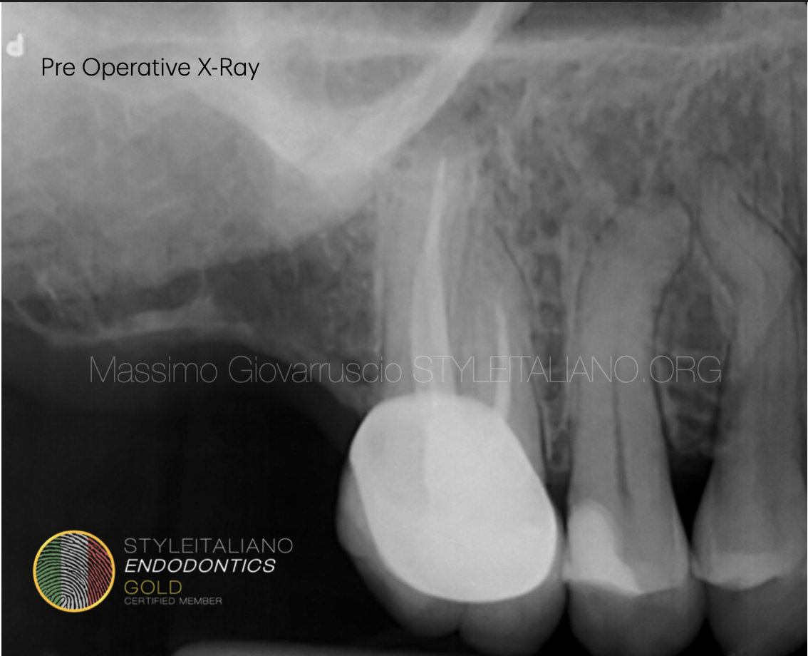
Fig. 1
X-Ray shows a mesial apical canal curvature.
Root Canal Treatment of an Upper Premolar with an Apical Curvature
Root canal treatment of an upper premolar with an apical curvature presents unique challenges due to the anatomy of the tooth. The procedure involves the following key steps:
1. Diagnosis and Treatment Planning
A clinical examination and periapical radiograph (or CBCT if necessary) help assess the degree and direction of the apical curvature.
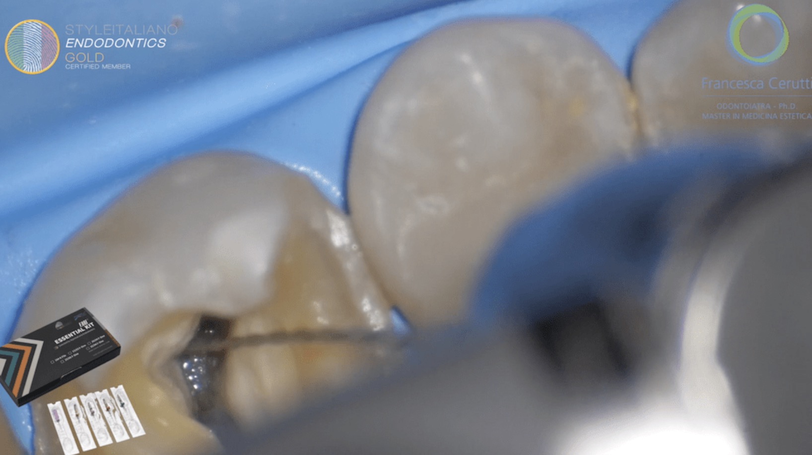
2. Access Cavity Preparation
A conservative access cavity is created using ultrasonic diamonds tips
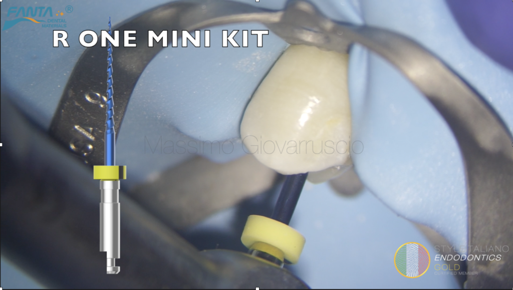
The pulp chamber is carefully explored to locate the root canals orifice and enlargement has been performed using R ONE MINI KIT Fanta Reciprocating files
3. Working Length and Apical Foramen Patency
The reciprocating file 17/04% connected to the apex locator, has been used into both MB and Palatal canal, until reaching the apical third.
Rotary glide path and apical Patency, has been confirmed using the same reciprocating file 17/04%.
A pre-bent manual K-File 10 connected to electronic apex locator (EAL) is used to determine the working length
4. Canal Preparation
FANTA R ONE MINI KIT Reciprocating File 25/06% is used with a crown-down or progressive enlargement technique, ensuring minimal stress on the apically curved region.
Copious irrigation with sodium hypochlorite (NaOCl 5,25%) is performed to remove debris and prevent ledge formation or file separation
5. Apical Management
A #20 NiTi file may be required to recapitulate the patency of the apex
6. Cavity Refining
The Palatal and Buccal orifices are been refined using diamonds ultrasonic tips. The isthmus should be cleaned properly as some residual pulp tissues can be hidden
7. Irrigation and Cleaning
Continuous irrigation with NaOCl (5,25%)
Sonic system irrigation activation (Eddy) enhance effectiveness
8. Obturation
A well-fitted single cone with Hydraulic Calcium Silicate sealer has been used
9. Coronal Seal and Restoration
A permanent Composite coronal seal has been placed to prevents microleakage
A partial-coverage restoration (e.g., Overlay) is recommended to reinforce the structurally compromised premolar
10. Follow-up
Post-operative radiographs verify the quality of obturation and the presence of a severe apical curvature
The patient has bee reviewed (3 months) to assess healing of the periapical tissues
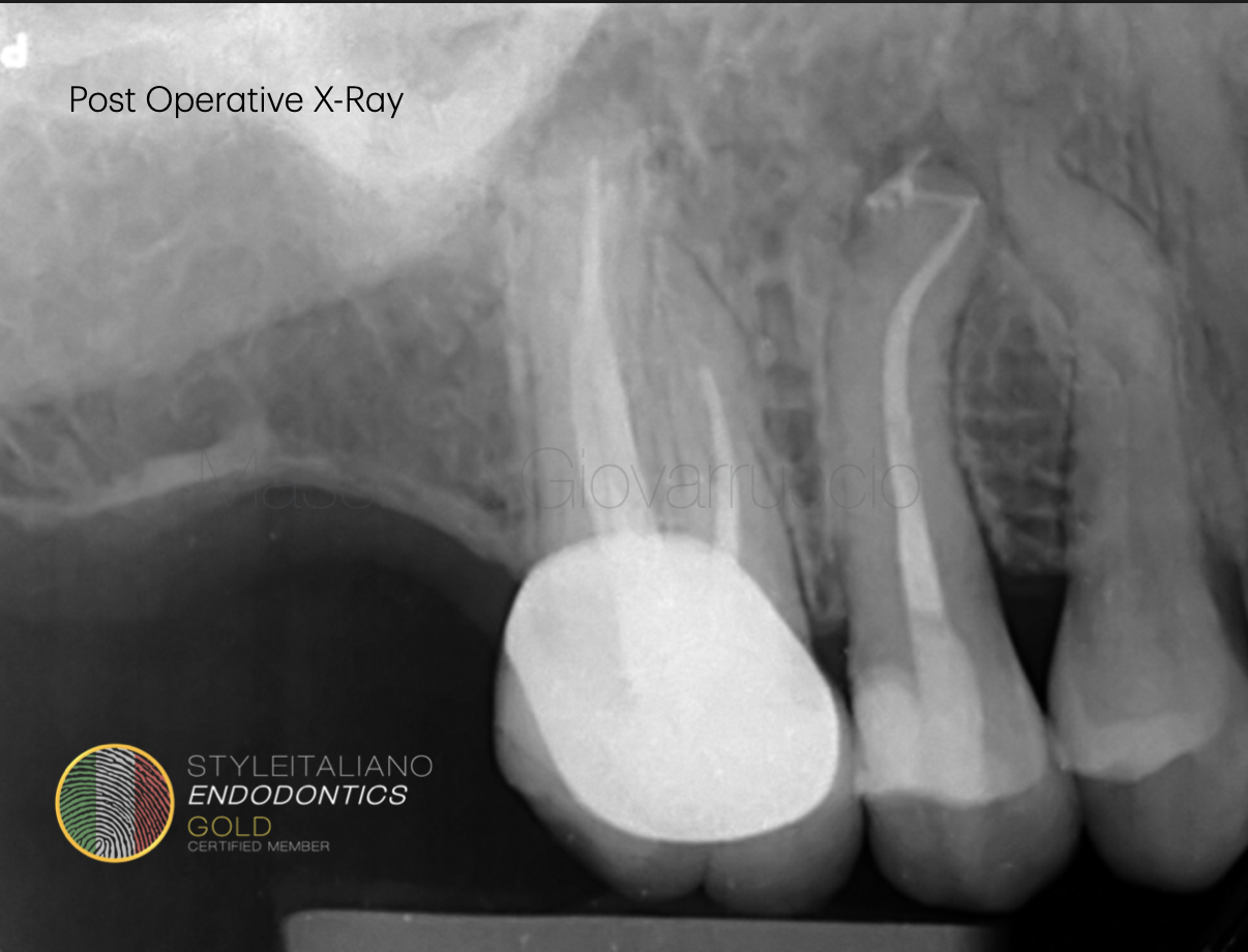
Fig. 2
Post operative X-ray
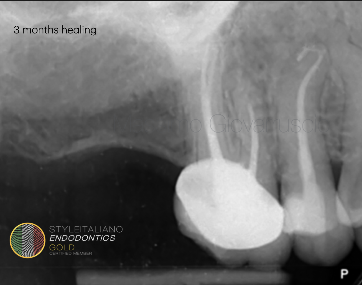
Fig. 3
3 months follow up
Conclusions
Challenges and Considerations
Sometimes is difficult to recognise a severe apical curvature. Manual Stainless Still instruments don’t have the capability to reach and negotiate the apical third.
Reciprocating files (R ONE Mini Kit) used in this case, demonstrated a great fitting into the complex apical anatomy.
0.19 tip and 05% variable taper design, has been used as the OPENER.
Straight line access can be avoided when using the latest generation files.
Minimall invasive protocol preserve Peri-cervical Dentin Structure (PCD)
0.17 tip and 04% variable taper design, has been used as a pre shaping and glider file.
First instrument into the canal, using a reciprocating glide path technique connected to an Electronic Apical Locator (EAL).
AF-L alloy, the less martensitic FANTA DENTAL property thermal treatment.
This confers two advantages: the flexibility and the proper torsional resistance that does not make the instrument too soft
0.12 tip and 06% variable taper design, with a maximum diameter of nearly 1mm.
This allows to preserve more cervical dentine (PCD)
Last instrument of the sequence