Simplified MTA apical plug
21/09/2024
Fellow
Warning: Undefined variable $post in /home/styleendo/htdocs/styleitaliano-endodontics.org/wp-content/plugins/oxygen/component-framework/components/classes/code-block.class.php(133) : eval()'d code on line 2
Warning: Attempt to read property "ID" on null in /home/styleendo/htdocs/styleitaliano-endodontics.org/wp-content/plugins/oxygen/component-framework/components/classes/code-block.class.php(133) : eval()'d code on line 2
Root resorption is a pathological process that may occur after surgical mechanical, chemical or thermal insult. Generally, it can be classified as internal and external root resorption. Depending on the diagnosis, biocompatible material like MTA can be used in management of these cases.
General dental practitioners can face difficulties in diagnosis and treatment planning for cases with root resorption. An understanding of the pathogenesis of root resorption is critical for diagnosis, effective management and improves outcome.
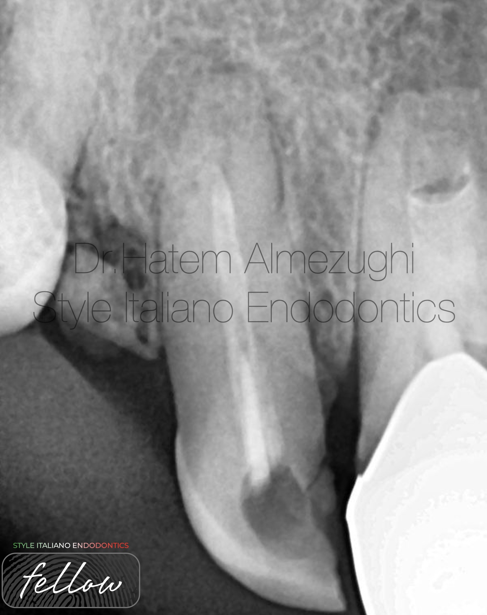
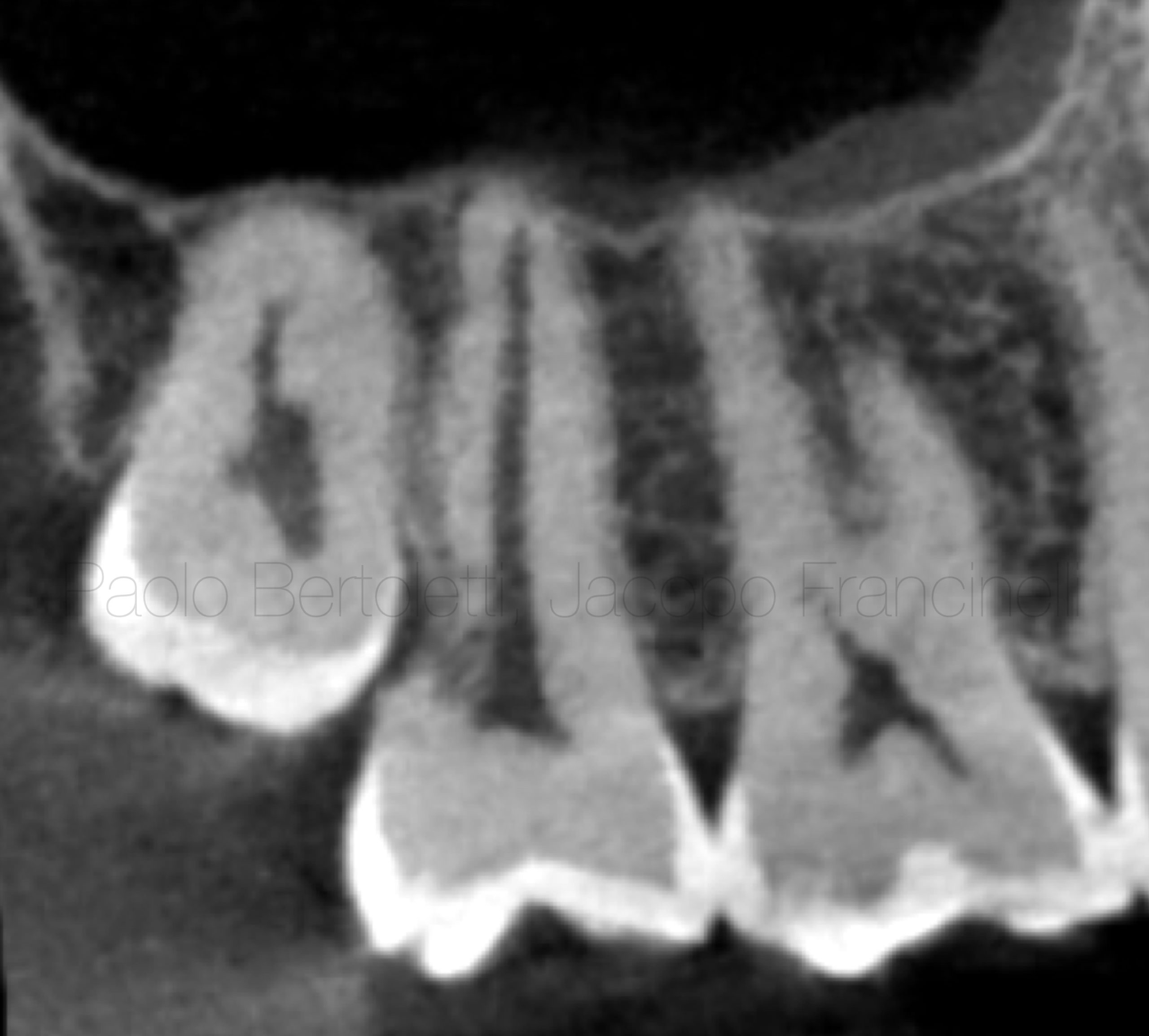
Fig. 1
PRE-OPERATIVE X-RAY
Shows tooth 12 with old under extended fill and large apical radiolucency.
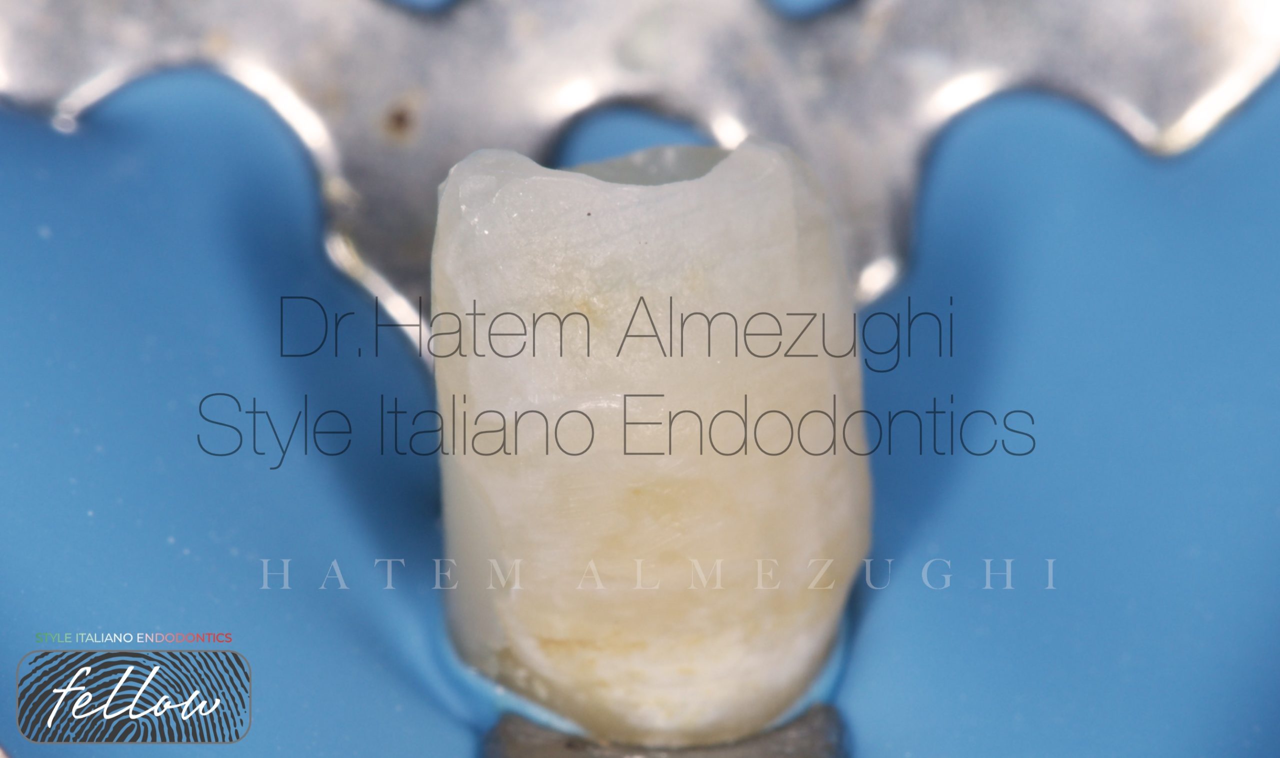
Fig. 2
BUILD UP
Build up done to obtain satisfactory isolation, now tooth ready to subject to the therapy protocol.
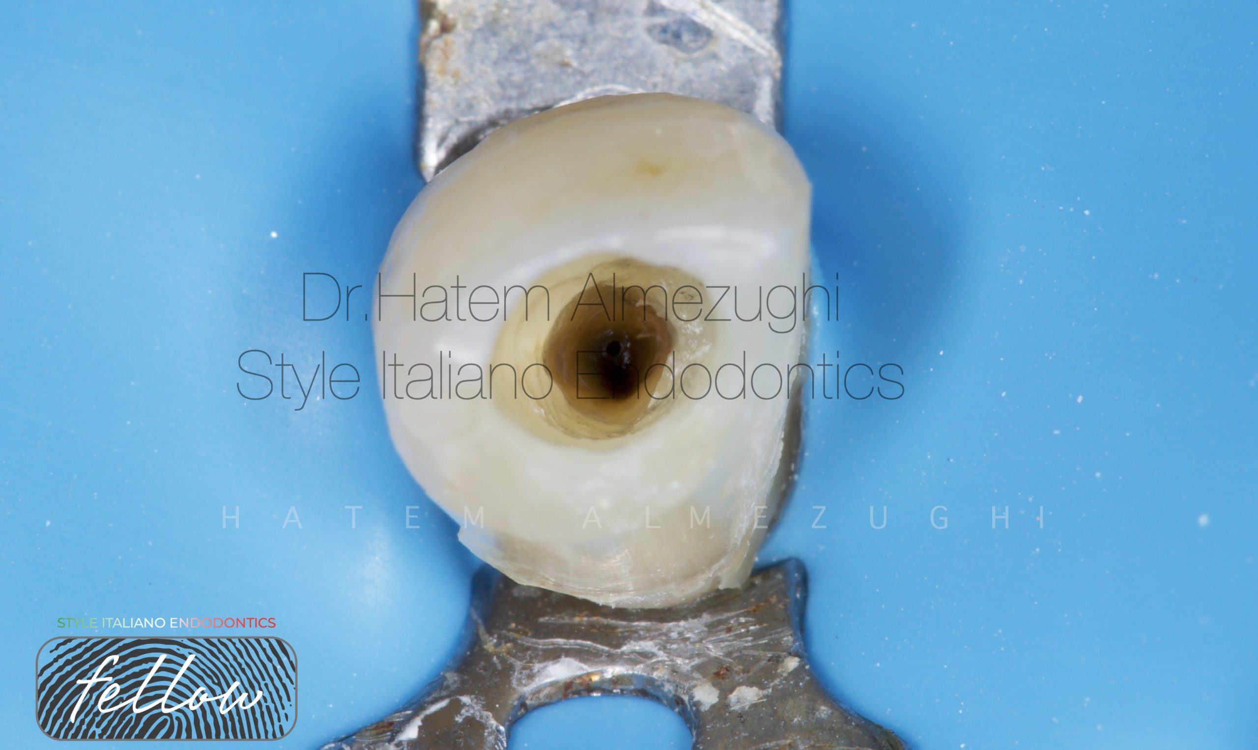
Fig. 3
INTRA ORAL PHOTO
Shows clear view of orifice
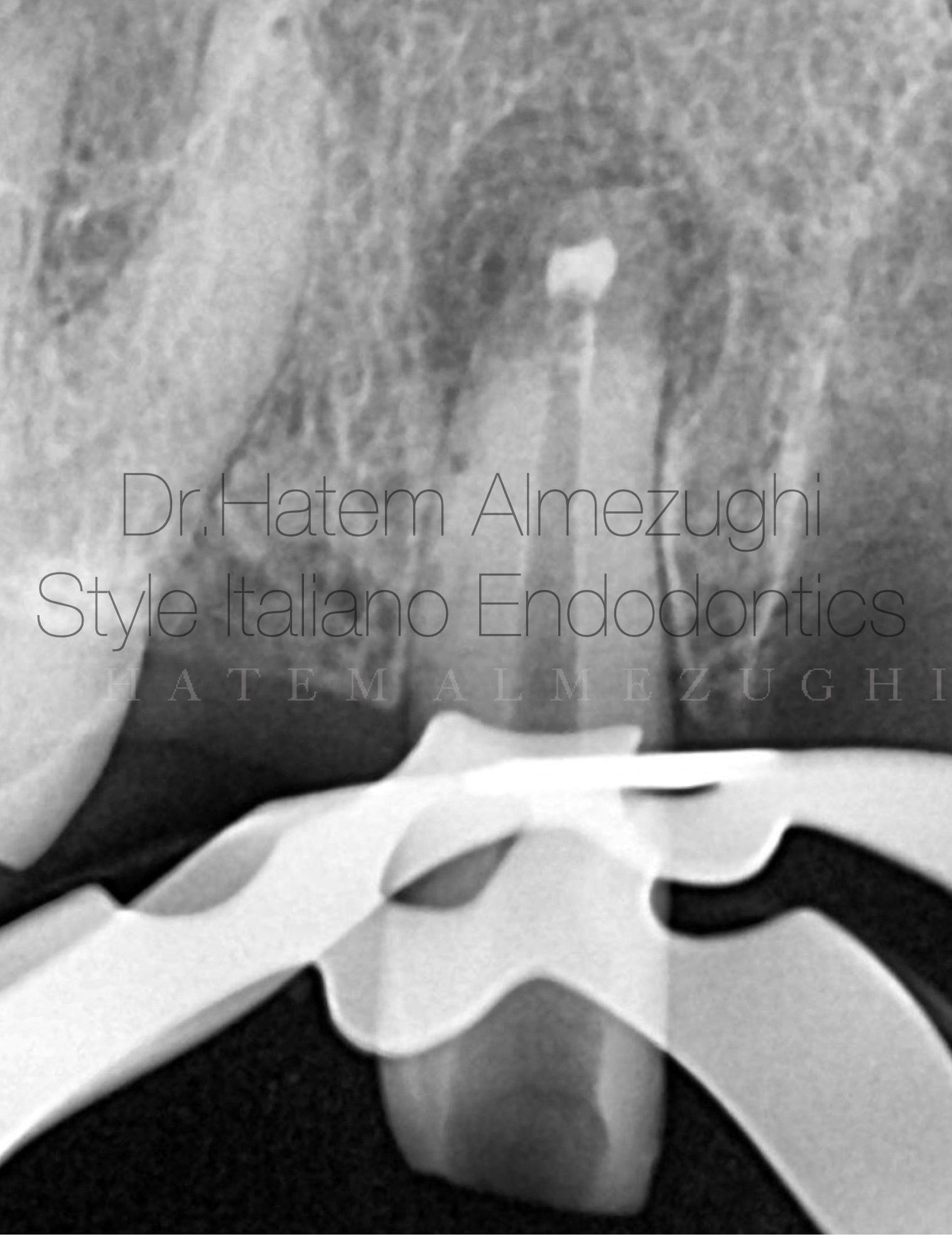
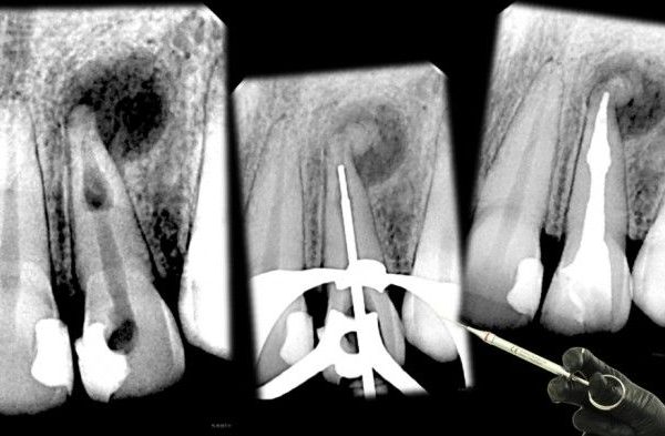
Fig. 4
Shows introducing the first increment of amount of MTA and delivering it up at the apical part
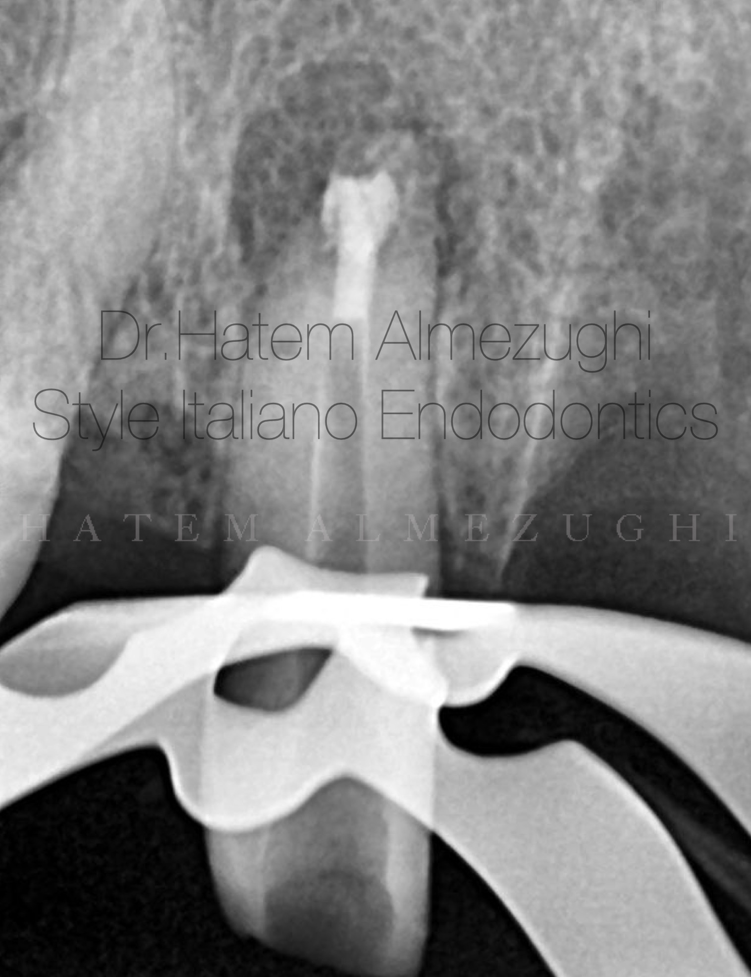
Fig. 5
After adding another amount of MTA with proper adaptation , another xrays was taken
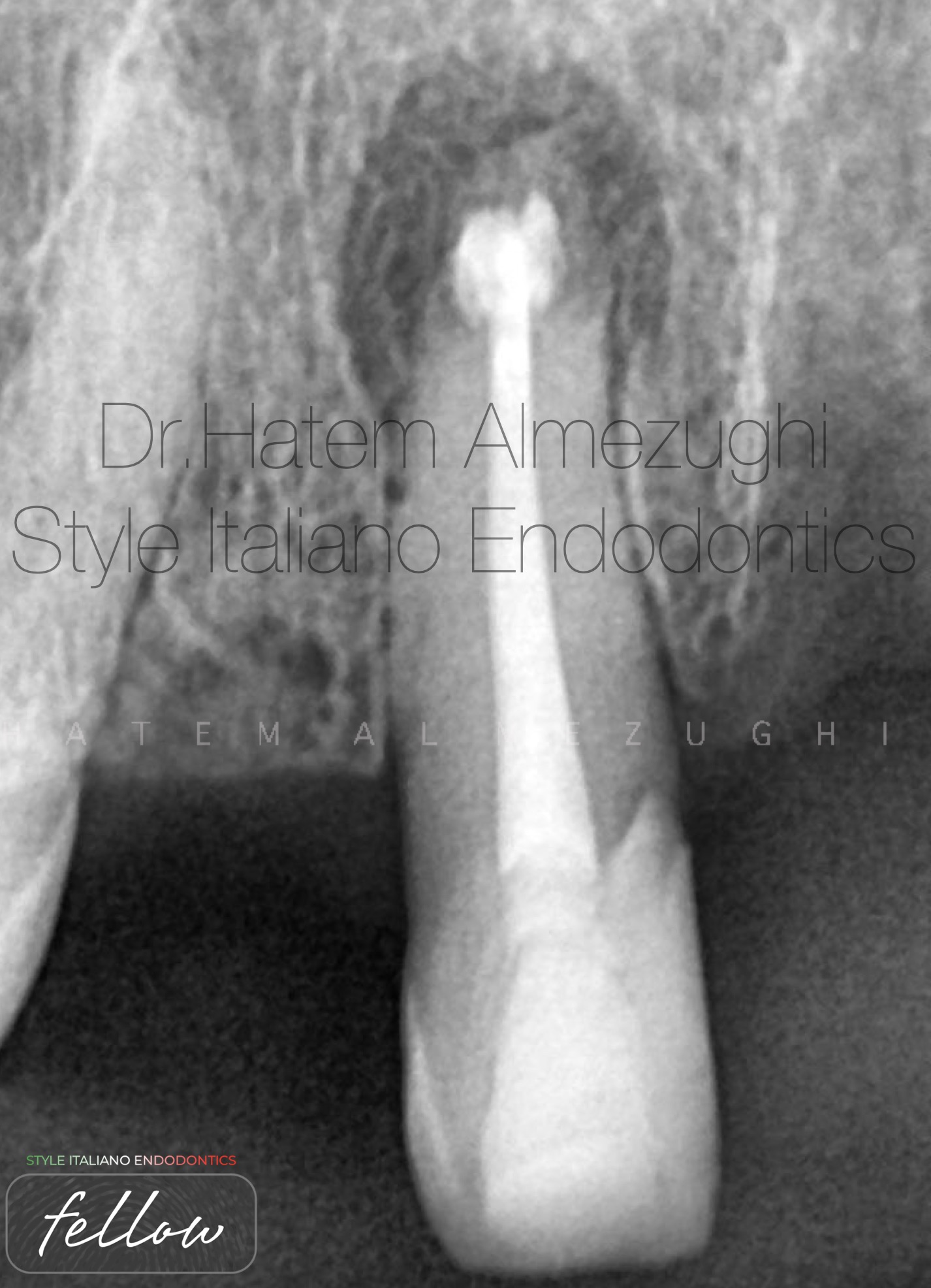
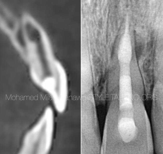
Fig. 6
Obturation done, and the large radiolucency clearly noticed, and follow up will reveal if there is any remarkable healing
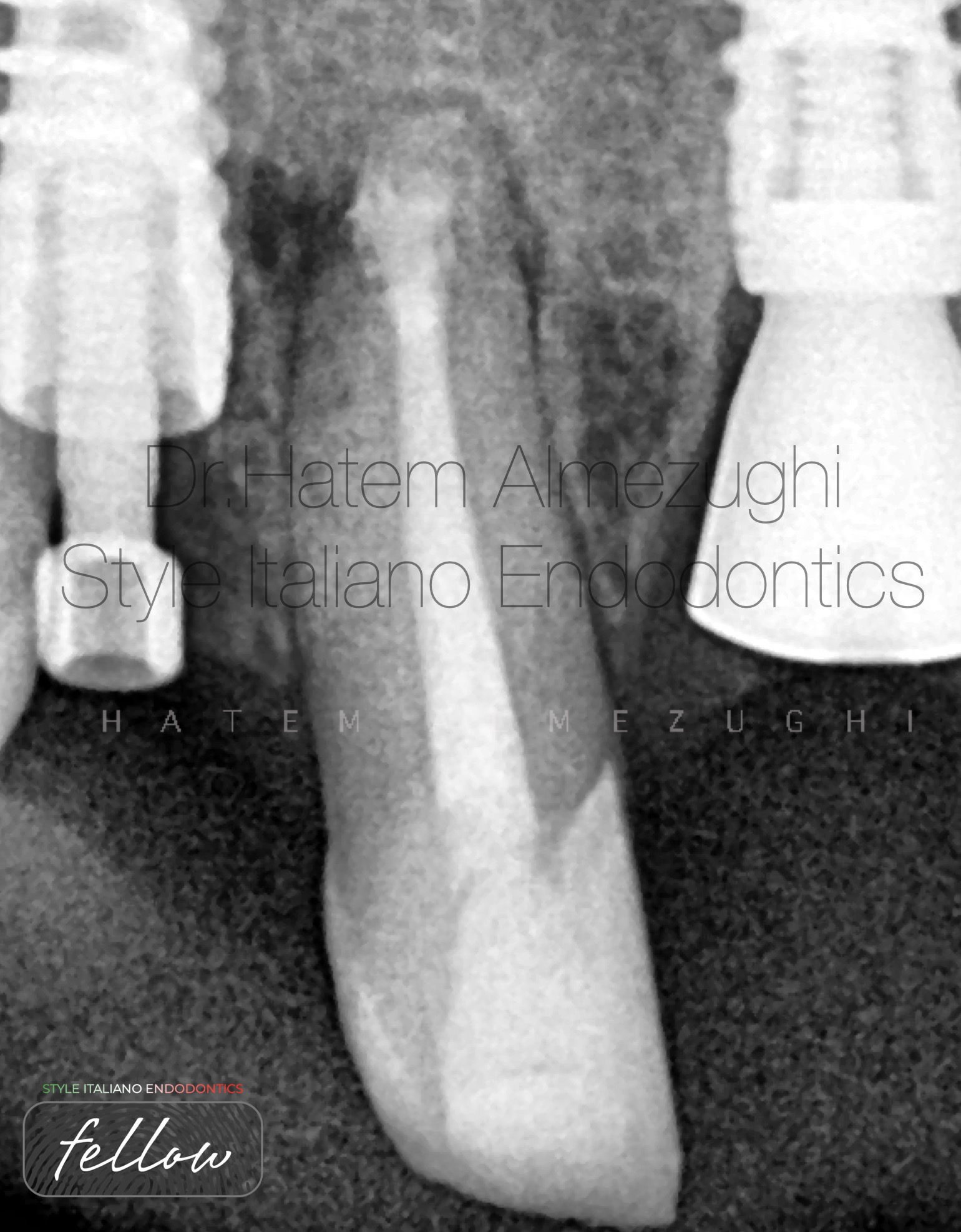
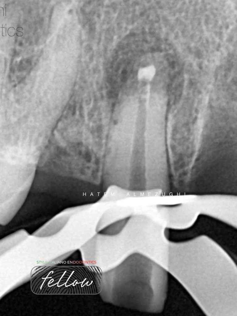
Fig. 7
This figure shows months follow-up, with promising healing.

Fig. 8
About Dr.Hatem Almezughi
- graduated from Tripoli university, Libya.
- Local and international speaker in different conferences and forums .
- Dr. Hatem Almezughi (BDS)
- instructor in many endodontic courses.
- Dental practice limited to endodontics and restorative .
- Fellow member in style Italiano endodontics.
Conclusions
Biocompatible materials are likely to impact the management of ITR & ETR cases and treatment outcomes.
however, ITR & ETR cases aren’t very common but encountered every once and a while.
It is therefore important that case reports, such as this one, are shared to inform clinicians on the management, materials and techniques that can be employed
in efforts to ensure good-quality and lasting treatment outcomes in such cases.
Selecting high quality material “MTA” is prioritized to ensure delivering quality treatment , not to mention the the tools and instruments like MAP 1 system for instance.
Bibliography
Patel S, Ricucci D, Durak C, Tay F. Internal root resorption: a review. J Endod 2010; 36: 1107-1121.
Levin L, Trope M. Root Resorption. In Hargreaves K M, Goodis H E (eds) Seltzer and Bender's Dental Pulp. pp 425-448. Chicago: Quintessence Publishing, 2002.
Patel S, Krastl G, Weiger R et al. ESE position statement on root resorption. Int Endod J 2023; 56: 792-801.
Wedenberg C, Zetterqvist L. Internal resorption in human teeth - a histological, scanning electron microscopic, and enzyme histochemical study. J Endod 1987; 13: 255-259.
Mattison G D, Delivanis H P, Delivanis P D, Johns P I. Orthodontic root resorption of vital and endodontically treated teeth. J Endod 1984; 10: 354-358.
Patel S, Dawood A, Wilson R, Horner K, Mannocci F. The detection and management of root resorption lesions using intraoral radiography and cone beam computed tomography - an in vivo investigation. Int Endod J 2009; 42: 831-838.
Nilsson E, Bonte E, Bayet F, Lasfargues J-J. Management of internal root resorption on permanent teeth. Int J Dent 2013; 2013: 929486.
Krastl G, Weiger R, Filippi A et al. Endodontic management of traumatized permanent teeth: a comprehensive review. Int Endod J 2021; 54: 1221-1245.
Andreasen F M, Andreasen J O. Resorption and mineralization processes following root fracture of permanent incisors. Endod Dent Traumatol 1988; 4: 202-214.
Silveira F F, Nunes E, Soares J A, Ferreira C L, Rotstein I. Double ‘pink tooth' associated with extensive internal root resorption after orthodontic treatment: a case report. Dent Traumatol 2009; 25: 43-47.
Patel S, Kanagasingam S, Pitt Ford T. External cervical resorption: a review. J Endod 2009; 35: 616-625.