Silver cones still alive…
11/10/2023
Fellow
Warning: Undefined variable $post in /home/styleendo/htdocs/styleitaliano-endodontics.org/wp-content/plugins/oxygen/component-framework/components/classes/code-block.class.php(133) : eval()'d code on line 2
Warning: Attempt to read property "ID" on null in /home/styleendo/htdocs/styleitaliano-endodontics.org/wp-content/plugins/oxygen/component-framework/components/classes/code-block.class.php(133) : eval()'d code on line 2
Retreatments in endodontics are always a challenge. Nowadays above all for Endodontist retreatments are a daily routine.
Each case is different and it’s important have the right tools and the right strategy to be able to face them.
In this article I am describing all the phases step by step to retreat a difficult case with the presence of silver cones as obturation materials.
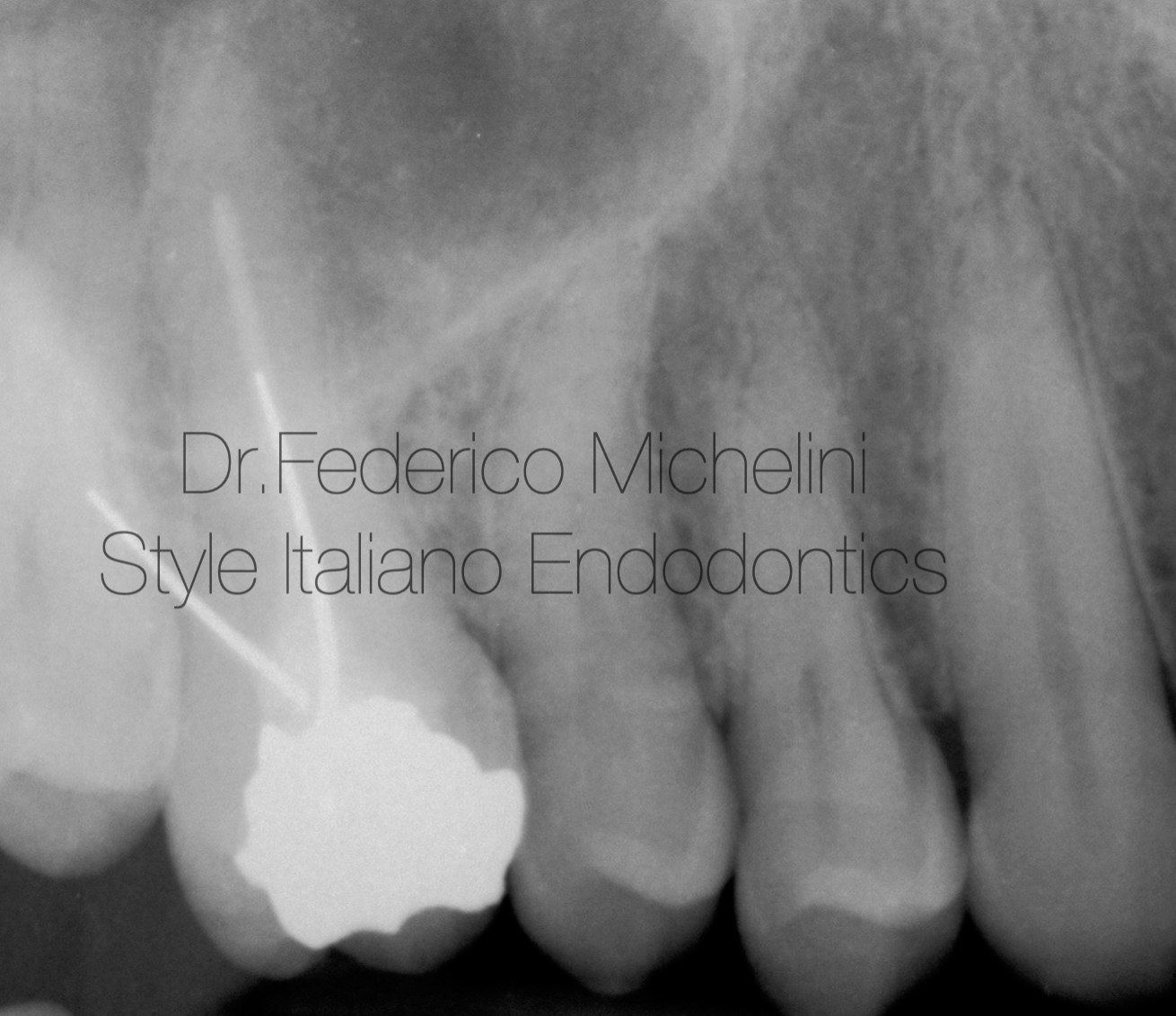
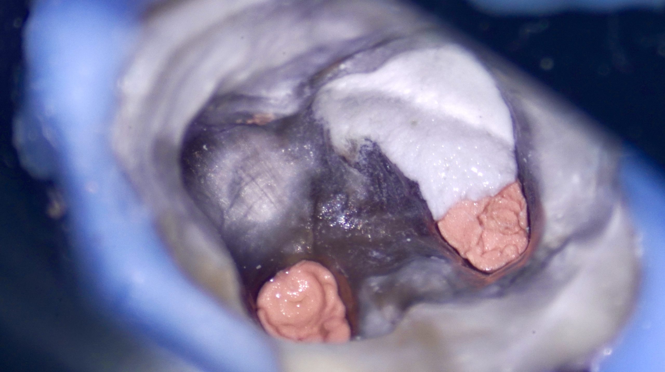
Fig. 1
Pre-operative X-ray
A patient came to my clinic for a breakage of an old obturation in amalgam and pain in a first quadrant especially to pressure and chewing.
After radiographic examination we see a poor root canal treatment and the presence of 2 silver cones.
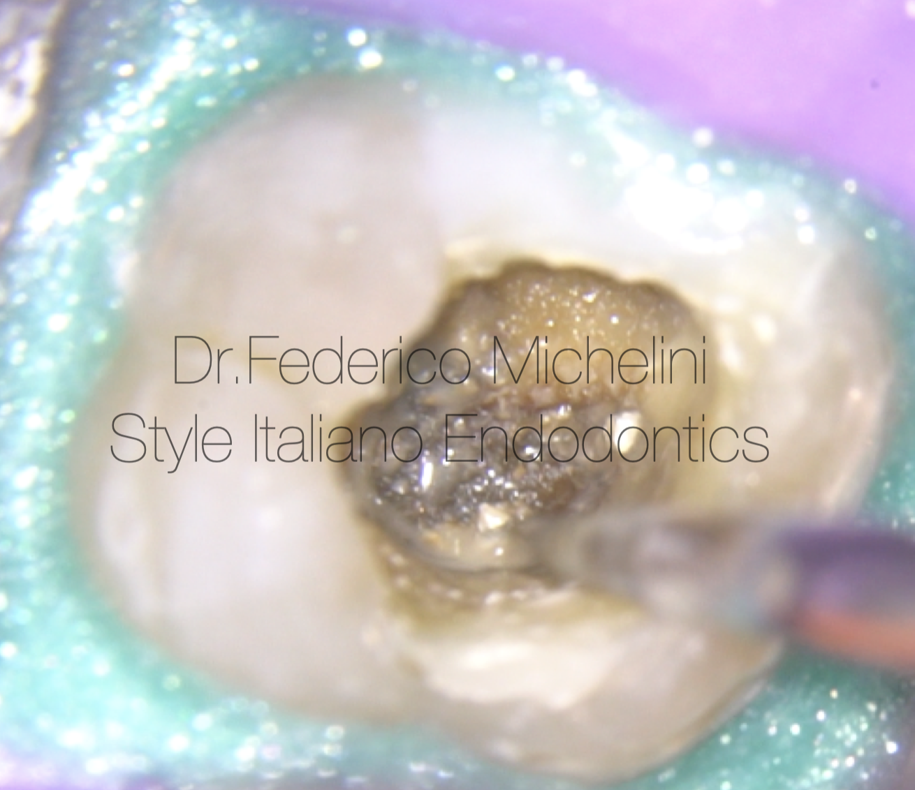
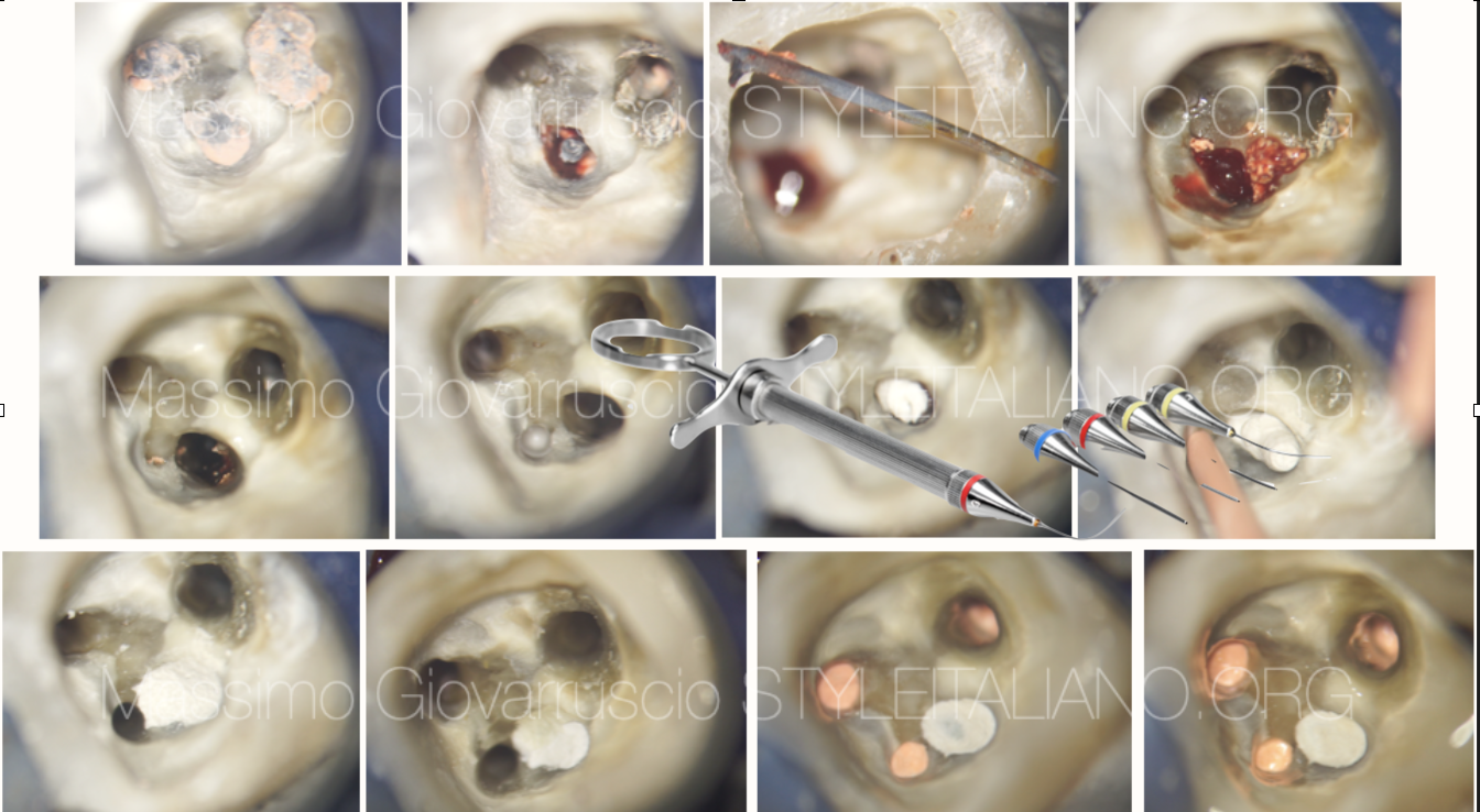
Fig. 2
Re-access and sonic tip for the removal of the first silver cone in disto-vestibular canal.
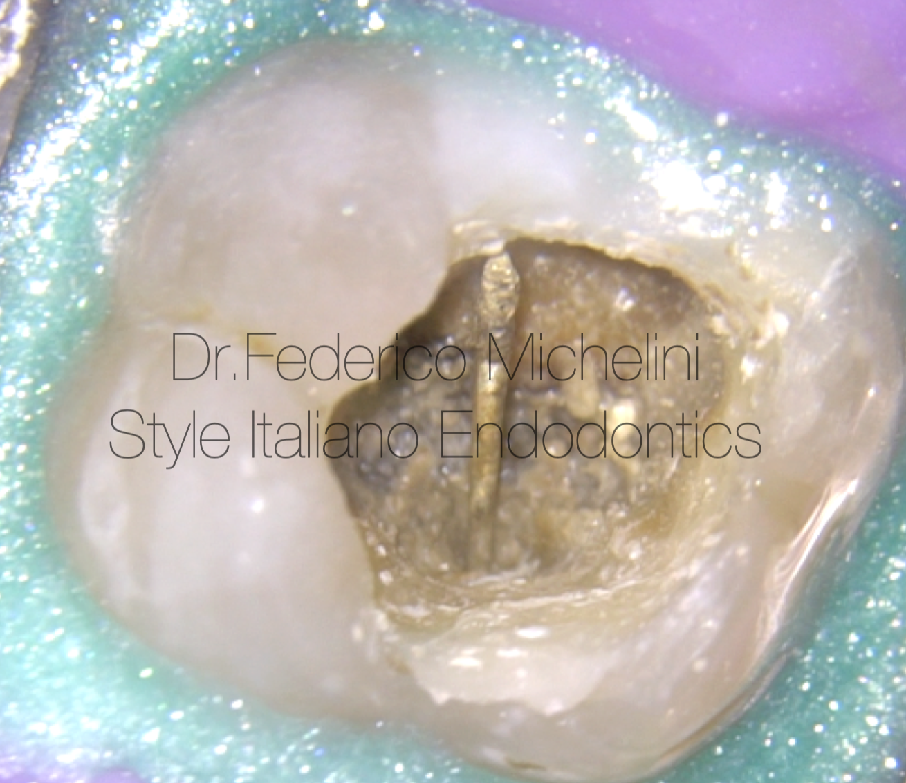
Fig. 3
First silver cone removed.
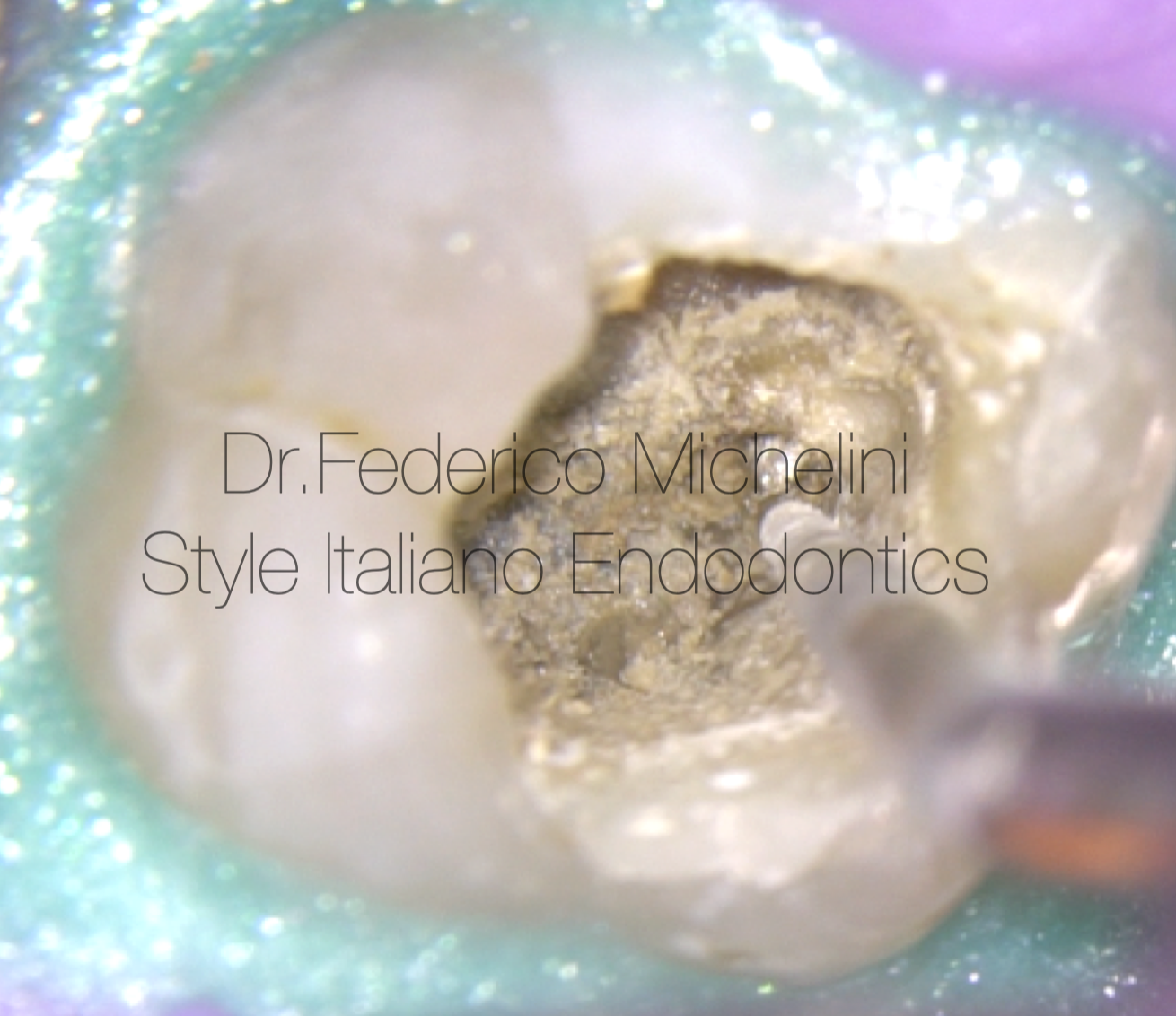
Fig. 4
Second silver cone in mesio-vestibolar canal.
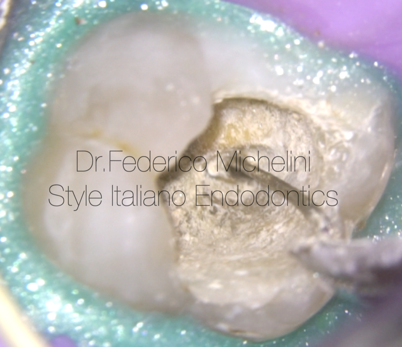
Fig. 5
Second silver cone removed.
The video shows the steps for removing silver cones:
It’s important works in a very delicately way for not break them.
If this happen we have to treat them as if they were broken instruments.
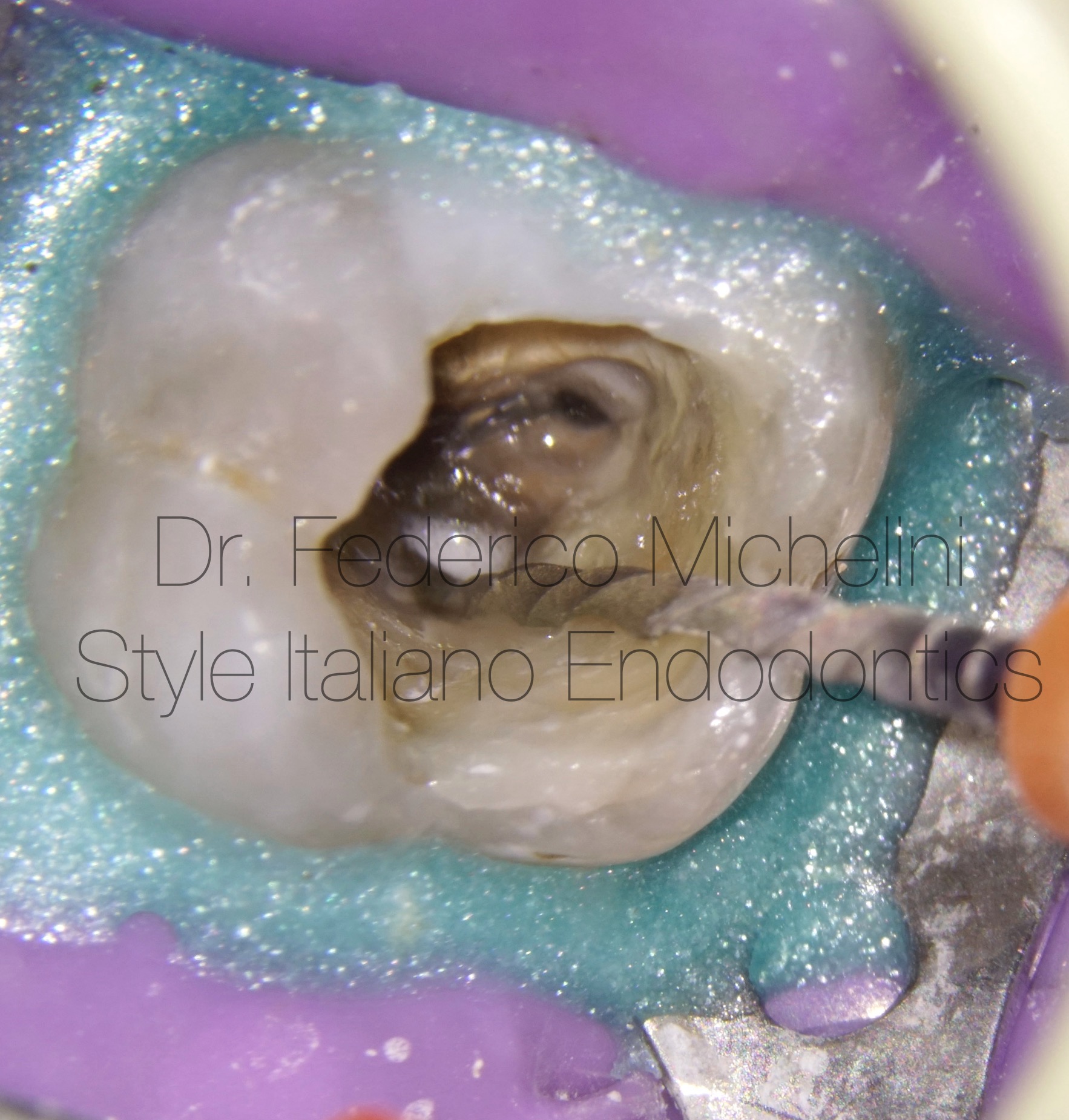
Fig. 6
Mechanical retreatment of the 3 main canals.
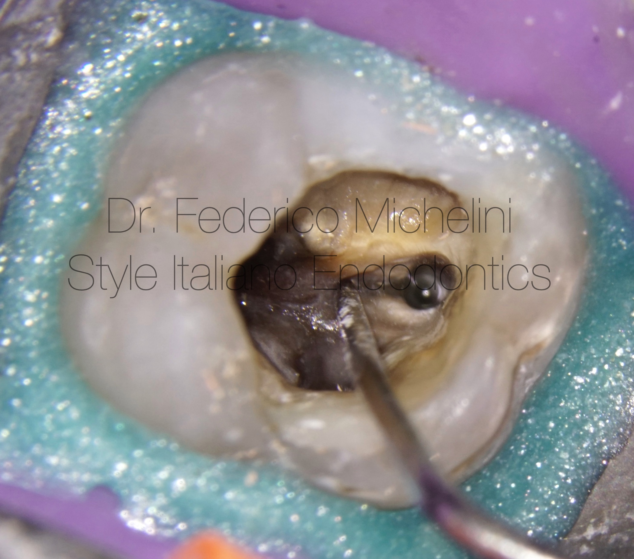
Fig. 7
Main canals re-treated and the presence of MB2 not treated.
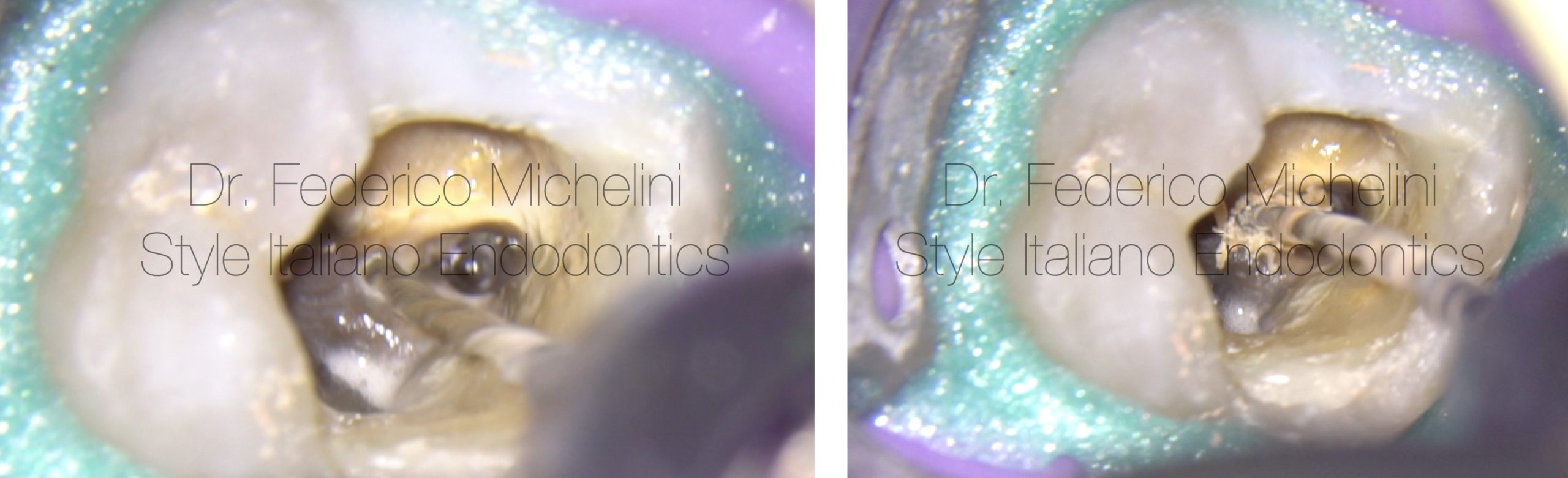
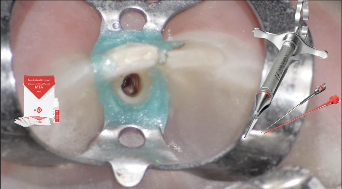
Fig. 8
Mechanical shaping of MB2.
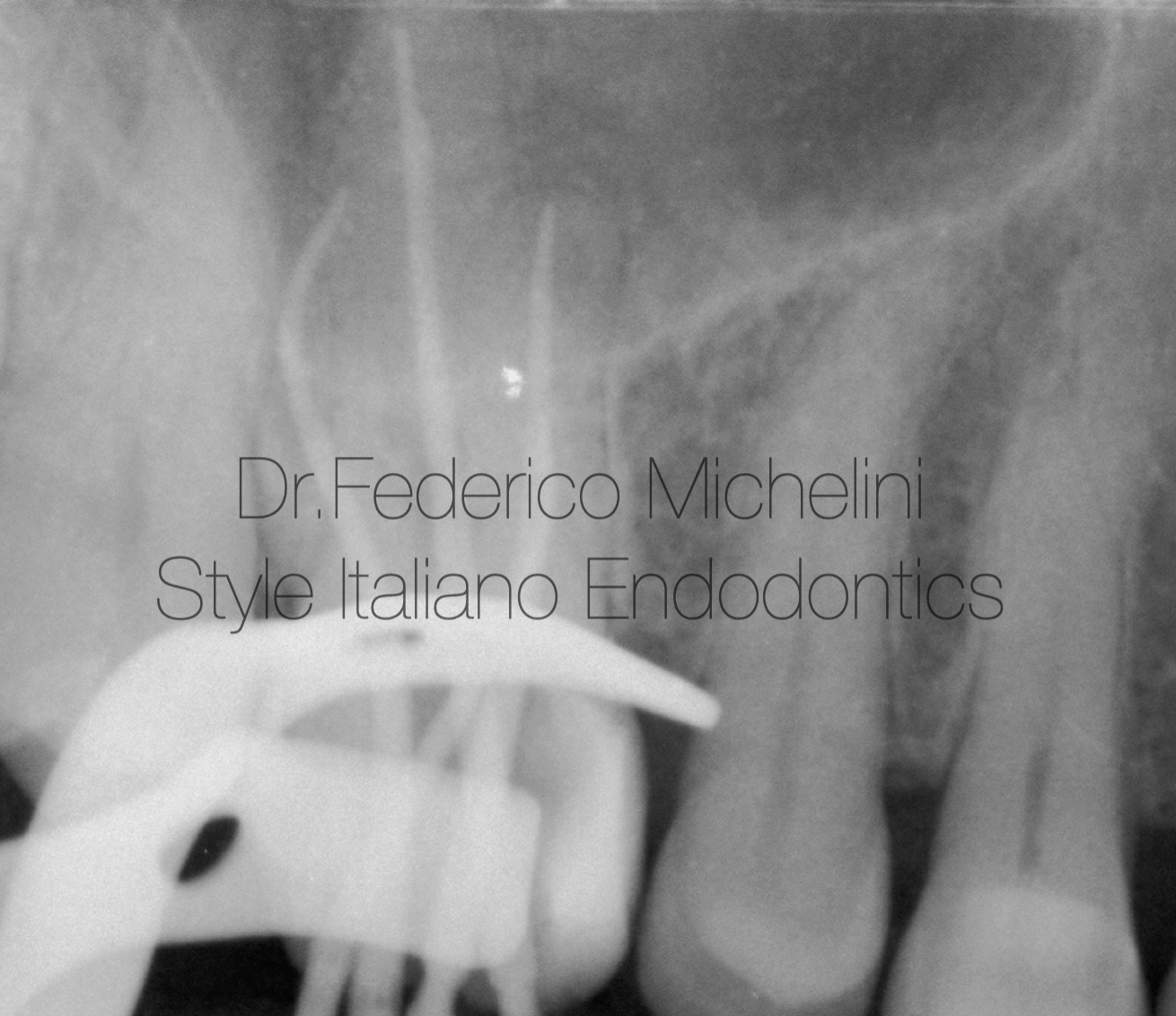
Fig. 9
Rx for checking the working lengths.
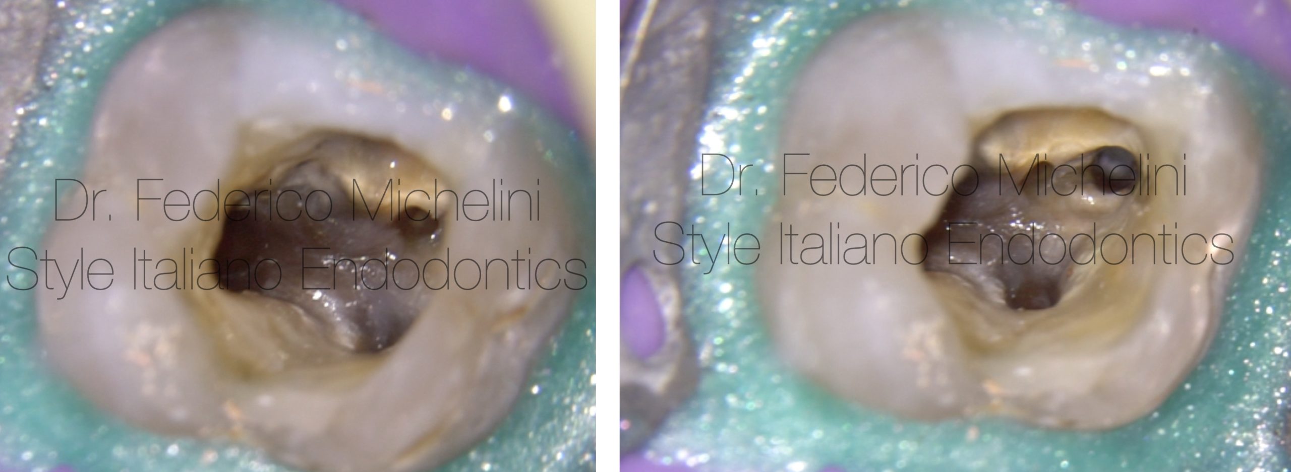
Fig. 10
Fig-8 4 Canals ready for obturation.
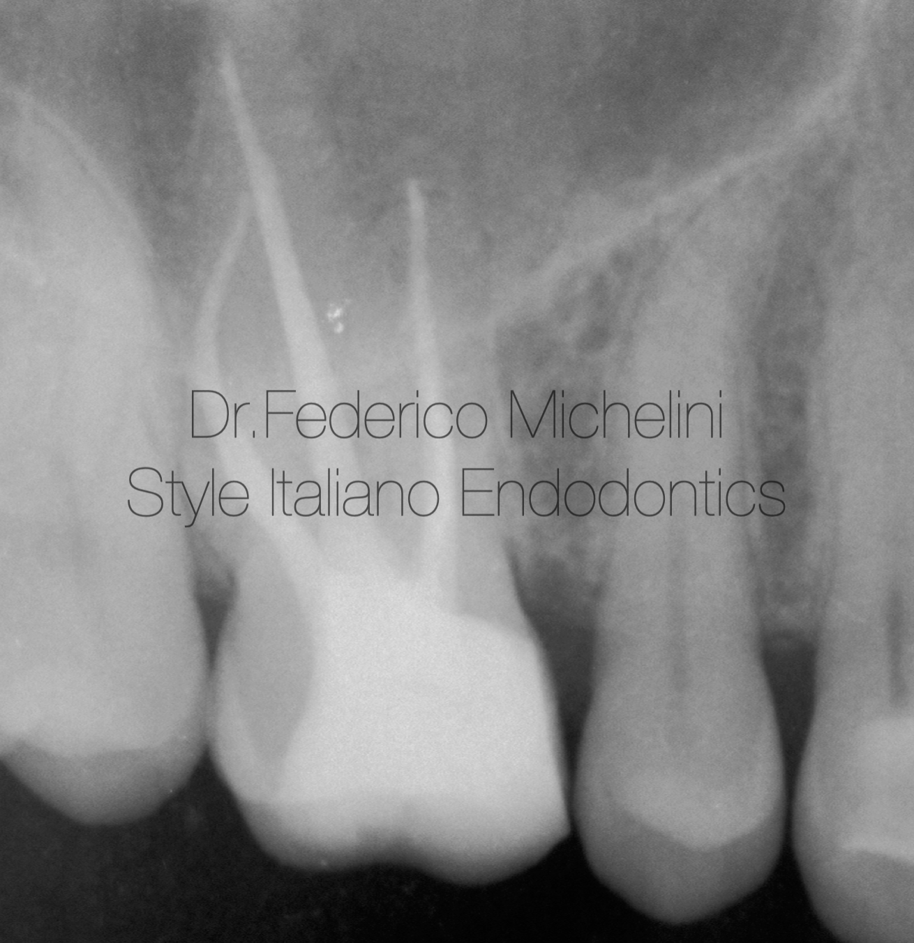
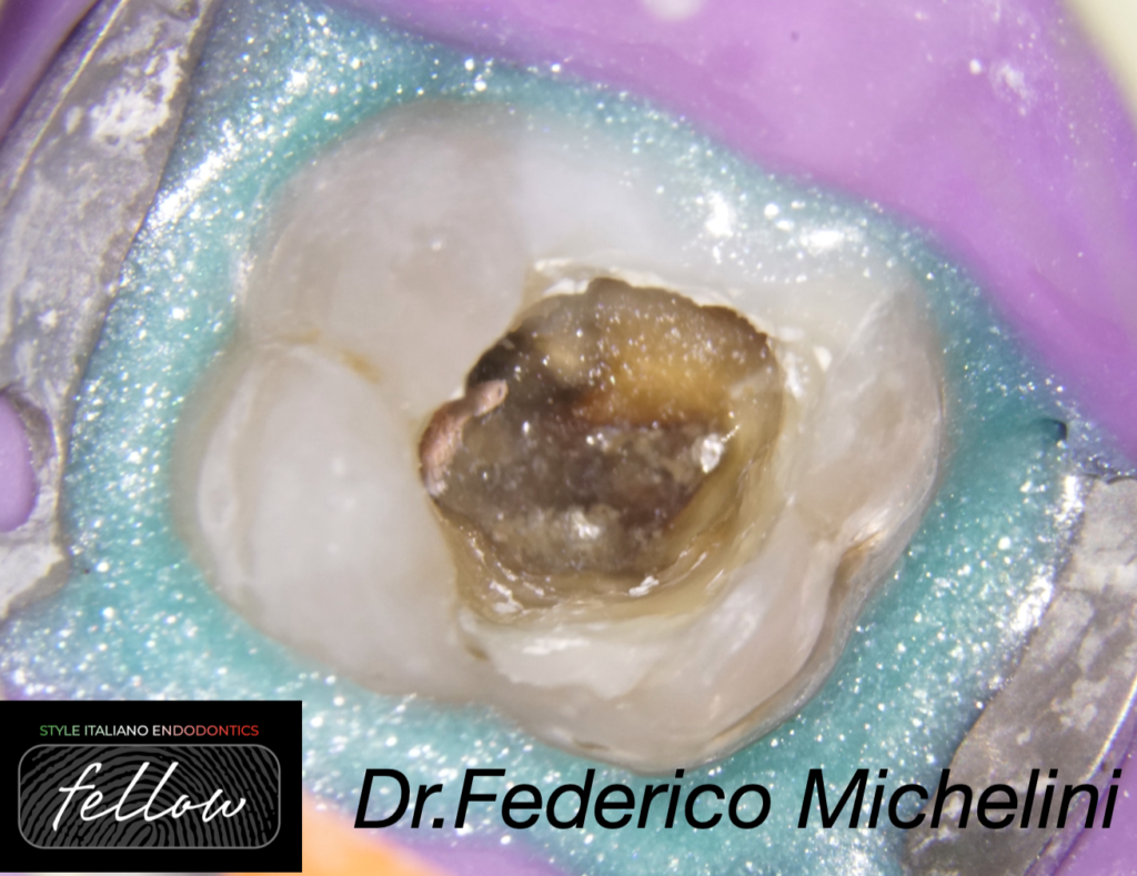
Fig. 11
X-ray for checking the obturation of the 4 canals and the pre-prosthetic reconstruction.
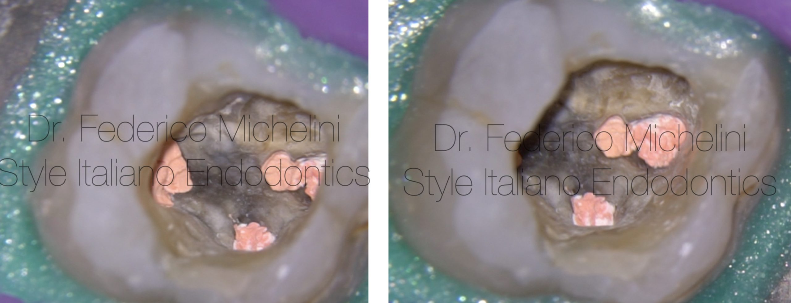
Fig. 12
Obturation of the 4 canals.
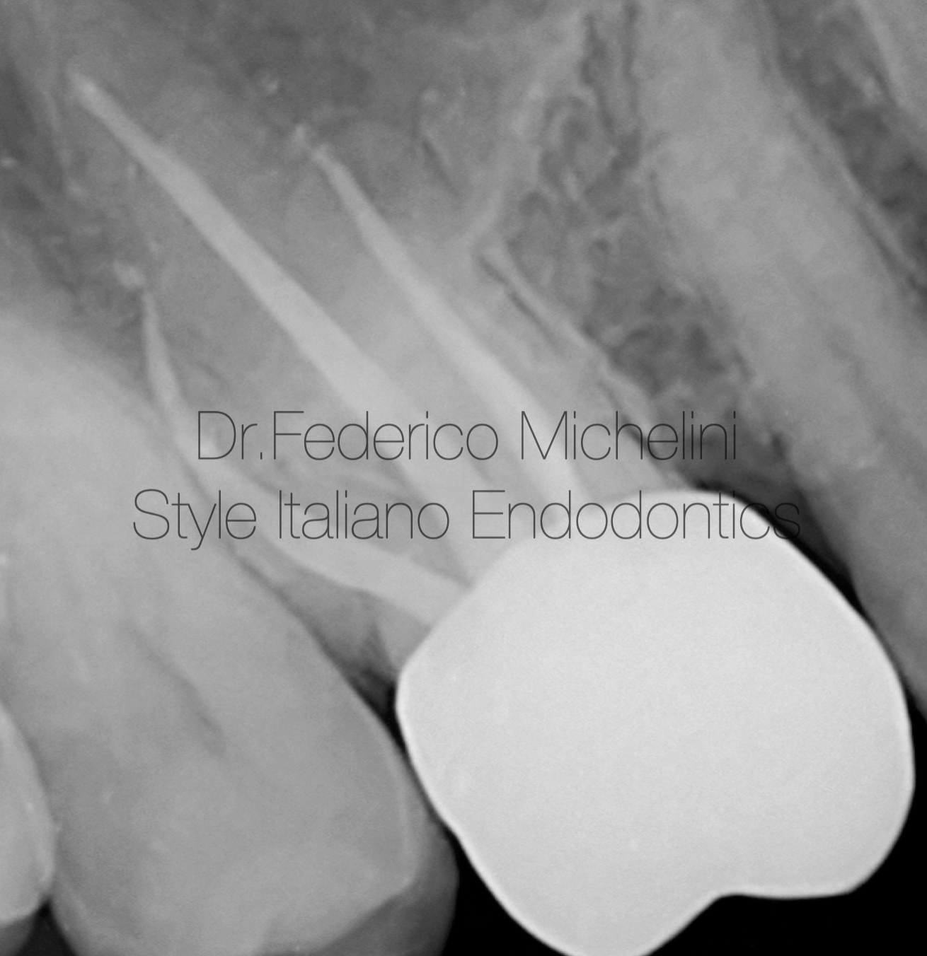
Fig. 13
1 years follow up.
Fig. 14
Dr. Federico Michelini
2015: Graduated in dentistry from the University of Valencia.
2017: Master in Endodontics from the University of Bologna.
Federico Michelini has been a speaker in national and international congresses regarding endodontics.
Tutor of the Master of Endodontics and of the Degree Course of Dentistry and Dental Prosthesis for the University of Bologna.
He won the Closed Meeting of Italian Society of Endodontics in 2019.
He attended the advanced course of retreatments and endodontics surgery of Italian Academy of Endodontics.
He attended the course of microscopy by Dott. Fabio Gorni.
Member of the Italian Society of Endodontics
Member Fellow of Style italiano Endodontics.
Opinion Leader and Speaker for Morita, Coltene and Labomed.
He works in Parma and Piacenza dedicating to microscopic endodontics.
Conclusions
Today thanks microscope and different types of materials, as in this case MTA or Bioceramic Putty, it is possibile manage difficult cases, avoid extraction and give “new life” to dental elements.
The purpose of this article is to give at all the clinicians a right and simple protocol to use in case of perforations following the philosophy of the group: feasible, teachable, repeatable.
Bibliography
1- Koc C, Aslan B, Ulusoy Z, Oruçoğlu H. Sealing ability of three different materials to repair furcation perforations using computerized fluid filtration method. J DentRes Dent Clin Dent Prospects. 2021 Summer;15(3):183-187.
2- Gorni FG, Andreano A, Ambrogi F, et al. Patient and clinical characteristics associated with primary healing of iatrogenic perforation after root canal treatment: results of a long-term Italian study. J Endod. 2016;42 (2): 211-215.
3- Estrela C, Decurcio DA, Rossi-Fedele G et al. Root perforations; a review of diagnosis, prognosis and materials. Braz Oral Res. 2018 Oct 18;32 (suppl 1):e73.