Huge Perforation!
16/09/2021
Ibrahim El Naggar
Warning: Undefined variable $post in /home/styleendo/htdocs/styleitaliano-endodontics.org/wp-content/plugins/oxygen/component-framework/components/classes/code-block.class.php(133) : eval()'d code on line 2
Warning: Attempt to read property "ID" on null in /home/styleendo/htdocs/styleitaliano-endodontics.org/wp-content/plugins/oxygen/component-framework/components/classes/code-block.class.php(133) : eval()'d code on line 2
Retreatment is one of the biggest challenges for the clinician due to the unexpected surprises, prognosis and diagnosis which may be tricky in many situations.
In this case, this female patient was complaining about pain during biting, related to a previously treated tooth.
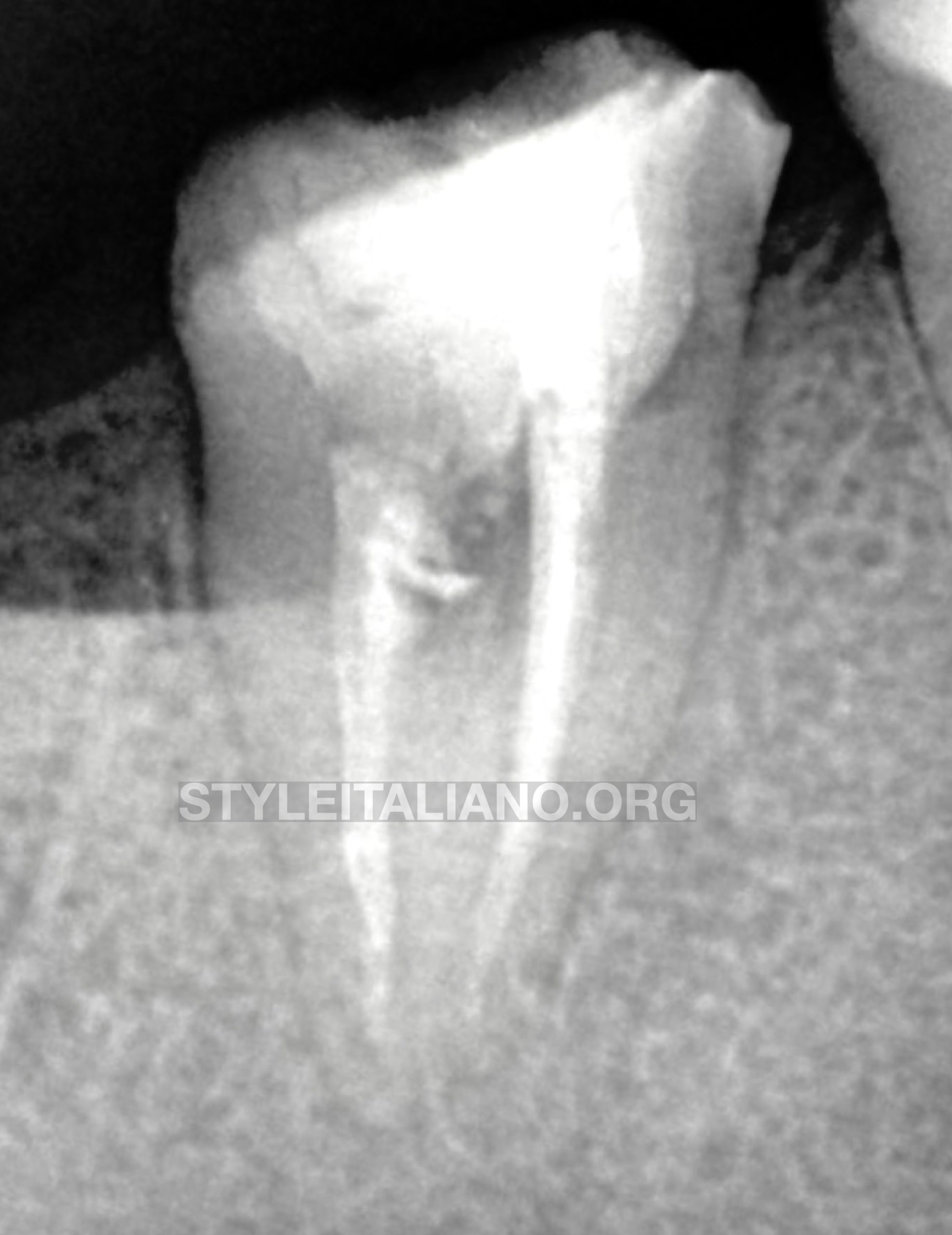
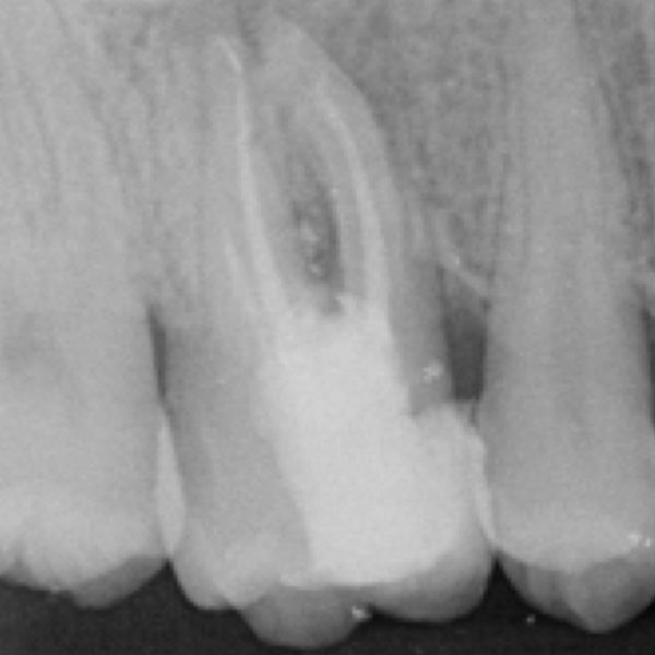
Fig. 1
Pre Op , X - Ray showing a poor quality obturation and radiolucency in the middle of the tooth.
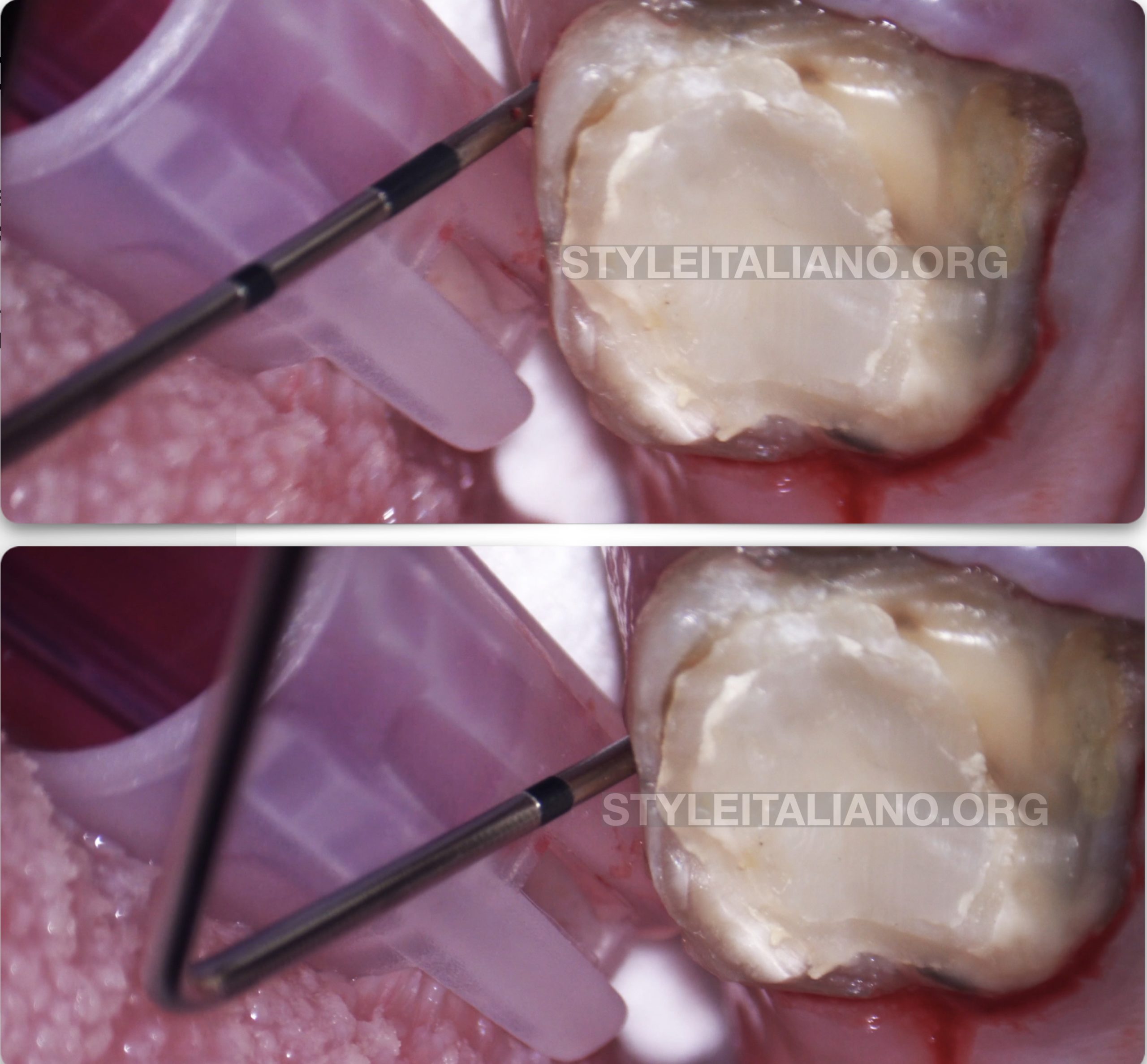
Fig. 2
In the Upper image we performed the testing before the tooth using the periodontal probe which gave normal sulcular depth.
In the lower image, while moving to the lingual furcation the probe penetrated deeper, indicating a problem in that site.
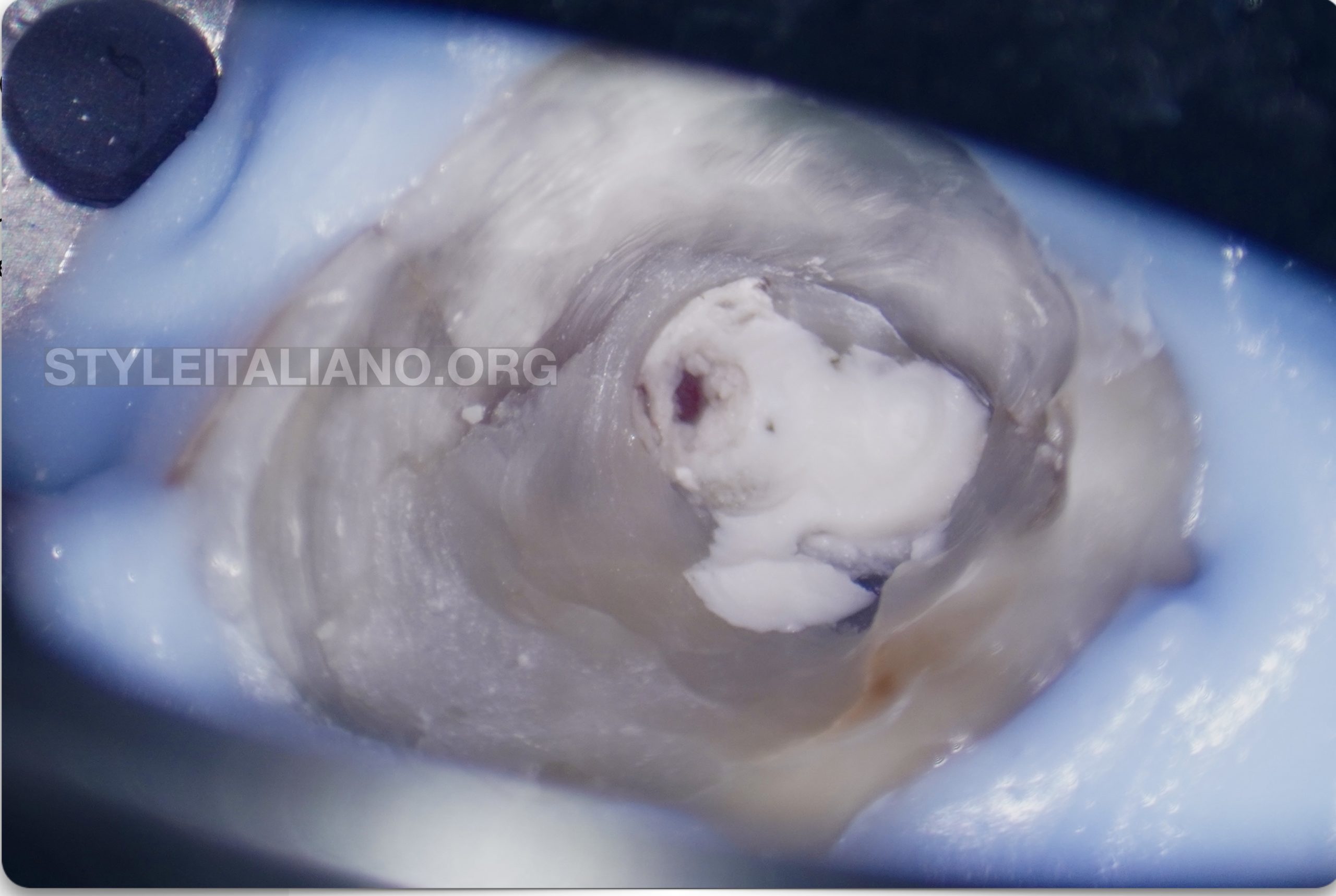
Fig. 3
Disassembling the restoration we found the presence of small Red Dot which may indicate a perforation which is covered by GIC in this situation.
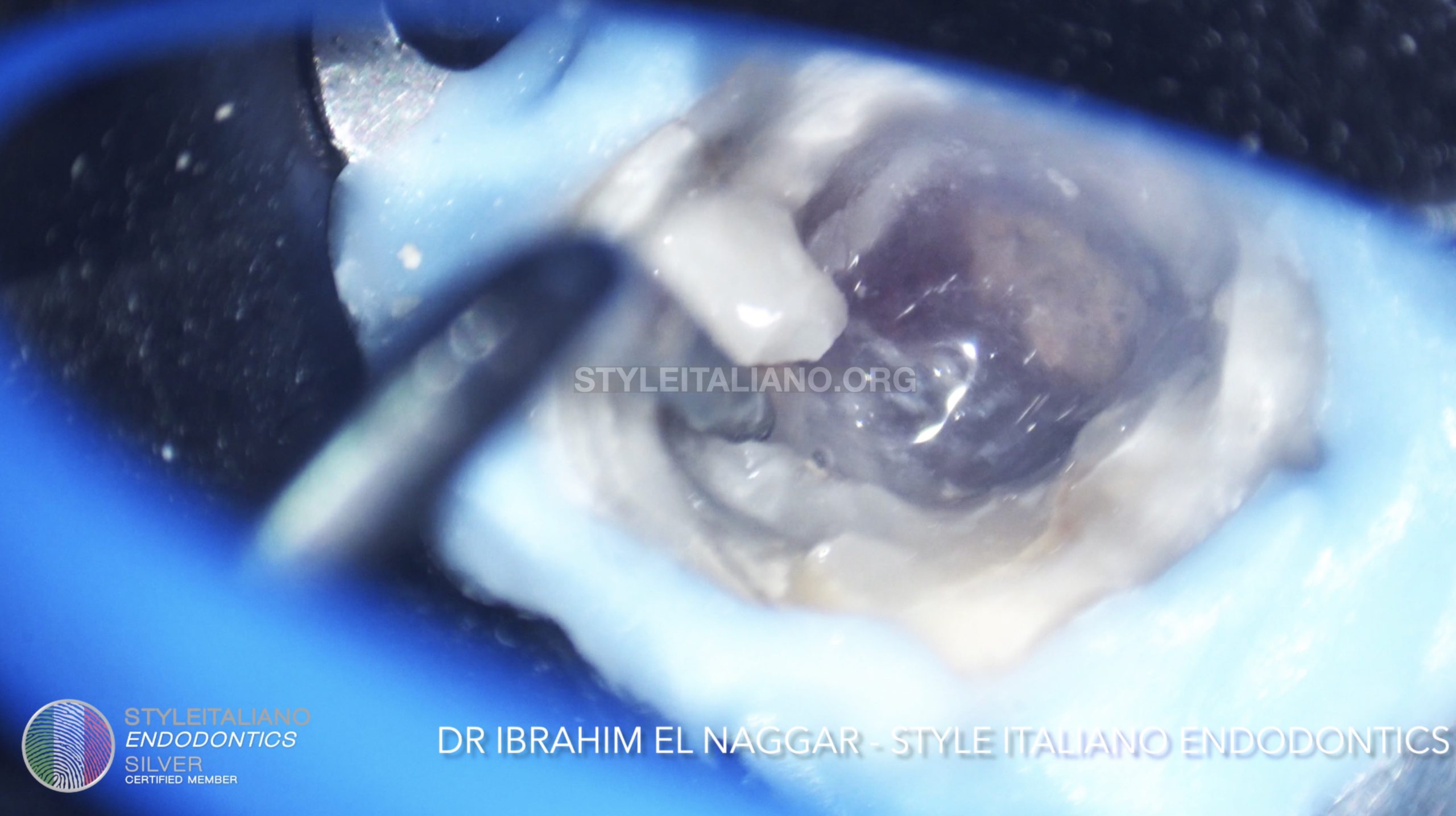
Fig. 4
Using the Ultrasonic the disassembling of the Composite around the canals was made easily.
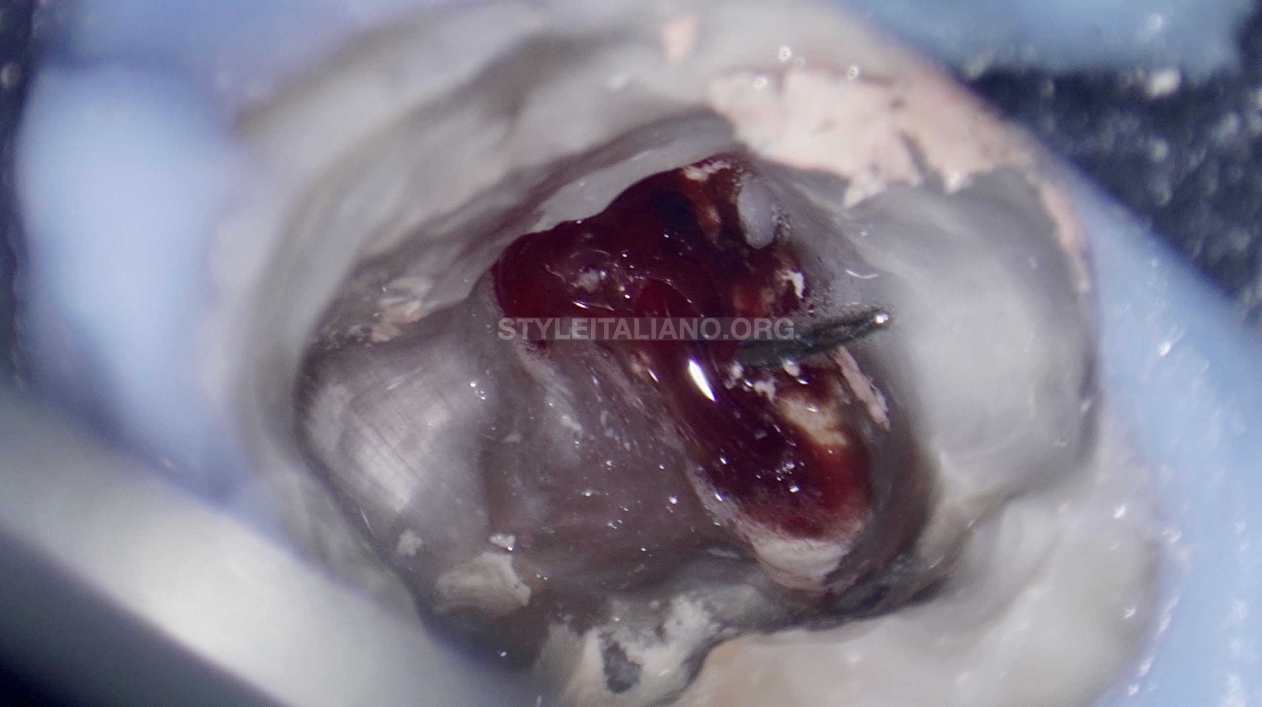
Fig. 5
The first surprise was the presence of a File underneath, moreover we had a good view of the perforation.
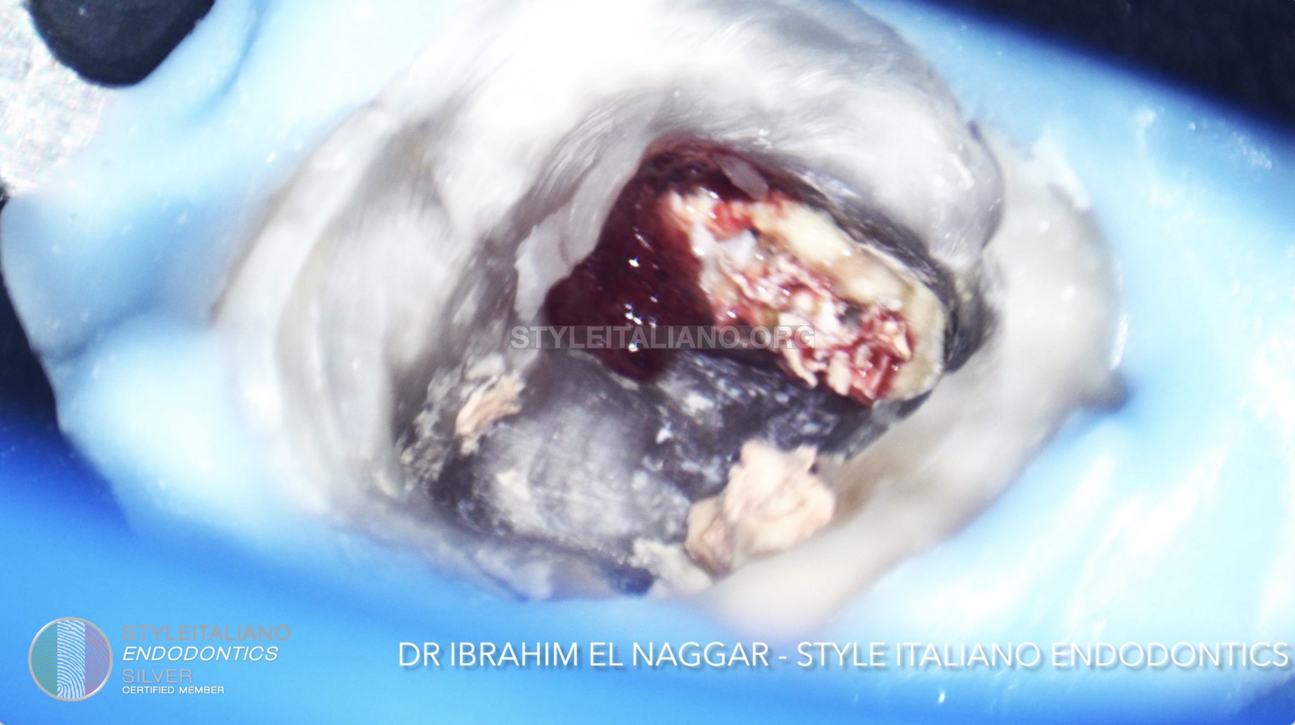
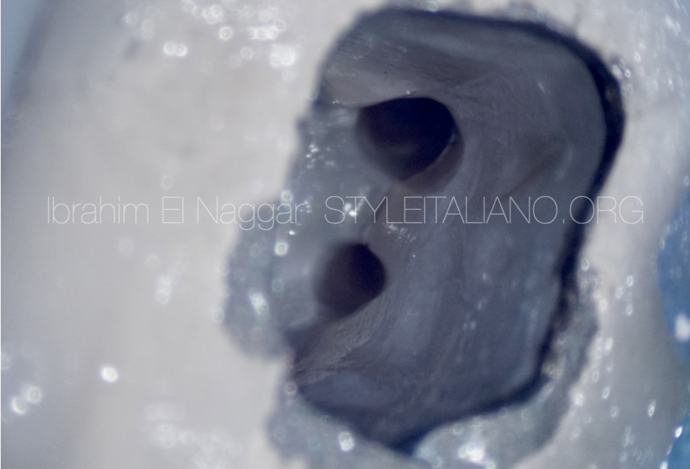
Fig. 6
Two canals and a huge perforation were visible
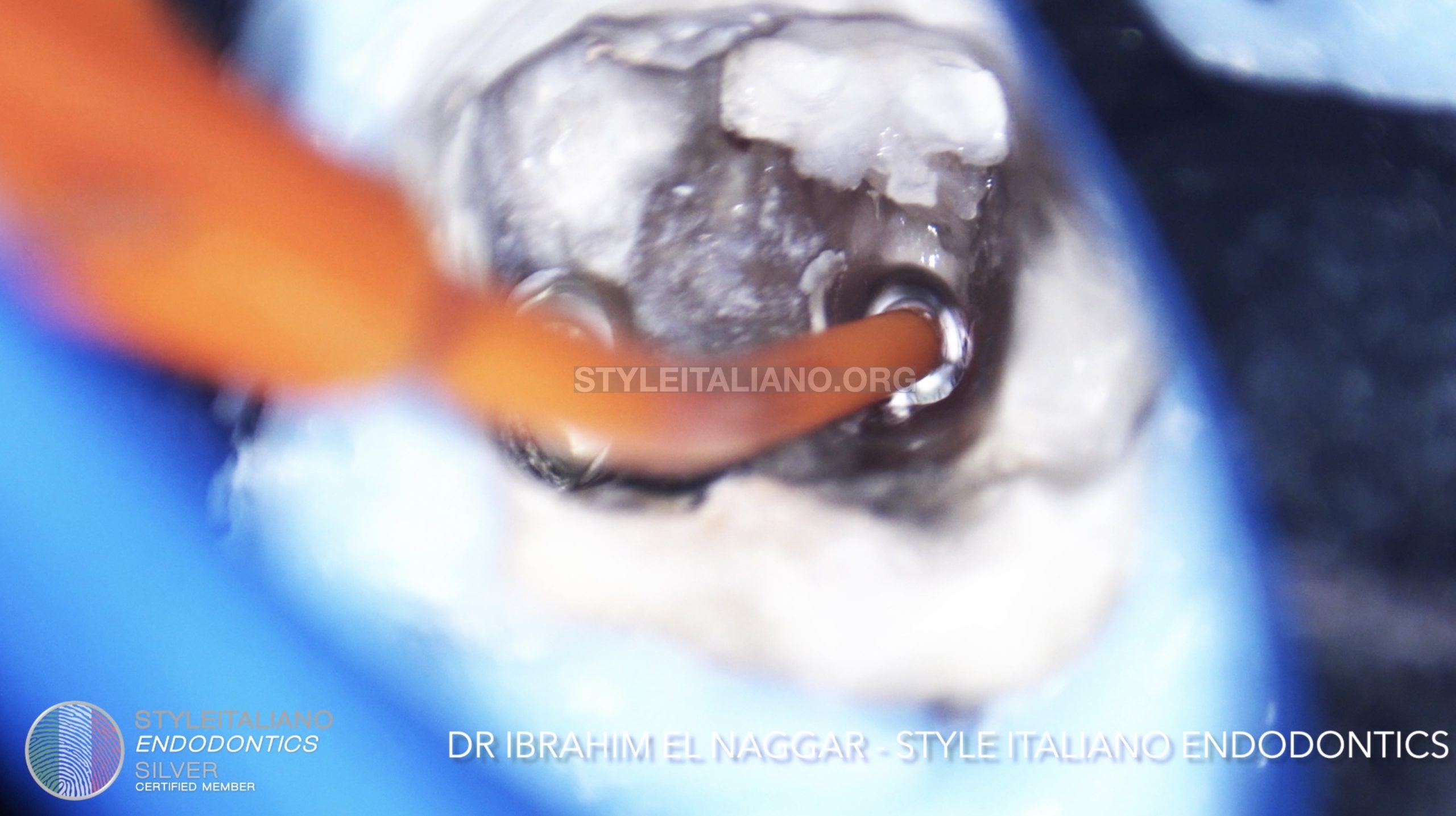
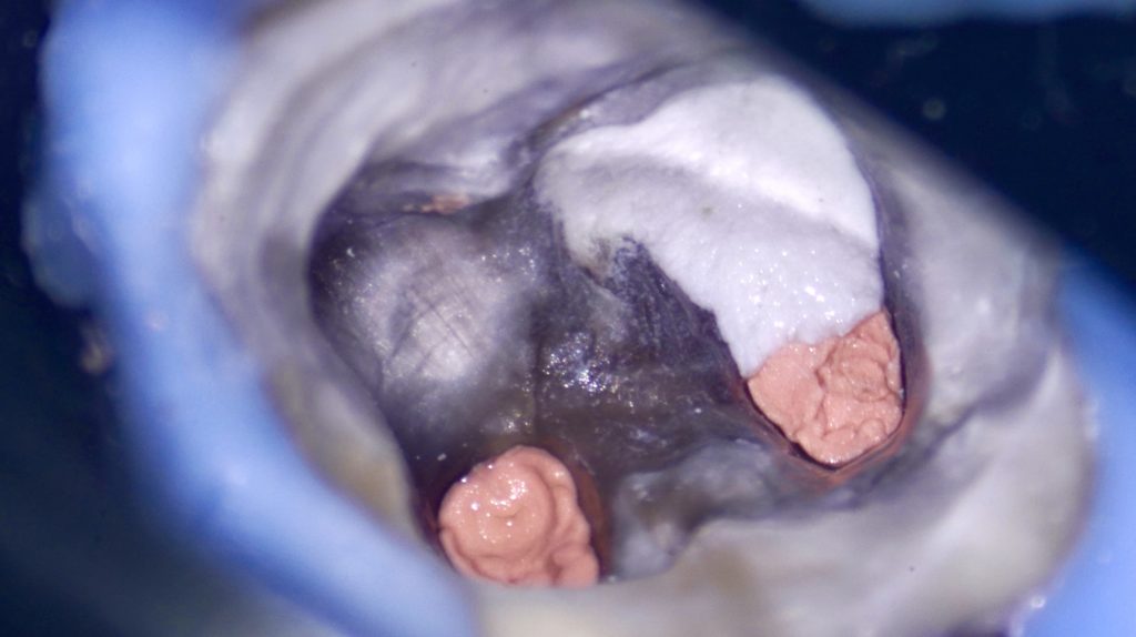
Fig. 7
The perforation site was closed with PTFE tape while starting with the retreatment of the two root canals
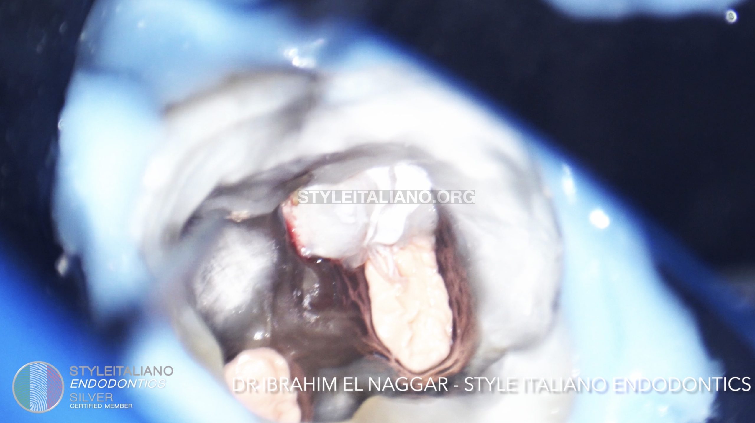
Fig. 8
Obturation done & PTFE in place.
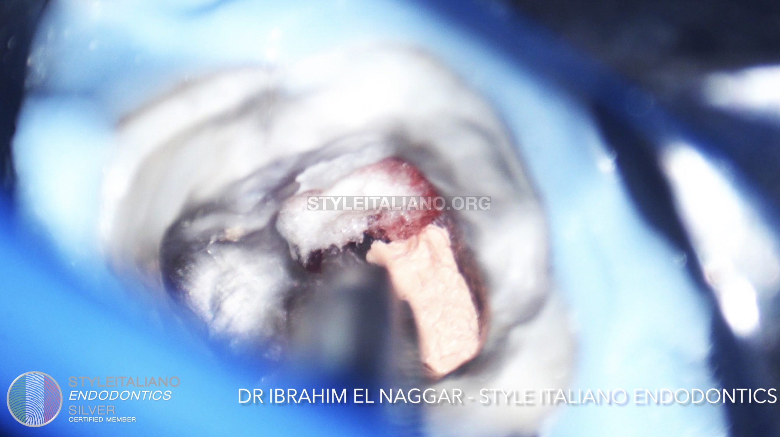
Fig. 9
A resorbable collagen foam was put in the perforation
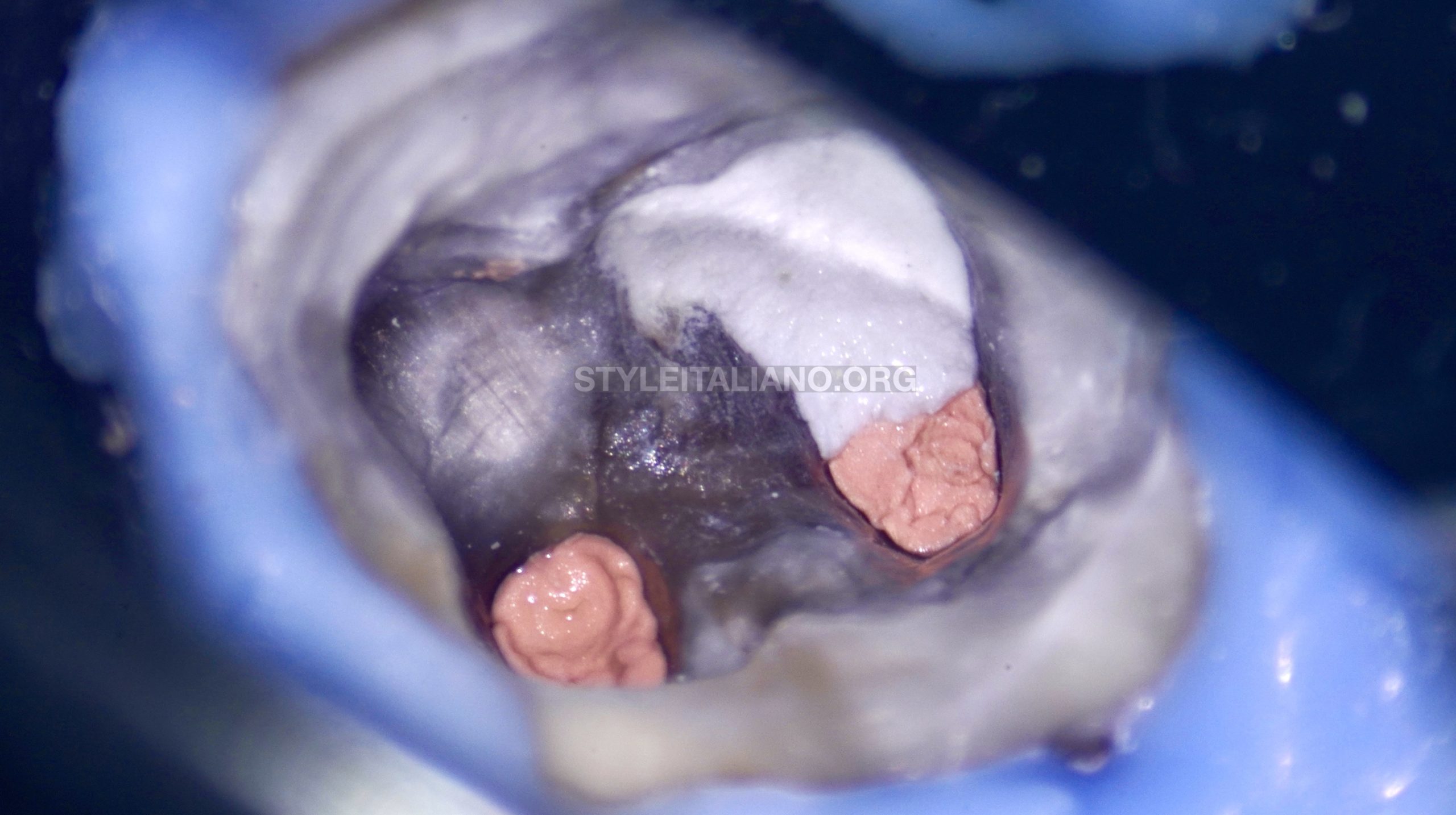
Fig. 10
A Bioceramic material was used to repair the perforation
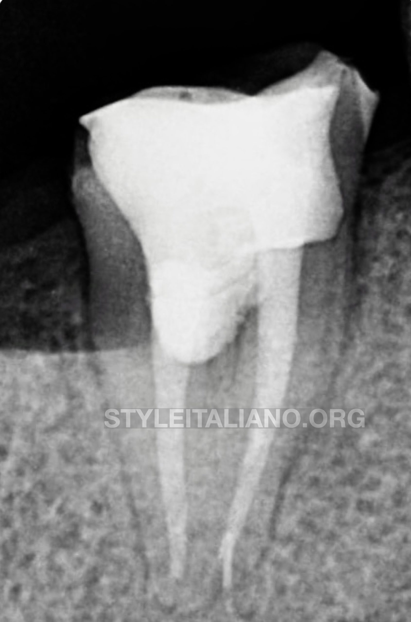
Fig. 11
Post Operative X-ray.
After retreating the tooth and repairing the perforation, the patient was sent back to the referring dentist.
Conclusions
The Clinician must be able to assess the current situation from diagnosis, treatment till prognosis and also to be able to deal with different clinical surprises during the Retreatment.
Bibliography
1. Endodontic Facts, American Association of Endodontists. Available at: http://www. aae.org/about-aae/news-room/endodontic-facts.aspx. Accessed June 1, 2015.
2. BorgesAH,BandecaMC,TonettoMR,etal.Portlandcementuseindentalrootper-
forations: a long term followup. Case Rep Dent 2014;2014:637693.
3. TsesisI,FussZV.Diagnosisandtreatmentofaccidentalrootperforations.Endodon-
tic Topics 2006;13:95–107.
4. Tsesis I, Rosenberg E, Faivishevsky V, et al. Prevalence and associated periodontal
status of teeth with root perforation: a retrospective study of 2,002 patients’ medical
records. J Endod 2010;36:797–800.
5. Gorni FG, Gagliani MM. The outcome of endodontic retreatment: a 2-yr follow-up.
J Endod 2004;30:1–4.
6. Clauder T, Shin S-J. Repair of perforations with MTA: clinical applications and
mechanisms of action. Endodontic Topics 2006;15:32–55.
7. Kvinnsland I, Oswald RJ, Halse A, Gronningsaeter AG. A clinical and roentgenolog-
ical study of 55 cases of root perforation. Int Endod J 1989;22:75–84.