Shaping Essentials and Furcal Canal Case Report
12/08/2021
Mohamad Zaafrany
Warning: Undefined variable $post in /home/styleendo/htdocs/styleitaliano-endodontics.org/wp-content/plugins/oxygen/component-framework/components/classes/code-block.class.php(133) : eval()'d code on line 2
Warning: Attempt to read property "ID" on null in /home/styleendo/htdocs/styleitaliano-endodontics.org/wp-content/plugins/oxygen/component-framework/components/classes/code-block.class.php(133) : eval()'d code on line 2
Endodontic anatomy is unpredictable, that’s why Preoperative radiographic analysis is very important step in managing endodontic challenges.
Some important data could be collected through good reading to preoperative radiograph like root length, canals number, estimated narrowness or canal sclerosis.
Once case difficulty is assessed, shaping technique and files sequence could be planned for safer canal shaping
Lesions in the furcation site could be of endodontic or periodontal origin so sufficient clinical examination like probing depth test will lead to more accurate diagnosis.
With normal probing depth an endodontic origin of the furcal lesion is more anticipated.
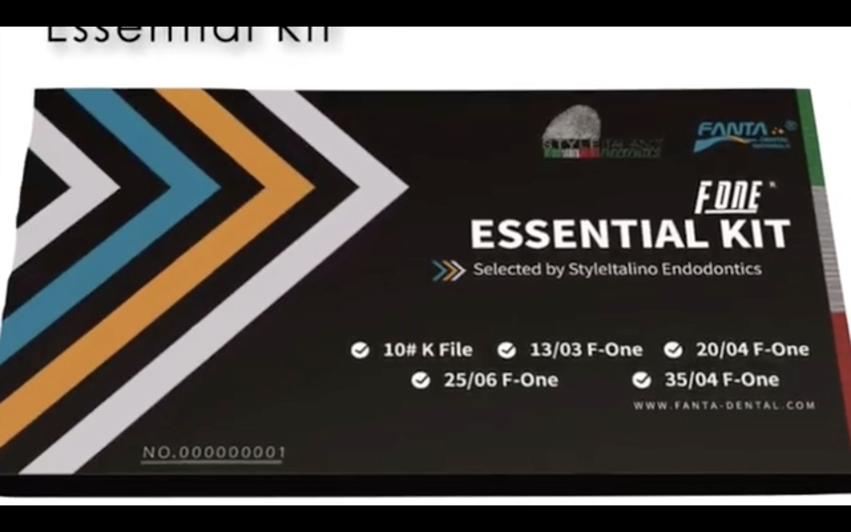
In this article I’ll describe shaping of long very narrow canals using the Essential kit from Fanta and report a rare condition of furcation canal in the floor of the pulp chamber.
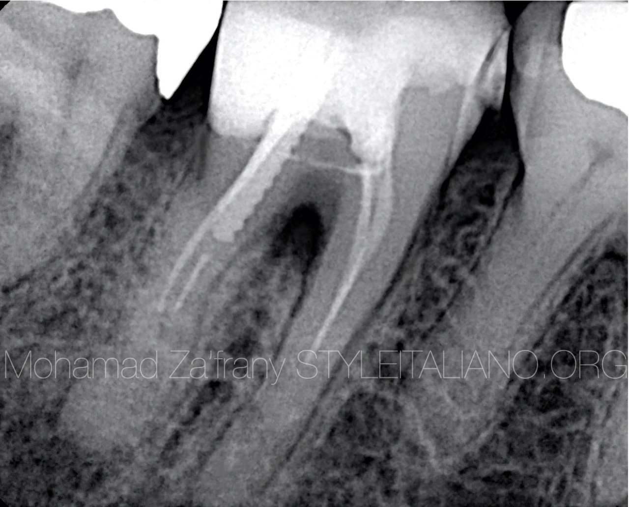
Fig. 1
Preoperative radiographic analysis
Tooth no. 46 was refereed for retreatment because of chronic abscess and repeated appearance of sinus tract.
Apical to old Gutta Percha, no tracing to canal space could be seen - that indicate narrow or sclerotic canals.
We can notice furcal lesion below roots trunk.
Lesion could be of periodontal origin: negative because of normal probing depth.
Lesion could be a result of crack or fracture specially with the presence of metal post in distal root
Lesion could be originated from lateral canal toward the furcation region.
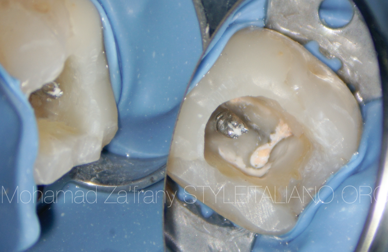
Fig. 2
After restoration removal, old Gutta Percha and old metal post exposed.
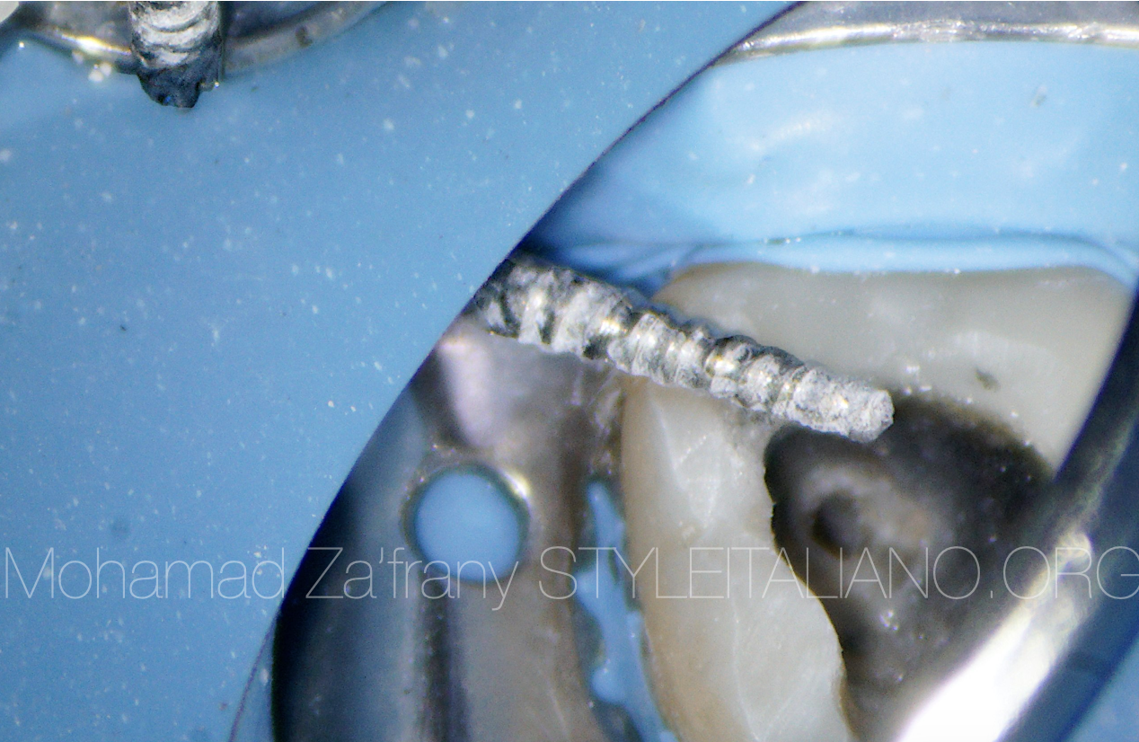
Fig. 3
With the use of ultrasonic vibration, the metal post was removed.
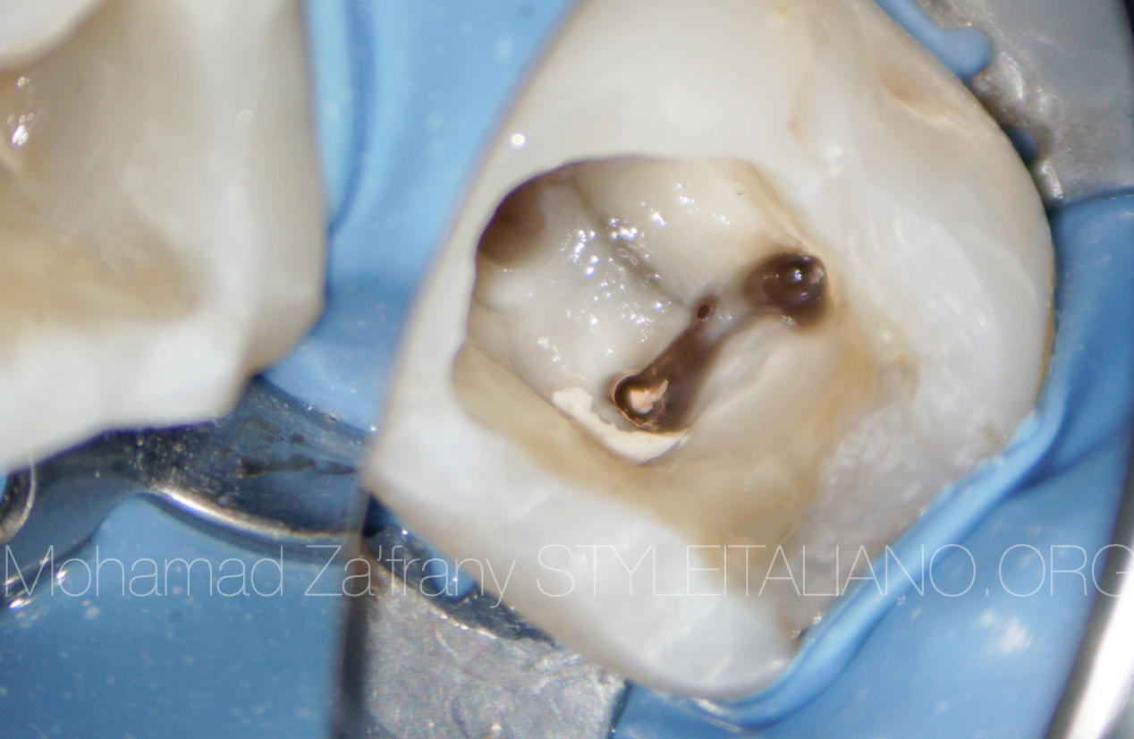
Fig. 4
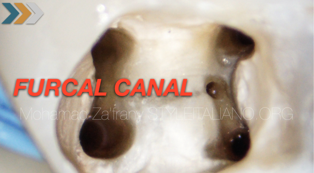
After removal of coronal Gutta Percha from the isthmus, a canal detected in MM region could be misdiagnosed as a perforation .
Thanks to color difference and floor map that recommend a normal anatomy rather than artificially made error.
Also with that kind of less care about quality of treatment it doesn’t seem to show a mismanipulated isthmus during canal locating procedures or shaping.
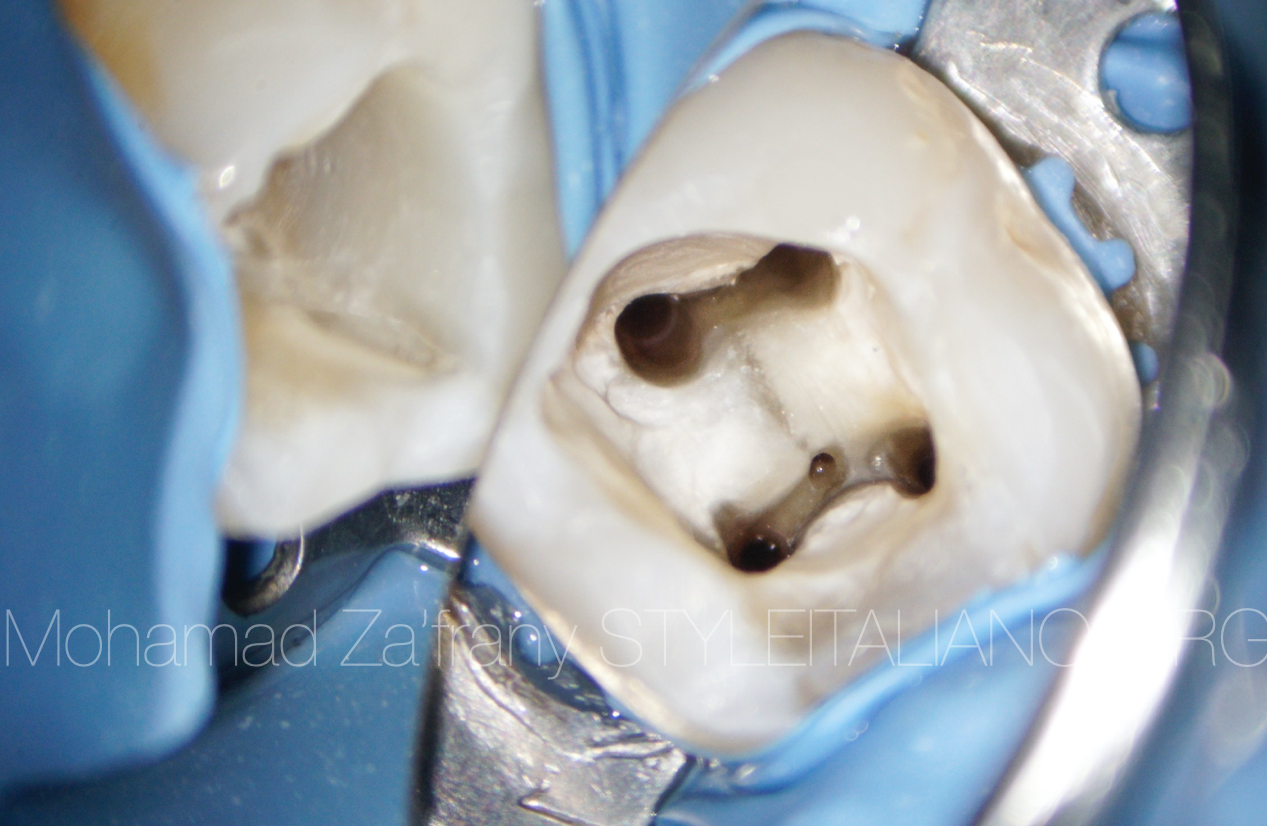
Fig. 5
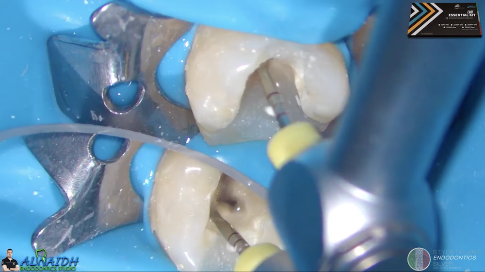
All canals cleaned from old GP and ready for managing the narrow mid and apical portions of the root.
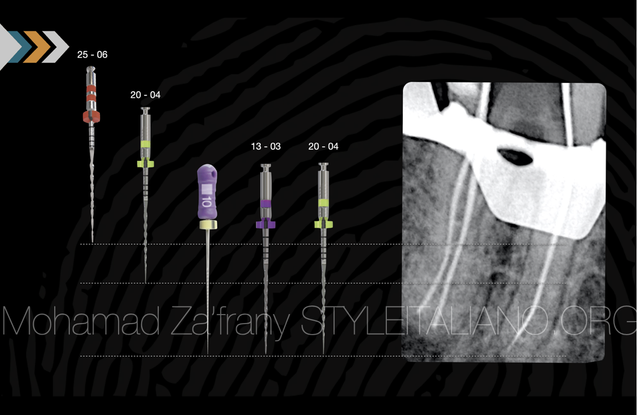
Fig. 6
After preflaring using 25 - 06 F1 design, 20 -04 used passively till resistance found.
Manual glide path using size 10 K file till full length.
Mechanical glide path done using 13 - 03 F1 file
Apical finishing and preparation done using 20 - 04 F1 till full length.
Irrigation activated sonically to avoid injury to root dentin structure using ultrasonics in skinny roots.
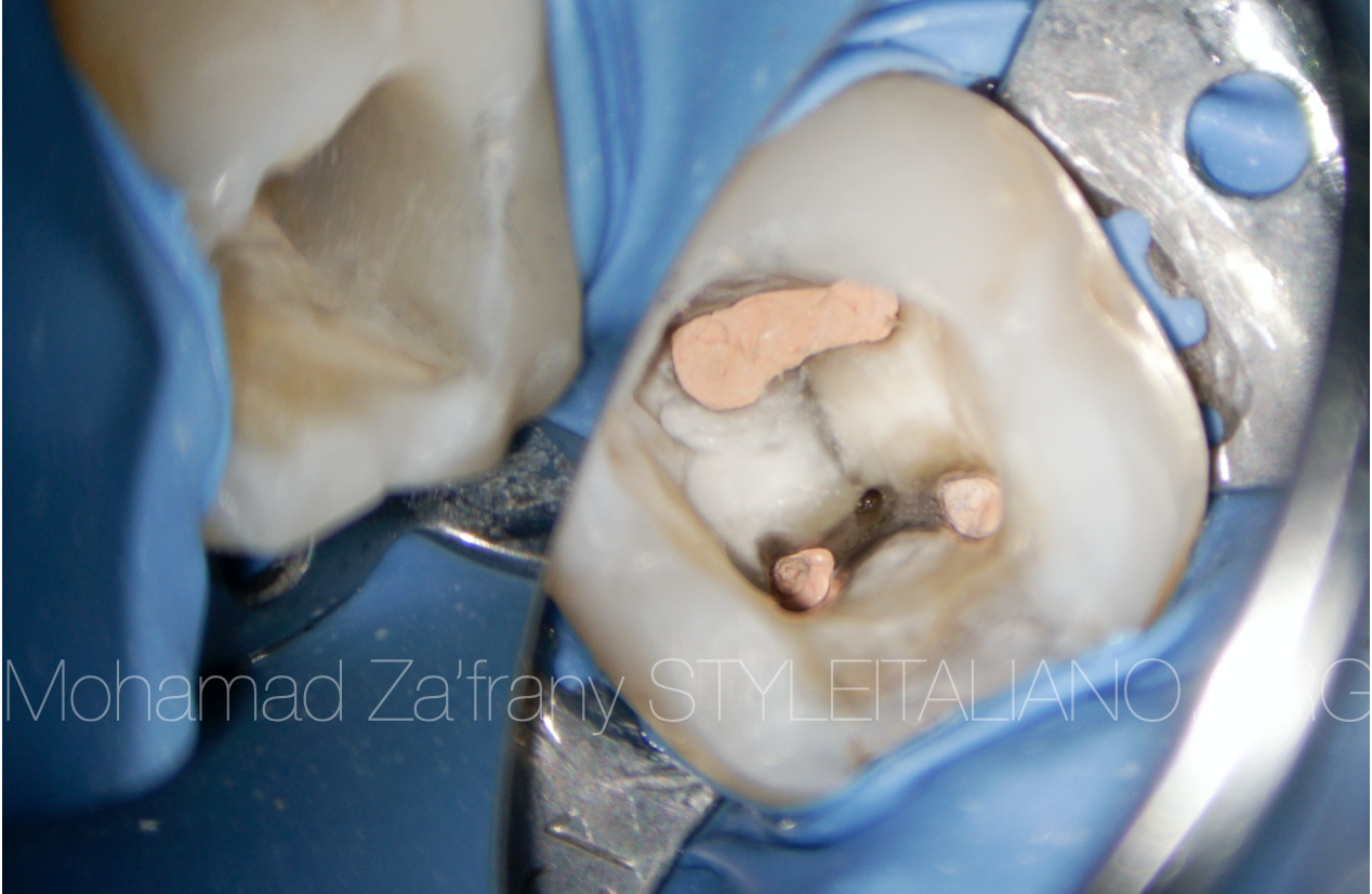
Fig. 7
Obturation done to the canals using GP and BC sealer in modified hot technique
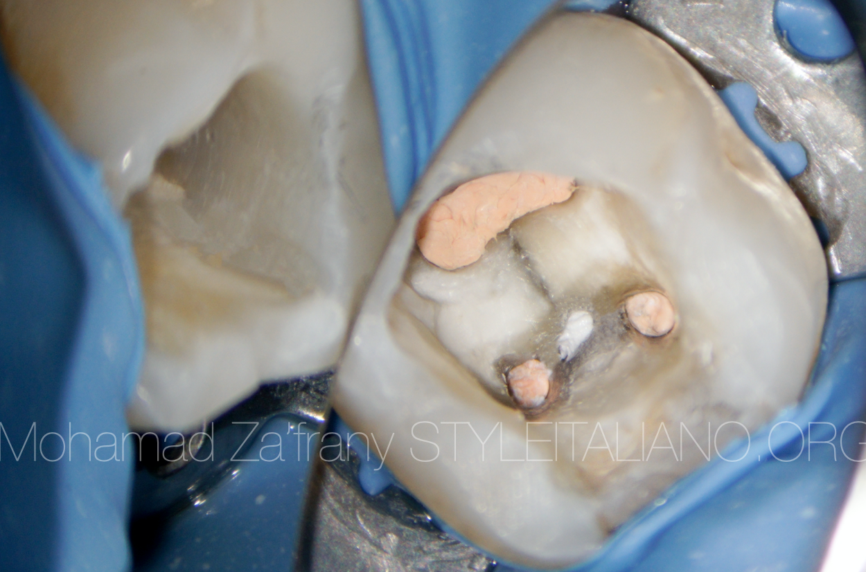
Fig. 8
Furcal canal obturated using Putty BioCeramic material
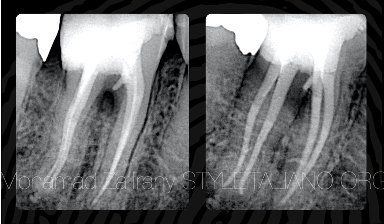
Fig. 9
Final radiographs showing obturation and filling to root canal system
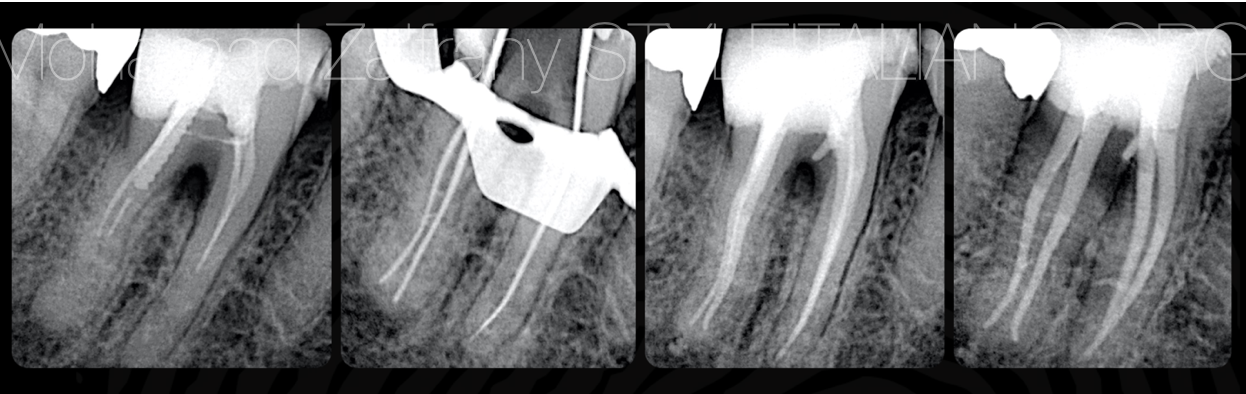
Fig. 10
Radiographic workflow
Case kept under follow up
Conclusions
Preoperative radiograph is very important before starting endodontic treatment
Case difficulty assessment will lead to successful treatment through good planning and selection of the approach technique and sequence of shaping files.
Essential kit files with flat design from Fanta and SIE show good performance during shaping of narrow long canals.
Knowlege of endodontic anatomy along with floor map guides and color difference is very important to distinguish between normal anatomy and iatrogenic errors.
Bibliography
Krasner P, Rankow HJ. Anatomy of the pulp chamber floor. JOE 2004; 30(1):5.
Cohen S, Hargreaves K. Pathways of the Pulp 9th ed. Mosby, St. Louis, MO, 2006.
Vertucci, F. J., & Williams, R. G. (1974). Furcation canals in the human mandibular first molar. Oral Surgery, Oral Medicine, Oral Pathology, 38(2), 308–314. doi:10.1016/0030-4220(74)90073-5
Trope M, Bunes A, Debelian G. Root filling materials and techniques: bioceramics a new hope? Endodontic Topics 2015, 32, 86–96