Modification in canal preparation for long and narrow canals of lower first molar
01/02/2021
Ahmed Alwaidh
Warning: Undefined variable $post in /home/styleendo/htdocs/styleitaliano-endodontics.org/wp-content/plugins/oxygen/component-framework/components/classes/code-block.class.php(133) : eval()'d code on line 2
Warning: Attempt to read property "ID" on null in /home/styleendo/htdocs/styleitaliano-endodontics.org/wp-content/plugins/oxygen/component-framework/components/classes/code-block.class.php(133) : eval()'d code on line 2
Molar teeth exhibit more difficulty in canal preparation, due to more complexities in their root canal system. The situation becomes more complex when the roots are long, and even more difficult if they are narrow and calcified. Many iatrogenic complications may encounter if there was not a good appraisal of the case and a systematic treatment plan, and sometimes, modification in the canal preparation.
A 26 years old female presented complaining of a moderate throbbing pain in her lower left first molar (LL6) that increased with food impaction. Clinically, LL6 had a deep distal decay. With the application of cold test, moderate throbbing pain started after few seconds and disappeared gradually after the removal of the cold test. LL6 was negative to tenderness.
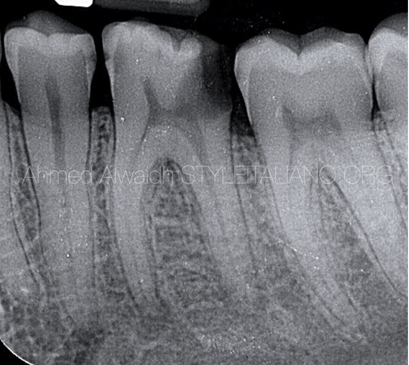
Fig. 1
Pre-operative IOPA of LL6 revealed a deep distal decay with evident pulp exposure. PA radiolucencies around the apices of LL6.
It was also evident from the radiograph that LL6 had long roots.
Diagnosis: LL6 with symptomatic irreversible pulpitis (SIP) and asymptomatic apical periodontitis (AAP).
Treatment plan: Non-surgical root canal treatment of LL6 with full coronal coverage to restore function.
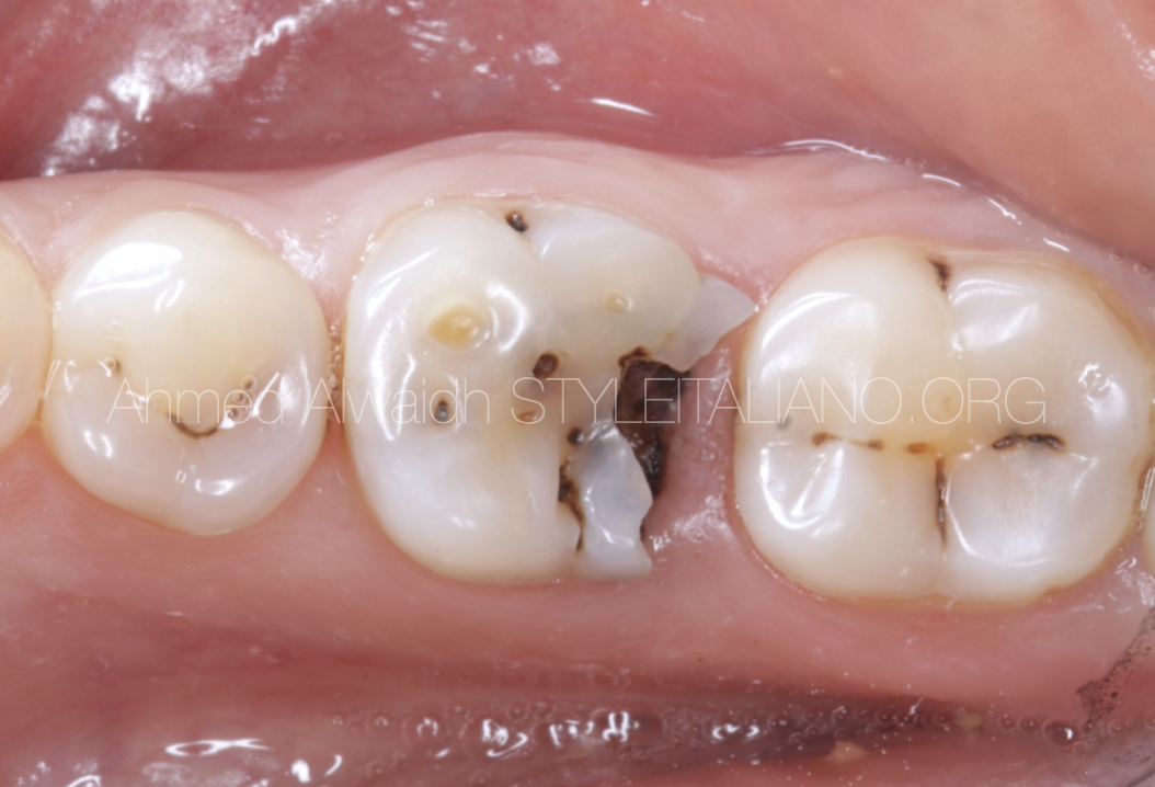
Fig. 2
Pre op image
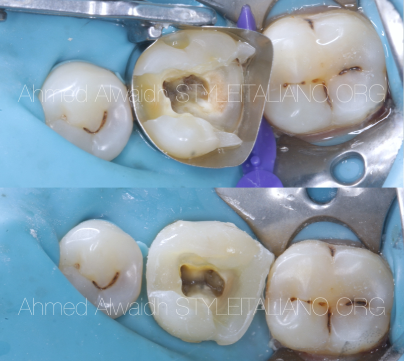
Fig. 3
After isolation with rubber dam and removing the decayed tissue, LL6 was converted into class I cavity to ensure restorability of the tooth and to enhance the irrigation procedure.
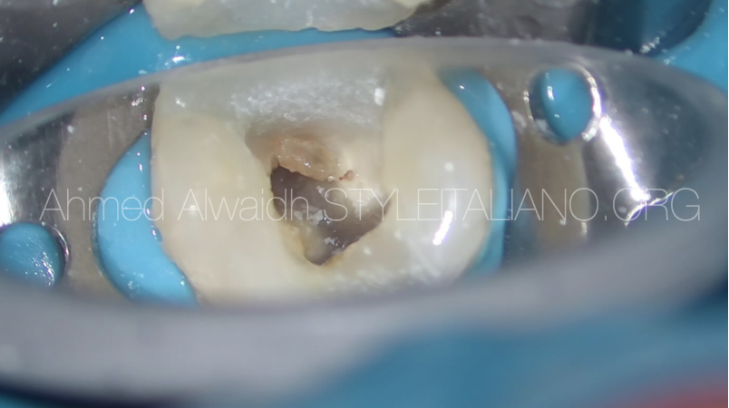
Fig. 4
Pulp stone on the distal canal orifice was removed using ultrasonic tip.
Due to long and narrow canals of this LL6, canals preparation was modified in order to prepare the canals safely. Besides, Endodontic files should be selected carefully that are safe, have good cutting efficiency and good flexibility too.
Step 1: Coronal preflaring:
- Orifices negotiation with k file #10/.02.
- F One file #25/.06 to a depth of 4-5 mm for straight line access.
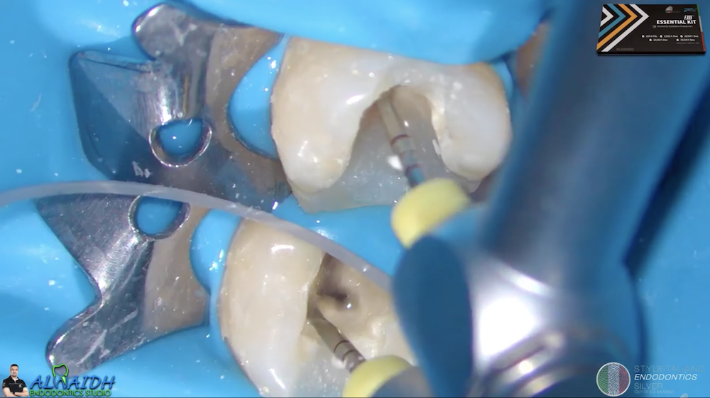
Step 2: Middle third preparation:
- Using F One GliderST file #13/.03 to a depth of 19 mm.
- Using F One #20/.04 file to a depth of 19 mm.
Step 3: Glide path of the canal to full working length.
- Negotiating the canals using K file #10/.02 with watch-wind motion to full working length with the aid of electronic apex locator. Working length was 24 mm. Mesial canals were very torturous, which was evident after the removal of the K file #10/.02.
- Using F One GliderST file (#13/.03) to full working length.
Step 4: Final preparation of the canals.
- Mesial canals were prepared with F One file #20/.04 to full working length.
- Distal canal was prepared with F One file #25/.06 to full working length.
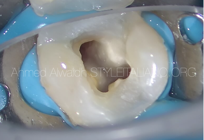
Fig. 5
Final irrigation was done with NaOCl 5.25% and EDTA 17%, agitated using both PUI (passive ultrasonic irrigation) and sonic (EDDY tips).
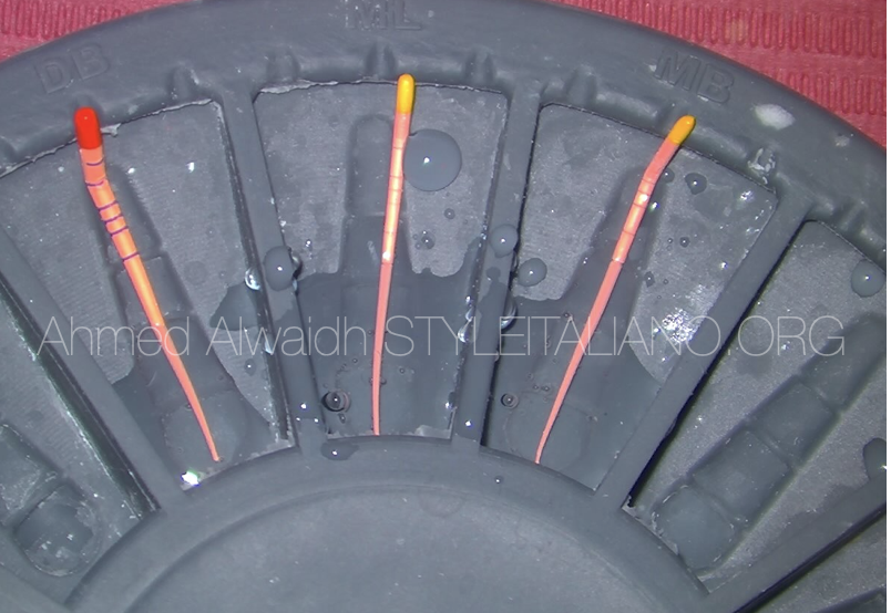
Fig. 6
After assessing apical gauges of the canals, the correspondent gutta percha cones were disinfected with NaOCl 5.25% for 3 min. Thanks to Plano which helps greatly in organising the master cones.
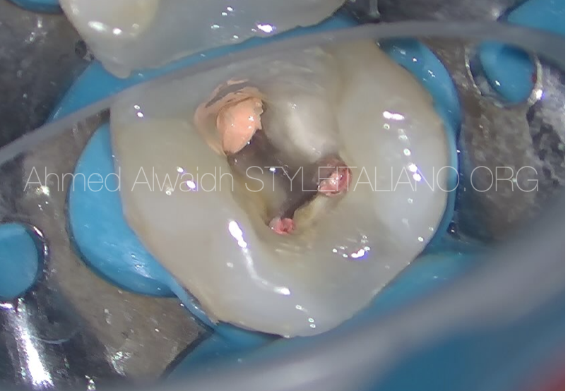
Fig. 7
Hydraulic obturation with pre-mixed bioceramic sealer.

Fig. 8
Final IOPA of LL6.
Patient been scheduled for full coverage crown.
Conclusions
The aim of Endodontic treatment is to cure or prevent apical periodontitis (Ørstavik & Pitt Ford 2008) which is mostly caused by bacteria (Kakehashi et al. 1965). To reach this goal, we need to remove remove vital and necrotic tissues, bacteria and their toxins from root canal system, and to fill this complex system with a root filling capable to produce a hermetic seal to prevent reinfection of the root canal space and prevent leakage of bacterial products into the periapical area. Yet, these objectives sometimes become difficult, especially in long, narrow and calcified canals, and iatrogenic complications may encounter like ledges, canal transportation, perforation…etc that may preclude complete chemo-mechanical debridement of the root canals.
Good interpretation of the pre-operative IOPA, making a good treatment plan of the case, and choosing the right files can decrease the incidence of the above-mentioned complications and promote higher successful procedure with more predictable outcomes.
F One files were chosen in this case due to their unique flat side design, which offers safety, higher cutting efficiency, in addition to their high flexibility (Alwaidh 2019). On the other hand, the Style Italiano Endodontics selection of F One files offers a great range to deal with even the most complicated cases, like this case report. Although I use F One files as a single file preparation in most of the cases, but there are some cases that needs our attention to make even modification of the sequence that is being set by the manufacturer, depending on the good appraisal of the case.
Bibliography
Alwaidh A (2019) Managing severely curved canal with Fanta AF F-One file, https://popdentistry.styleitaliano.org/articles/managing-severely-curved-canal-with-Fanta-AF-F-One-file.
Kakehashi S, Stanley HR, Fitzgerald RJ (1965) The effects of surgical exposures of dental pulps in germ-free and conventional laboratory rats. Oral surgery, oral medicine, oral pathology 20(3), 340-349.
Ørstavik D, Pitt Ford TR (2008) Apical Periodontitis: Microbial infection and host responses. In: Orsta Ørstavik vik D and Pitt Ford TR, Essential Endodontology 2nd edition. Oxford: Blackwell Science, 1-9.