Selective Root Canal Re-Treatement of Lower Left Pre-Molar
15/07/2024
Fellow
Warning: Undefined variable $post in /home/styleendo/htdocs/styleitaliano-endodontics.org/wp-content/plugins/oxygen/component-framework/components/classes/code-block.class.php(133) : eval()'d code on line 2
Warning: Attempt to read property "ID" on null in /home/styleendo/htdocs/styleitaliano-endodontics.org/wp-content/plugins/oxygen/component-framework/components/classes/code-block.class.php(133) : eval()'d code on line 2
In retreatment cases , the clinician has to deal with many mishaps such as underfilled, missed and obstructed canals as also with iatrogenic damages as separated instruments and perforations. Underfilled and missed canals are the main reasons for endodontic failures (>30%).Especially underfilling can be a real challenge since it can strongly reduces the success rate upto 3-4 % of endodontic failures and extraction of endodontically treated teeth might be required. Knowledge of Anatomical variations and is the main factor influencing the outcome of successful endodontic re-treatment.
i
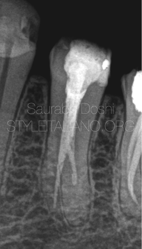
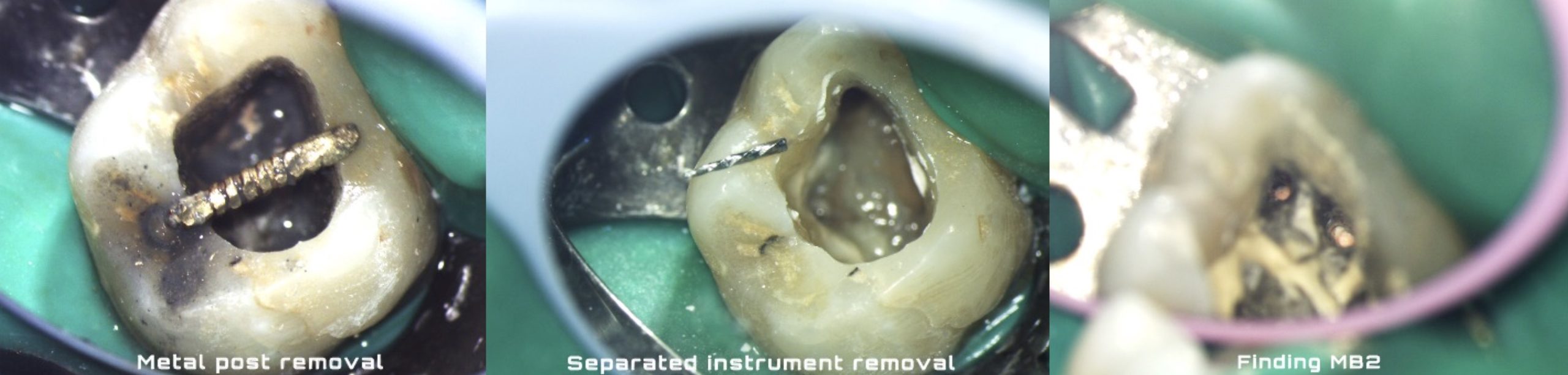
Fig. 1
The 46 years old patient was referred to our clinic by his general dental practitioner for endodontic retreatment. She complained about experiencing bitting tenderness on tooth 35.Underfilled canal was seen in the diagnostic radiograph suggestive of pulp remnants inside the canal.
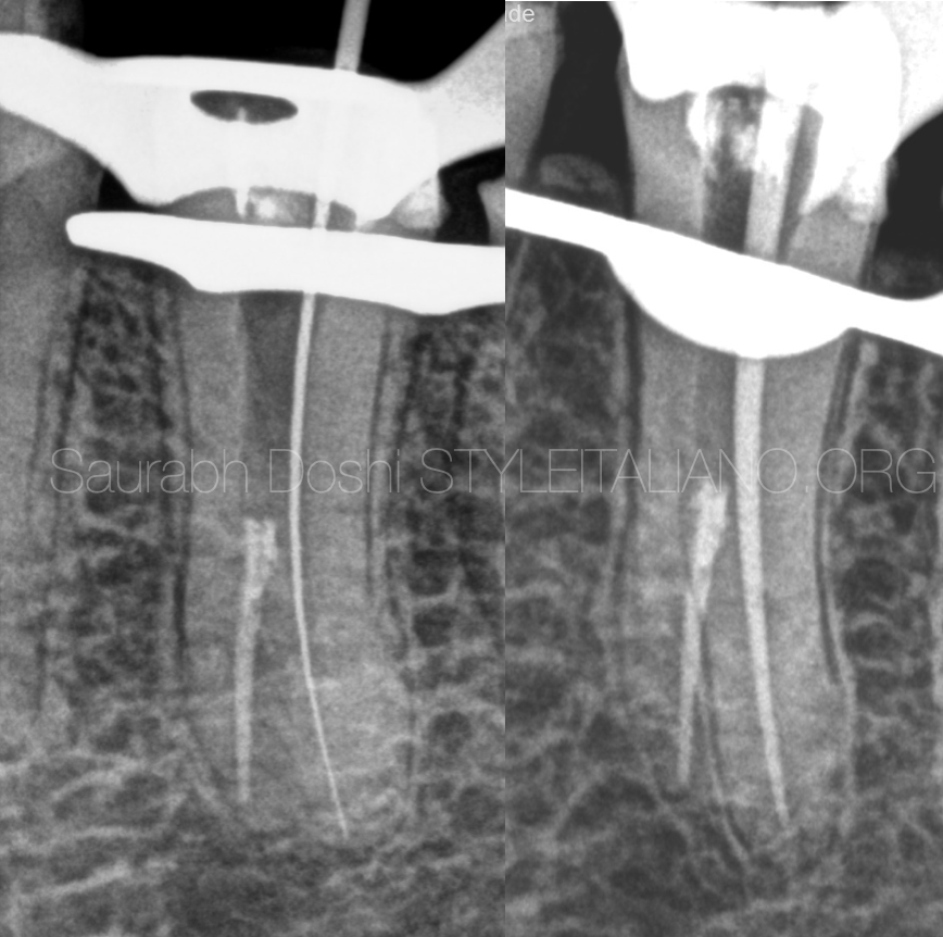
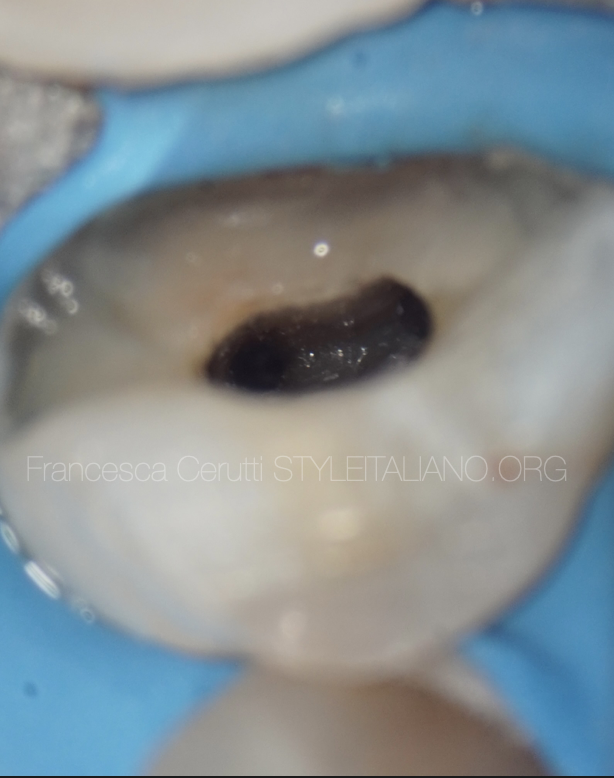
Fig. 2
The access cavity was driven by removing the coronal filling material and the infected dentine.
Old gutta percha was removed . Working length was established using #10K file only in distal canal.
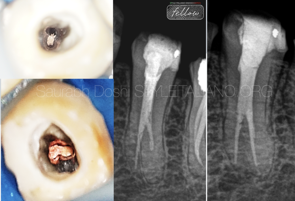
Distal canal was prepared and shaped with 25.04% azure file and master cone selection was done.
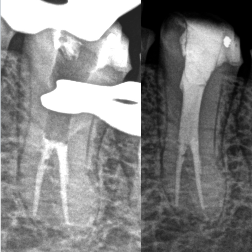
Fig. 3
The distal canal was obturated with single cone technique using bioceramic sealer followed by back fill in the remaining coronal third canal space.
Fig.5
Post endodontic restoration was completed using bulk fill restoration.
Fig. 4
About the author:
Dr Saurabh Doshi
2015: Diploma in Laser dentistry (Austria)
2017: Master with homer in Endodontics from M. A. Ragoonwala Dental College and research centre, Pune, India
2021: Diplomate of Indian Board of Endodontics from prestigious Indian Endodontic Society.
Author of the book "The root canal obturation - Past, Present and Future"
Speaker in various Indian Dental Association Forums.
A passionate clinician, avid sportsman and doting father.
Conclusions
Clinicians should be aware of the potential anatomical complexities of the mandibular premolars which possess the ability to implement their own knowledge in clinical and radiographic interpretation. Anatomical variation in premolars can be treated predictabily with proper diagnosis and treatment planning.
Bibliography
Vertucci F.J.,Seeling A.,Gillis R.(1974) Root canal morphology of the human mandibular second premolars. Oral Surgery.38:456-464
Yang L, Han J, Wang Q, Wang Z, Yu X, Du Y. Variations of root and canal morphology of mandibular second molars in Chinese individuals: a cone-beam computed tomography study. BMC oral health. 2022;22(1)
Siqueira JF. Aetiology of root canal treatment failure: why well-treated teeth can fail. Int Endod J. 2001;34(1):1–10.