Retreatment of Mandibular First Molar
05/08/2023
Fellow
Warning: Undefined variable $post in /home/styleendo/htdocs/styleitaliano-endodontics.org/wp-content/plugins/oxygen/component-framework/components/classes/code-block.class.php(133) : eval()'d code on line 2
Warning: Attempt to read property "ID" on null in /home/styleendo/htdocs/styleitaliano-endodontics.org/wp-content/plugins/oxygen/component-framework/components/classes/code-block.class.php(133) : eval()'d code on line 2
There have been many recent technical advances in modern endodontics that have the potential to affect treatment outcomes. The purpose of this article is to determine the success of nonsurgical root canal retreatment in molars using contemporary endodontic techniques.
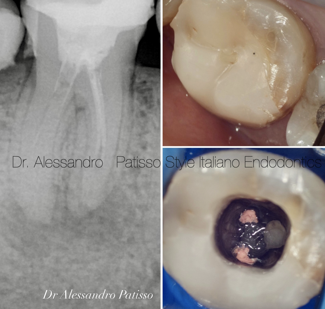
Fig. 1
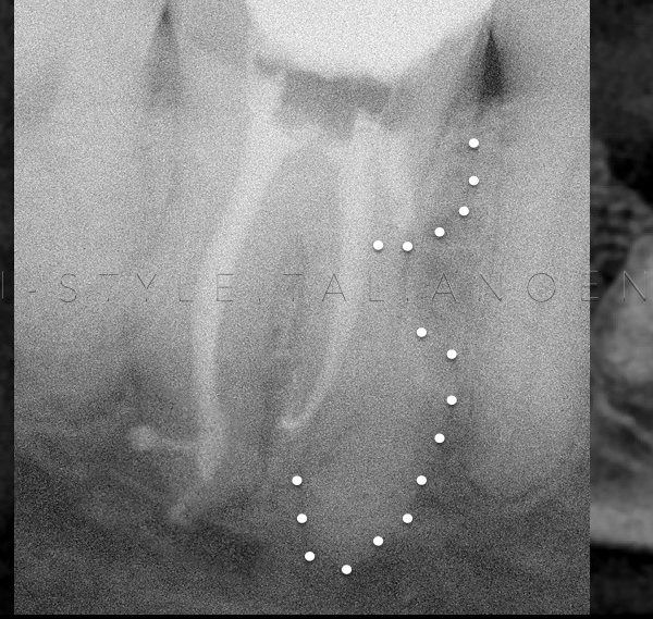
A 52-year-old patient presented himself who complained of severe pain in the fourth quadrant, the radiology examination revealed a periapical lesion affecting the dental element 46 previously already treated endodontically about 5 years earlier. The tooth had an endodontic pin in the distal root. The patient is explained that the old treatment was inappropriate and the tooth had to be re-treated.
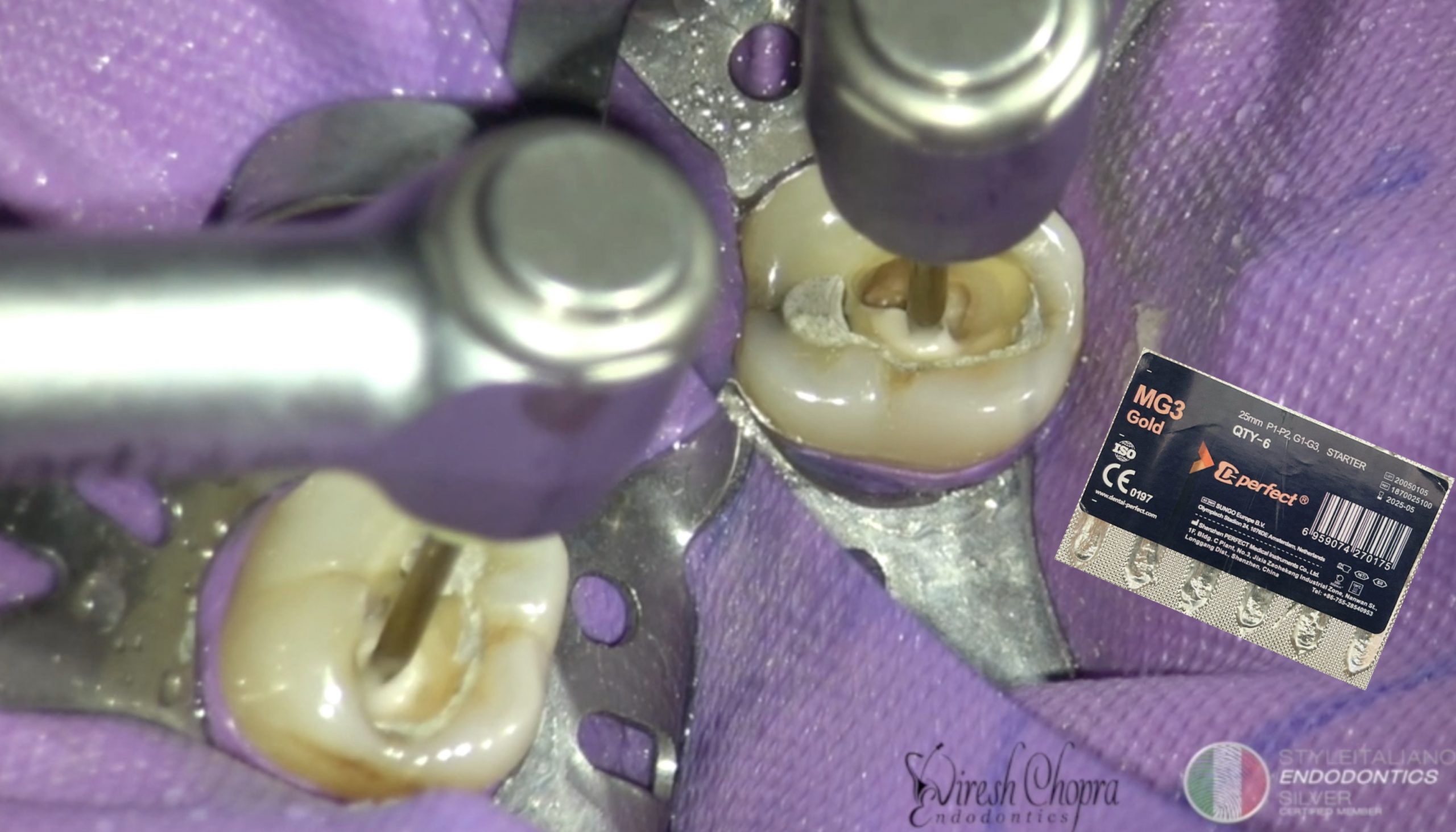
The use of ultrasonic tips in endodontics guarantees maximum precision,the tip works on the fiber post by consuming it, preserving dental tissue.The ultrasonic tip is used following the axis of the post to avoid creating perforations.
We continue to wear out the fiber post also trying to loosen it, trying to wear out the cement used to block it even with the ultrasonic tip, checking the mobility of the post. When the fiber post is not released, it needs to be consumed to the end.
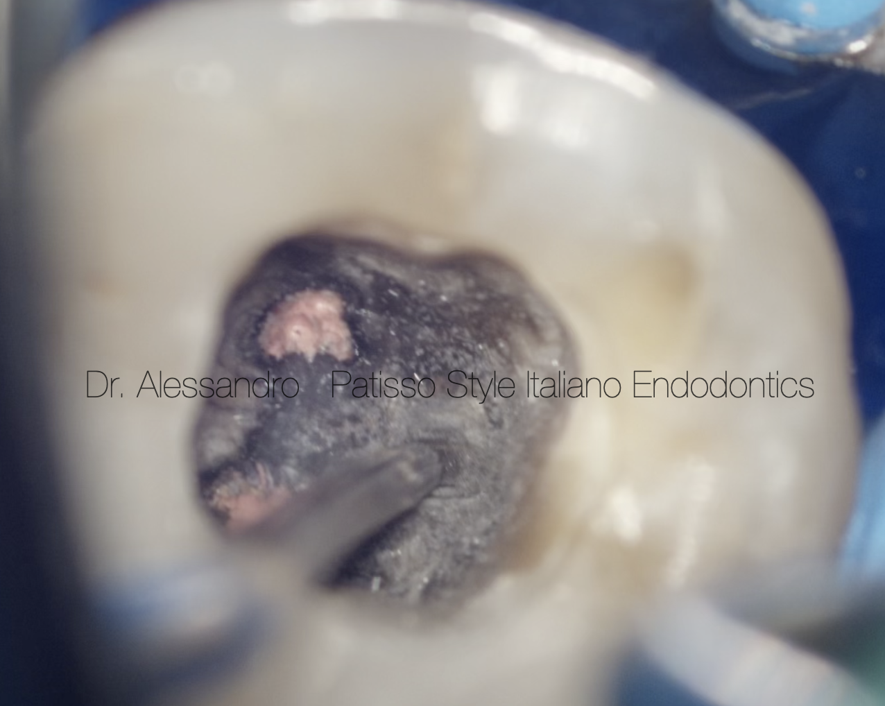
Fig. 2
The use of ultrasonic tips in endodontics guarantees maximum precision,the tip works on the fiber post by consuming it, preserving dental tissue.The ultrasonic tip is used following the axis of the post to avoid creating perforations.
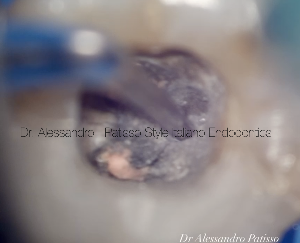
Fig. 3
The ultrasonic tips degrease the cement used to fix the post, this allows us to remove the fiber post without consuming it entirely.
Fig. 4
We continue to wear out the fiber post also trying to loosen it, trying to wear out the cement used to block it even with the ultrasonic tip, checking the mobility of the post. When the fiber post is not released, it needs to be consumed to the end.
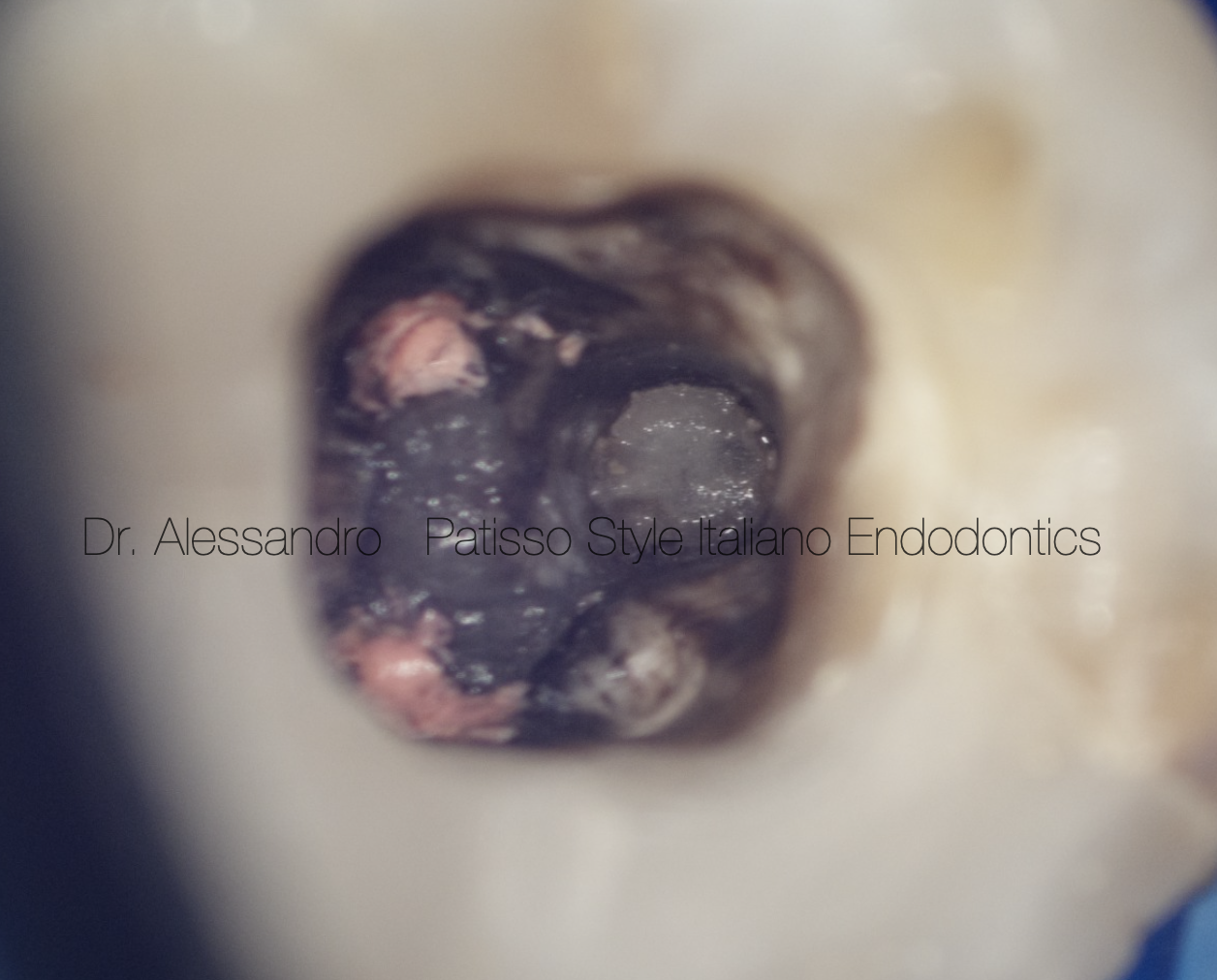
Fig. 5
The ultrasonic tips degrease the cement used to fix the post, this allows us to remove the fiber post without consuming it entirely.
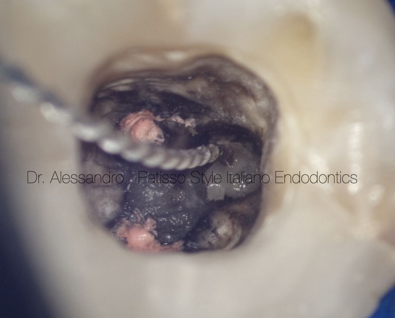
Fig. 6
With manual tools we try to go beyond the fiber pin gradually increasing the diameter, to facilitate the entry of other larger tools and make it even more mobile.
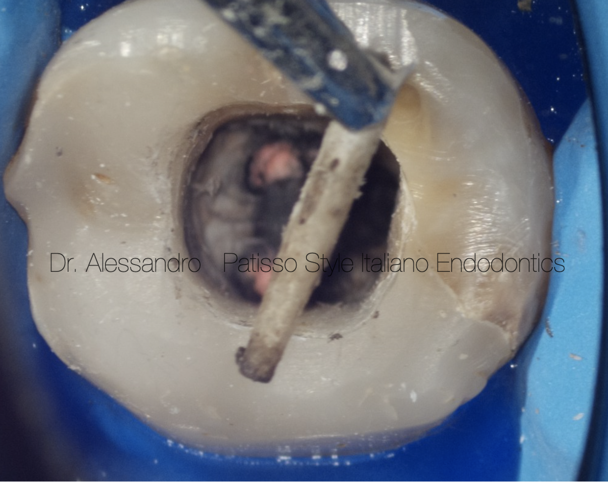
Fig. 7
Once the fiber post has been passed, use a hand tool with a larger diameter to hook it and remove it.
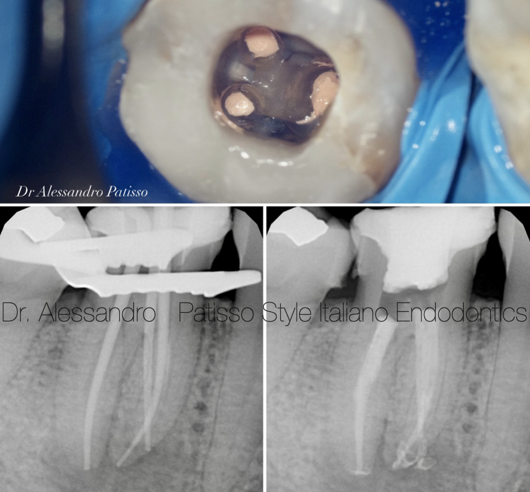
Fig. 8
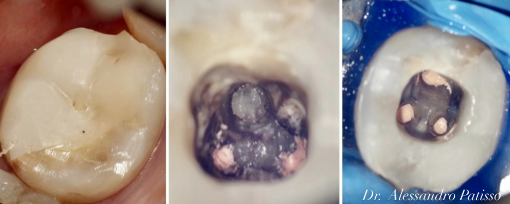
Once the post is removed, we focus on removing the rest of the cement and gutta-percha.
After cleaning and mechanically instrumenting the canals, we dedicate ourselves to the activation of irrigants such as sodium hypochlorite.
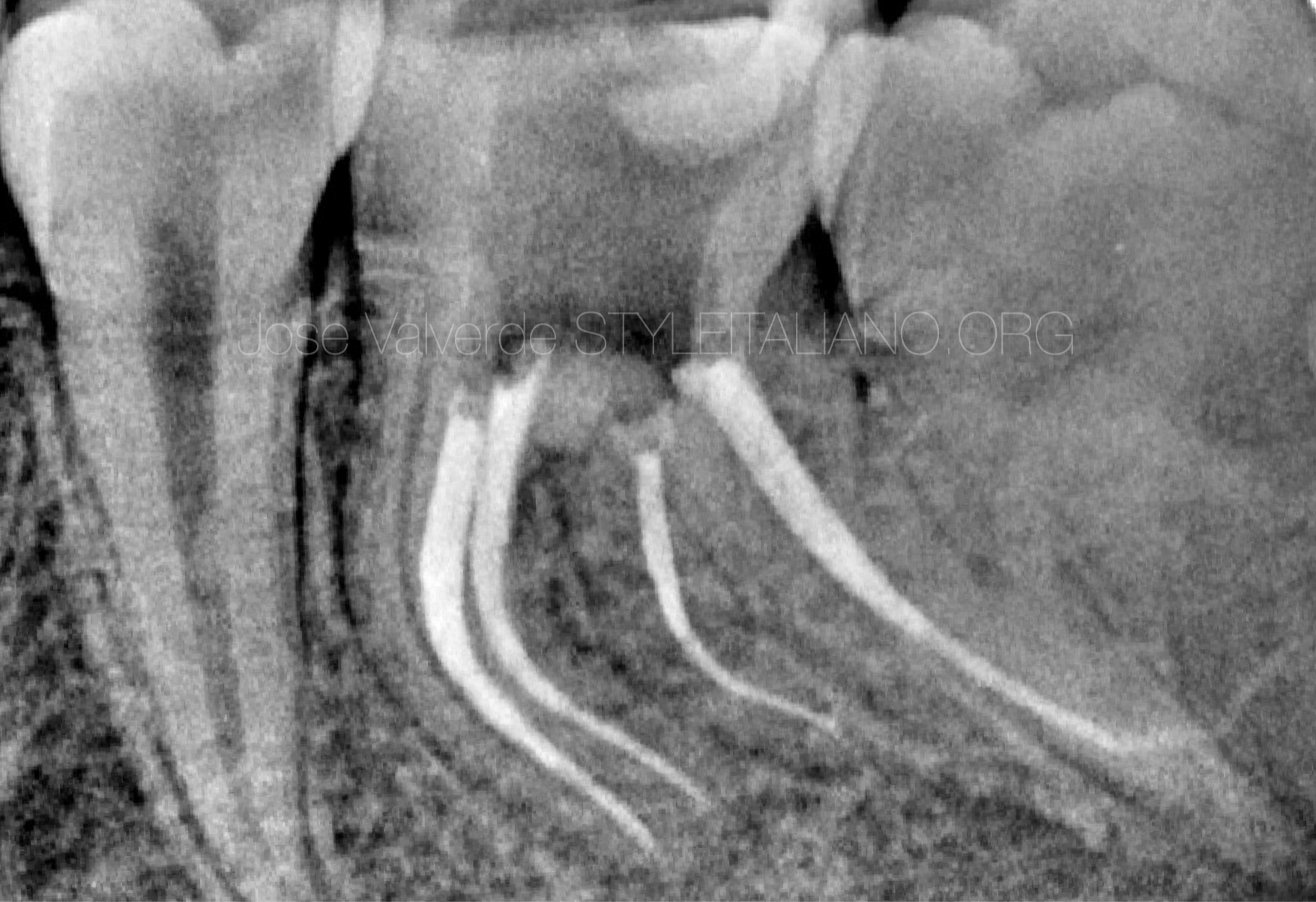
We verify the working length with an intraoperative X-ray and finally obturate the canals.
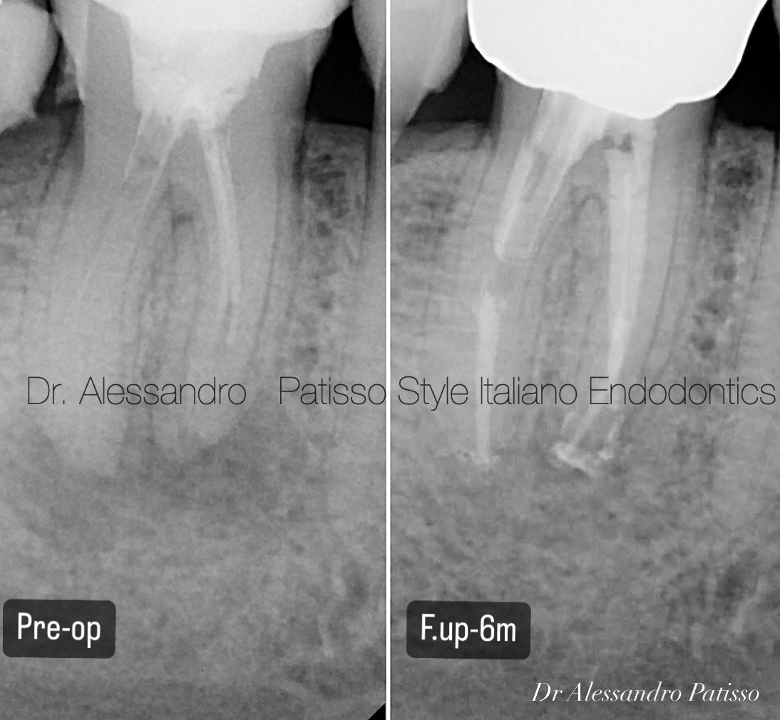
Fig. 9
Initial radiograph with a follow-up six months after completion of the tooth.

Fig. 10
About the Author
Graduated in Dental Hygiene at the University of Bari in 2009 and Dentistry and Dental Prosthetics at the Universidad Europea de Madrid in 2015.
From the beginning he has practiced exclusively Clinical Edodontics, perfecting himself through numerous courses around Italy. Among the most recent he attended the courses of Dr Giuseppe Carrieri, finishing first in the course.
Today he works as a freelancer using only the microscope.
Tutor in the courses of Dr Giuseppe Carrieri Fellows member of Styleitaliano Endodontics.
Conclusions
The use of the ultrasonic tips made the job of removing the fiber post easier, improving the visibility of the operating field the other hand the risks of over enlargement of the post space, or removal of healthy surrounding dentine was avoided by exclusively working on the post thanks to the better visibility given by the high magnification and light provided by the operatory microscope.
Bibliography
- Schilder H. Filling root canals in three dimensions. Dent Clin North Am. 1967; : 723-74Reit C. Grondahl H.G. Management of periapical lesions in endodontically treated teeth:ùstudy on clinical decision making. Swed Dent J. 1984; 8: 1-7
- American Association of Endodontists. Glossary of Endodontic Terms, 8th ed. Chicago: American Association of Endodontists; 2012.
- Hauman, C. H. J., Chandler, N. P., & Purton, D. G. (2003). Factors influencing the removal of posts. International Endodontic Journal, 36(10), 687–690.
- Siqueira JF, Barnett F. Interappointment pain: mechanisms, diagnosis, and treatment. Endodontic Topics 2004, 7, 93109