Retreatment of a three rooted maxillary second premolar
04/11/2021
Gabriele Ragucci
Warning: Undefined variable $post in /home/styleendo/htdocs/styleitaliano-endodontics.org/wp-content/plugins/oxygen/component-framework/components/classes/code-block.class.php(133) : eval()'d code on line 2
Warning: Attempt to read property "ID" on null in /home/styleendo/htdocs/styleitaliano-endodontics.org/wp-content/plugins/oxygen/component-framework/components/classes/code-block.class.php(133) : eval()'d code on line 2
The goal of endodontics is to clean, shape and filling 3D the endodontic space, eliminating most of the bacterias presents in the canals.
Many of the difficulties found in root canal treatment are due to variations in root canal morphology.
Endodontic failure may be associated with the persistence of infection because of a missed canal or incomplete elimination of microorganisms and necrotic pulp remnants during instrumentation. That is why it is very important to know perfectly anatomy and all its variables.
The percentage according to Vertucci is around 1% in second maxillary premolars.
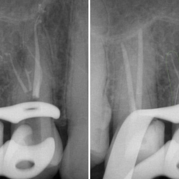
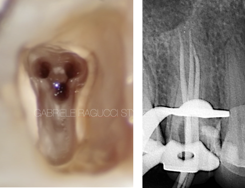
In this case we deal with the retreatment of a mandibular second premolar with a particular anatomy. We removed the gutta-percha with a ultrasonic tip an we localize and shape the 3 canals with reciproc r25 and protaper gold F3.
We obturate the tooth with warm vertical compaction and we restore it with a post and a composite overlay.
Full video of the procedure
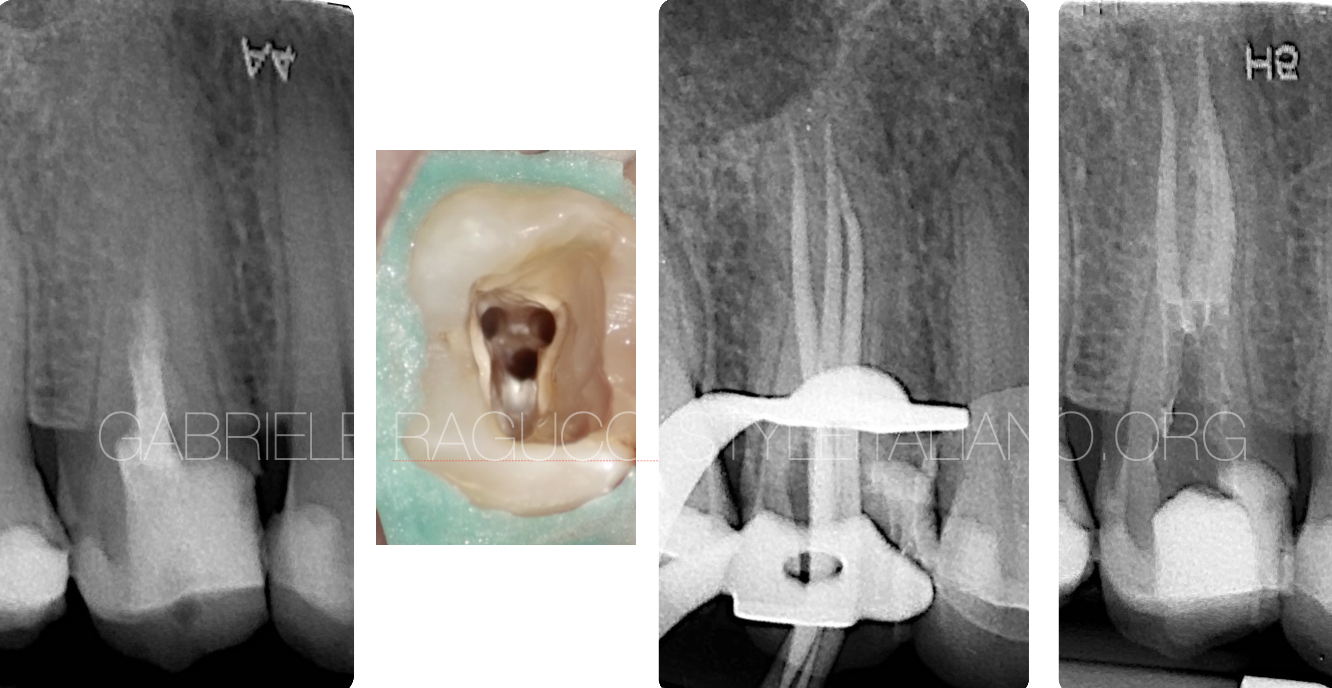
Fig. 1
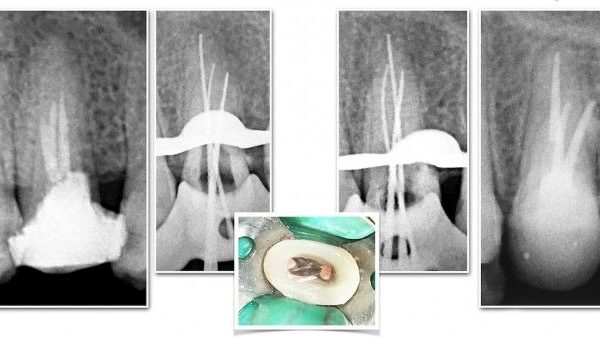
The retreatment steps
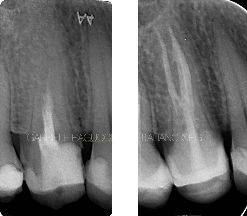
Fig. 2
Pre operative X-ray and 3 years follow up X-ray
Conclusions
A good knowledge of the anatomy can help solving difficult cases.
Bibliography
Soares JA, Leonardo RT. Root canal treatment of three-rooted maxillary first and second premolars-a case report. International Endodontic Journal 2003; 36: 705-710
F Vertucci, A Seelig, R Gillis. Root canal morphology of the human maxillary second premolar. 1974 oral sung oral.med oral path
Fichera G, Dinapoli G, Re D Restauri estetico adesivi indiretti; modello per diagnosi di configurazione cavitaria Dentista moderno 2006; 21-57


