Retreatment
08/10/2024
Fellow
Warning: Undefined variable $post in /home/styleendo/htdocs/styleitaliano-endodontics.org/wp-content/plugins/oxygen/component-framework/components/classes/code-block.class.php(133) : eval()'d code on line 2
Warning: Attempt to read property "ID" on null in /home/styleendo/htdocs/styleitaliano-endodontics.org/wp-content/plugins/oxygen/component-framework/components/classes/code-block.class.php(133) : eval()'d code on line 2
Patient: Male , aged 33 years
Chief complaint: Swelling and pain in the right upper first molar for 1 days.
Presenting symptoms: 1 days ago, the right upper tooth felt painful with gingiva swollen, pain at night,and unable to chew.
Past history: Respiratory system: No history of respiratory disease.
Infectious history: No history of severe infectious disease.
Allergic history: Not allergic to penicillin.
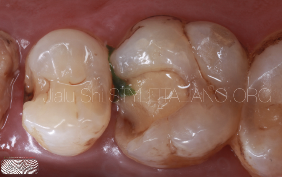
Fig. 1
Examination: On examination, imfitting filling material was visible and secondary caries could be seen around it.
The 16# tooth had no deep periodontal pockets, no excessive mobility, and no
fluctuating buccal swelling.
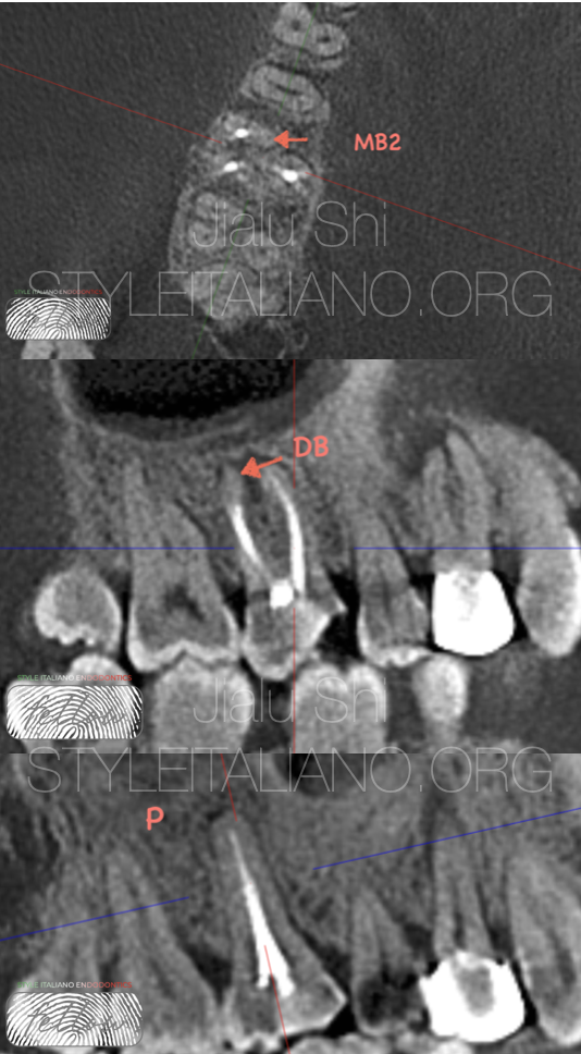
Fig. 2
Pre-operative CBCT
CBCT can clearly show that MB2 is missing, and the working lengths and prepared numbers of DB and P are not enough
The diagnosis of 16# apical periodontitis.
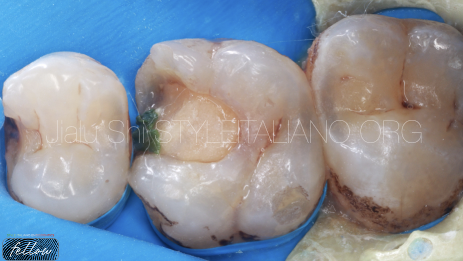
Fig. 3
The patient was informed regarding the guarded overall prognosis of this tooth.
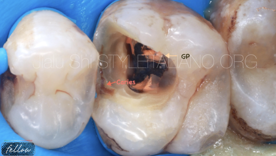
Fig. 4
By removing the original uncohesive filling, it can be clearly seen that there is still some caries in the mesial margin, which we must pay attention to and remove before entering the root canal system. This is very important and the key to successful retreatment
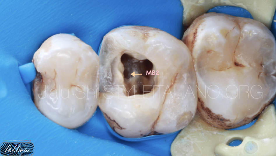
Fig. 5
After removing all the infected tissue, we found MB2 and removed the original GP .
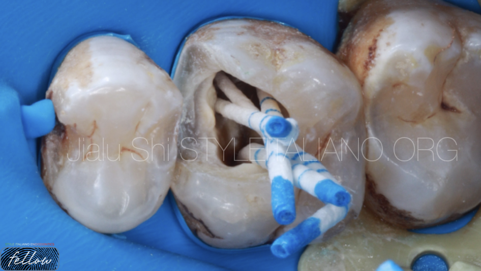
Fig. 6
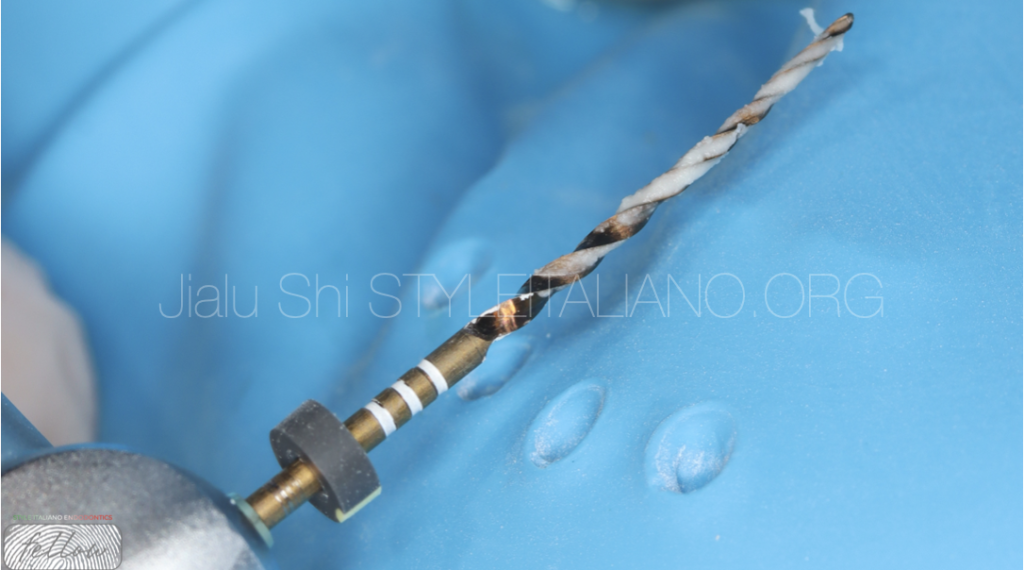
Preparation the root canal by Protaper Gold rotary file and irrigation by 3% NACLO.Irrigation + activation for 15 minutes, strictly following the flushing regimen in SIE's book "Retreatments"
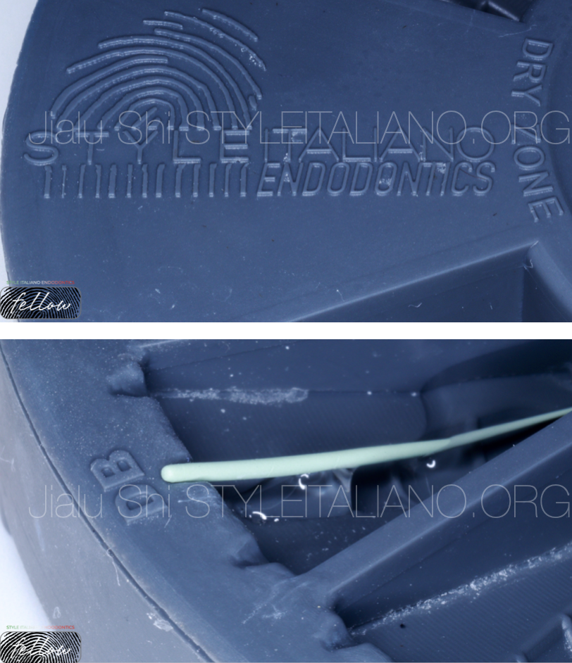
Fig. 7
Sterilize GP with 1% NACLO
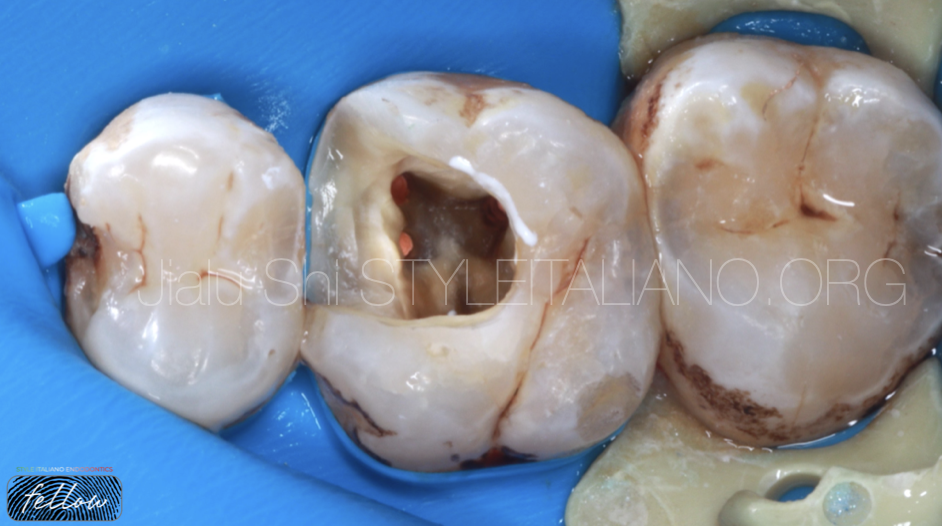
Fig. 8
BC sealer(croot sp)+GP obturated the root canal systems by single-cone technique.
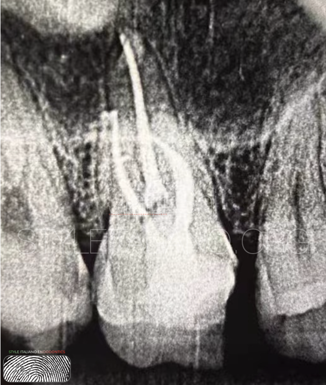
Fig. 9
Post operative X-ray
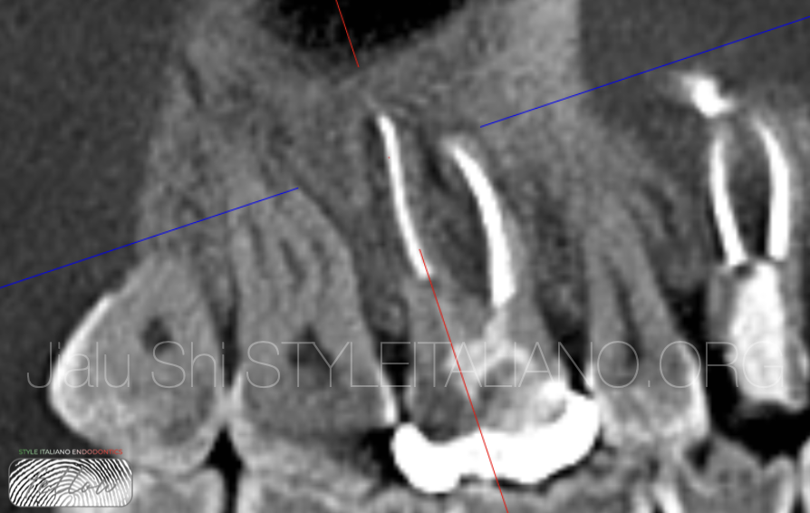
Fig. 10
50 days CBCT Sagittal DB.and MB
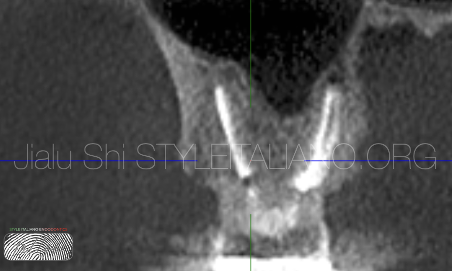
Fig. 11
Coronal DB
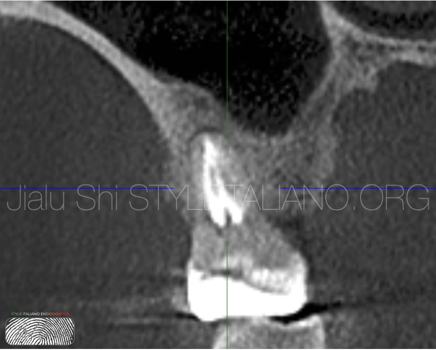
Fig. 12
50 days CBCT Coronal MB and MB2
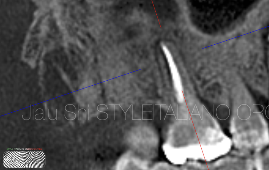
Fig. 13
50 days CBCT Sagittal P
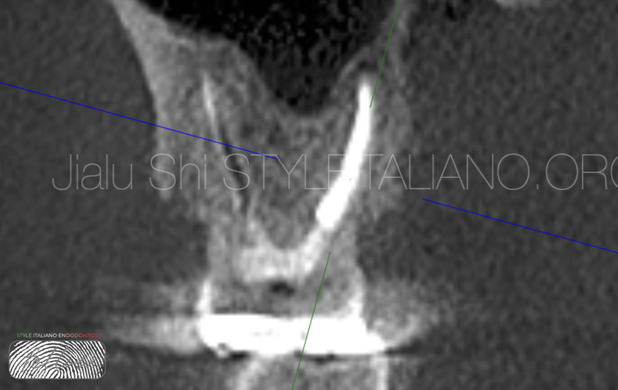
Fig. 14
Coronal P
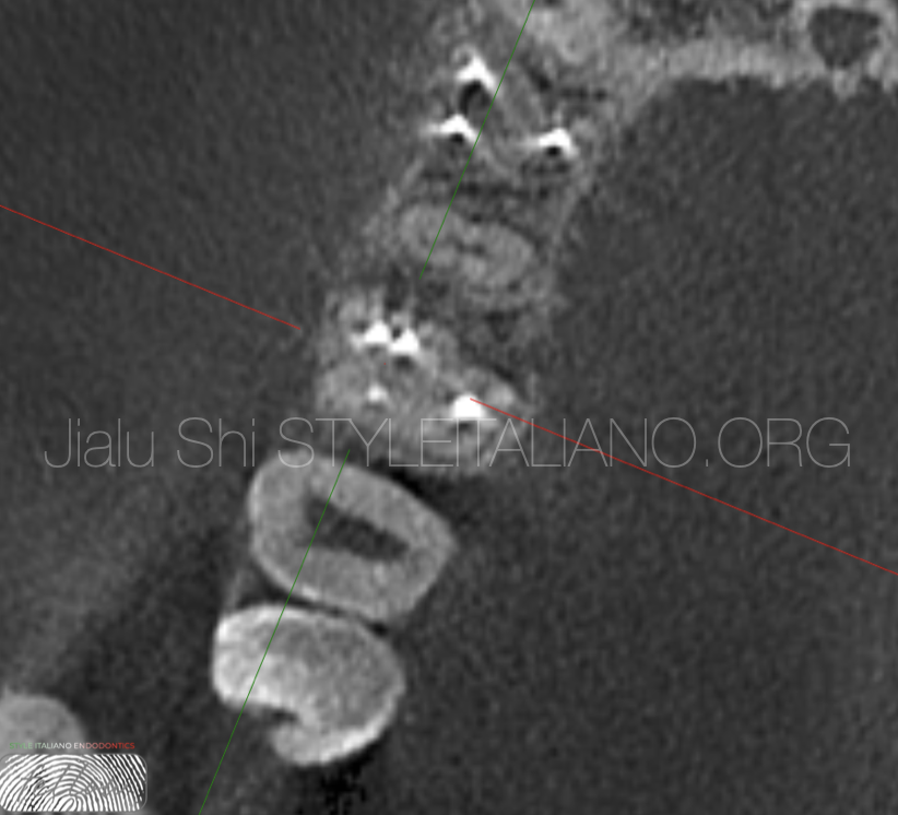
Fig. 15
50 days CBCT Axial

Fig. 16
About the author:
Dr Jialu Shi
Fellow member of Style Italiano Endodontics
Conclusions
A very important factor in determining the effectiveness of retreatment is whether the cause of failure can be accurately identified. Only through logical analysis can we develop a program that is effective and good for the patient. Finding the source of the infection and addressing it accurately often provides great convenience for diagnosis and treatment. I hope every patient will recover.
Bibliography
1, Cristina Bucchi1 | Eyal Rosen2 | Silvio Taschieri (2022) Non-surgical root canal treatment and retreatment versus apical surgery in treating apical periodontitis: A systematic review
2, Pio Bertani | Fabio Gorni| Massimo Gagliani |Francesca Cerutti | Riccardo Tonini (2019). Retratments
3, Stefano Corbella1,2 | Clemens Walter 3 | Igor Tsesis4(2022). Effectiveness of root resection techniques compared with root canal retreatment or apical surgery for the treatment of apical periodontitis and tooth survival: A systematic review
4,Schilder H. Cleaning and shaping the root canal. Dent Clin North Am. 1974; 18: 269-296S


