Post endodontic restoration of Class I cavities
14/06/2022
Massimo Giovarruscio
Warning: Undefined variable $post in /home/styleendo/htdocs/styleitaliano-endodontics.org/wp-content/plugins/oxygen/component-framework/components/classes/code-block.class.php(133) : eval()'d code on line 2
Warning: Attempt to read property "ID" on null in /home/styleendo/htdocs/styleitaliano-endodontics.org/wp-content/plugins/oxygen/component-framework/components/classes/code-block.class.php(133) : eval()'d code on line 2
The loss of tooth structure caused by the Endodontic access cavity results in a decrease of rigidity by 5%.
After root canal treatment the remaining tooth structure could be restored using different restorative options.
The simplest cavity is “Class I. A molar with a “Class I” cavity should be restored with conservative approach and the thickness of remaining walls should be more than 2,5mm for using direct composite restorative procedures.
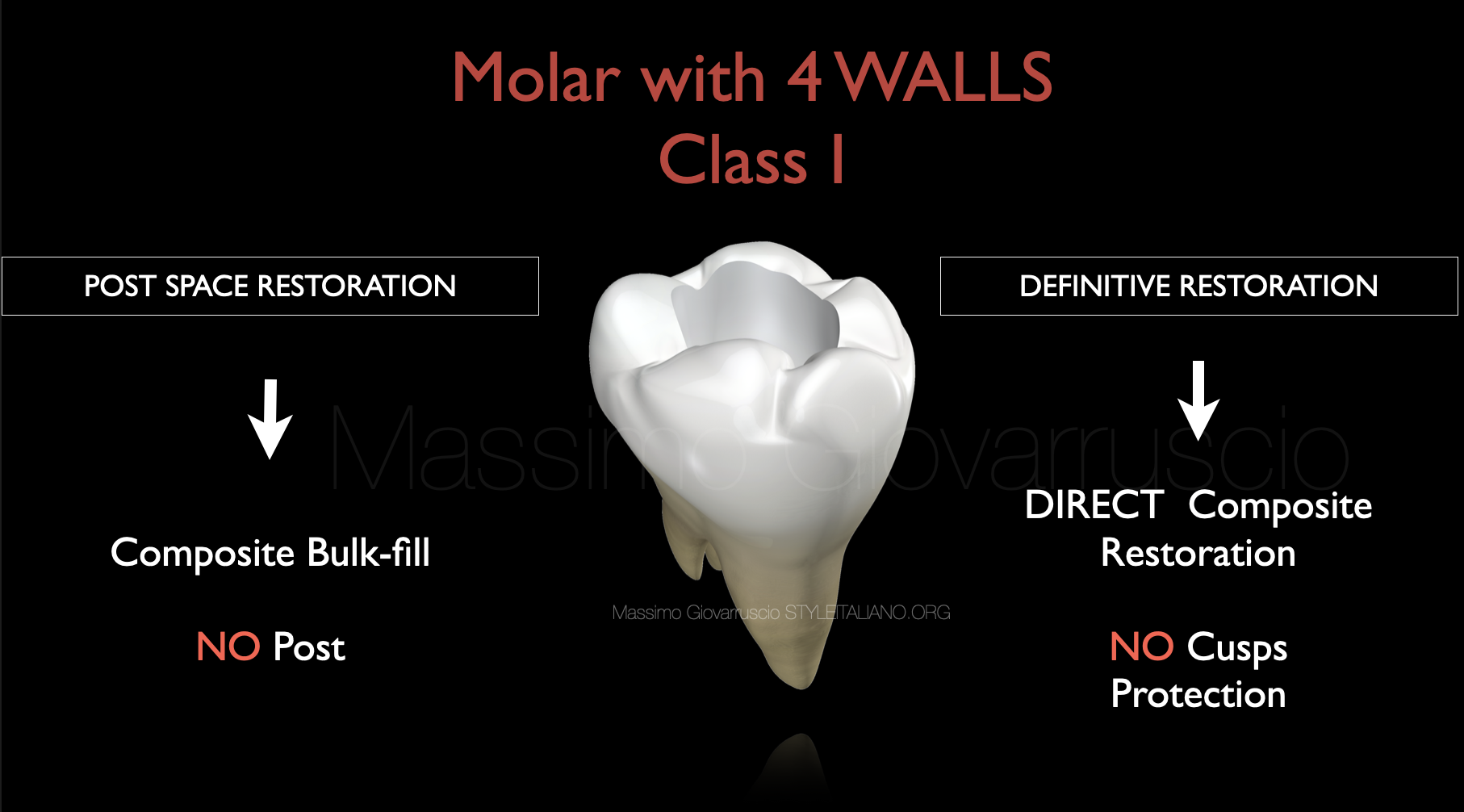
Fig. 1
Nowadays, to restore Class I cavities, we can use Bulk Fill and conventional Composite materials.
Flowable base bulk-fill composites seem suitable for narrow, deep cavities and Class I cavities deeper than 4mm, such as a post-endodontic restorations. The lower viscosity facilities adaptation in less accessible spaces due to plastic flow.
In general, composite resin restorations cannot be regarded as definitive restorations in functional root- filled posterior teeth, except in cases where there has only been a minimal loss of tooth structure and the thickness of remaining walls is more than 2,5mm (for example, where both marginal ridges remain intact). This situation is far more likely to occur in primary root canal treatments than in teeth undergoing root canal re-treatment.
Endodontically treated posterior teeth restored with direct composite restorations have a survival percentage which is similar to that of teeth restored with indirect restorations up to 5 years.
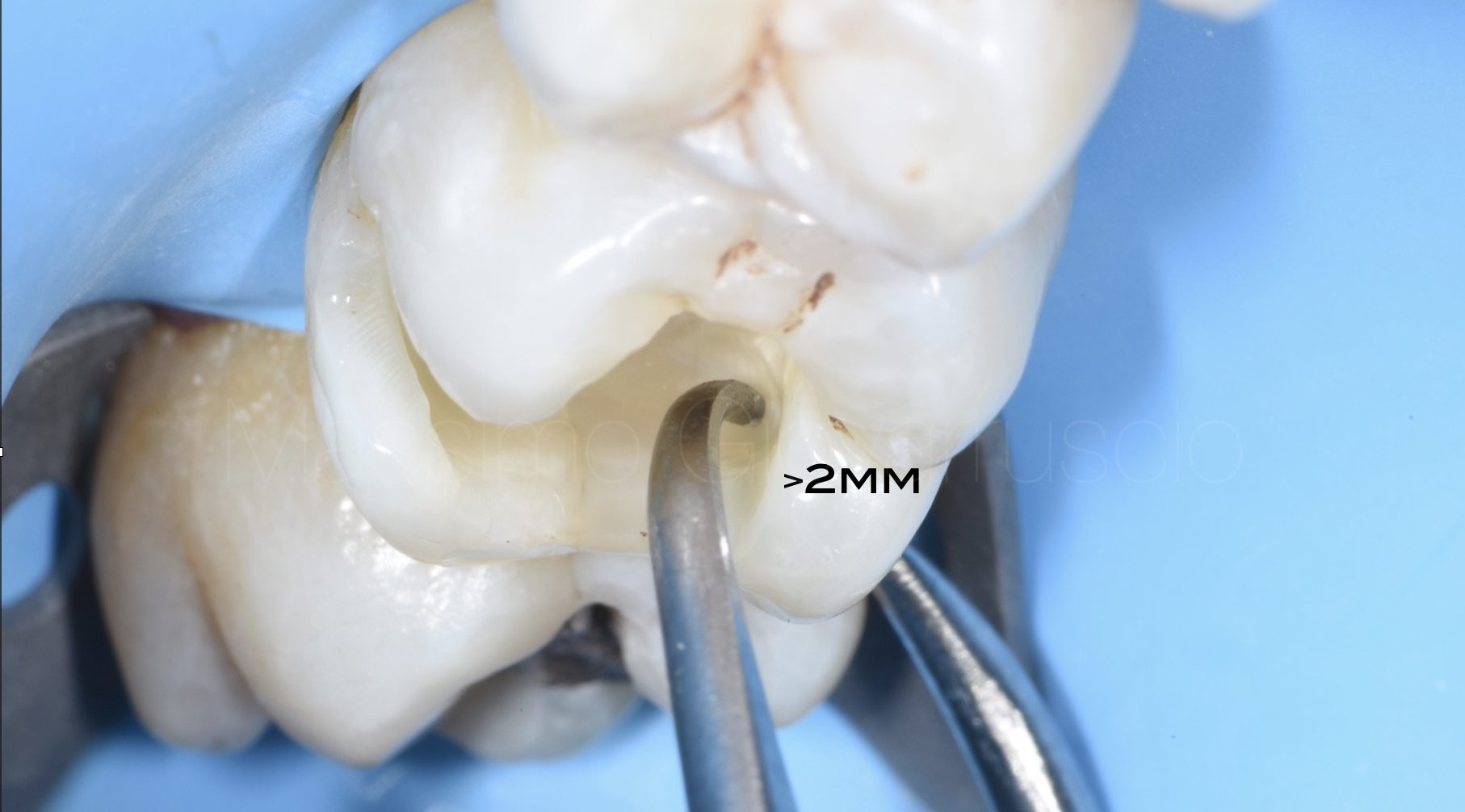
Fig. 2
The volume of residual tooth structure suggests a strong relationship between remaining tooth tissue and survival. Digital intra-oral scanning offers a measurable and reproducible way to assess the restorative predictability of root filled teeth. This a valid tool which could be applied easily to future outcome studies.
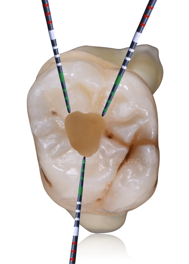
If one does not have a Digital intra-oral scanner, it is possible to measure the residual tooth volume using a Dental Calibro in all dimensions, and also to include the peri-cervical dentine. The minimum remaining thickness recommended should be no less than 2,5mm.
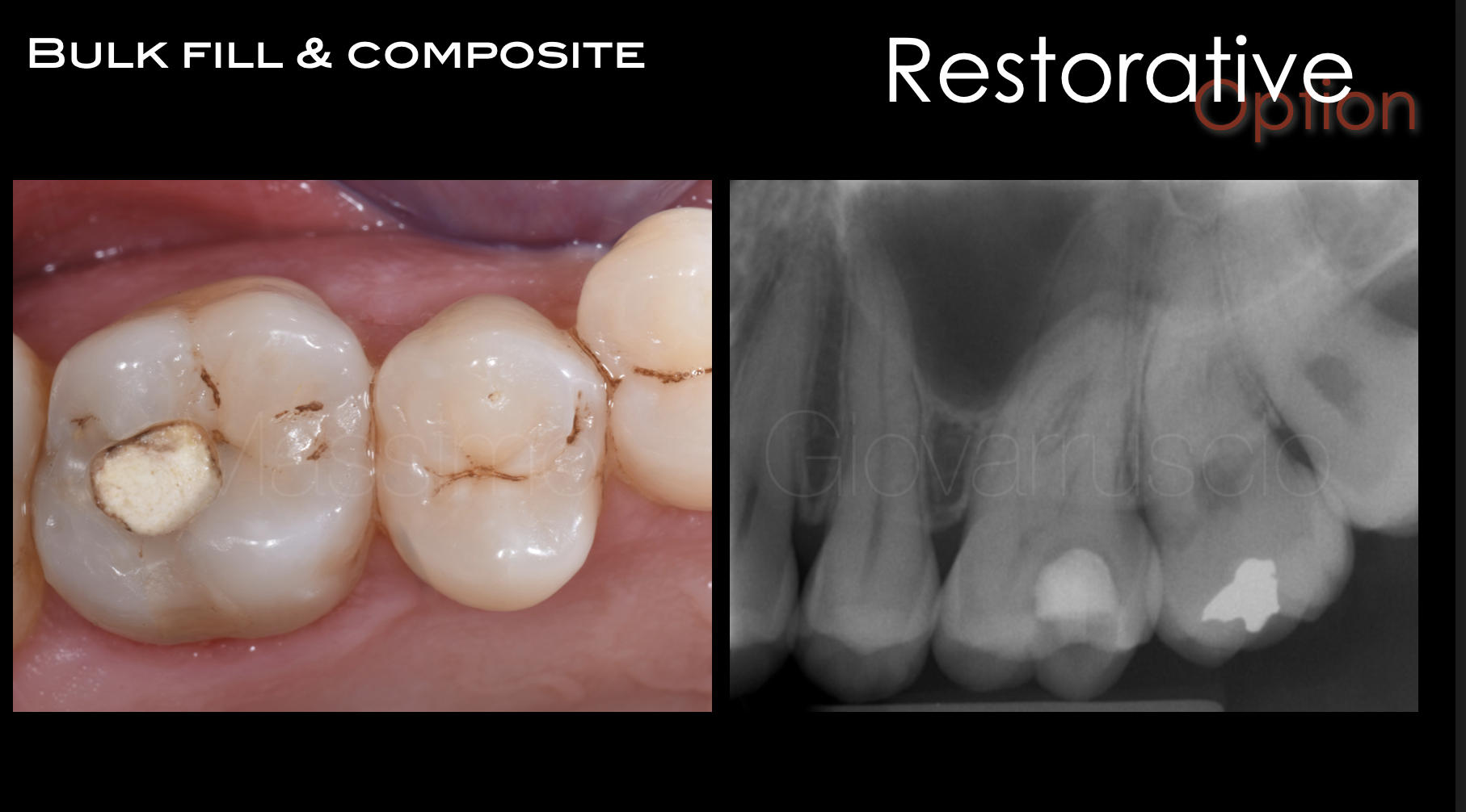
Fig. 3
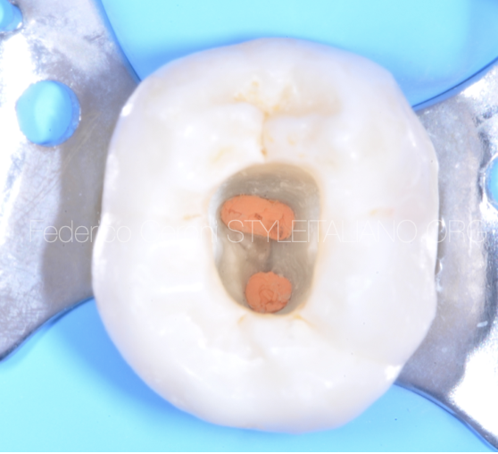
The upper molar has been accessed by referral dentist for an irreversible pulpitis. The dentist performed an emergency access to release the pain. Ledermix paste has been used in the emergency management of the patients. Temporary GIC filling has been placed.
The loss of tooth structure caused by the Endodontic access cavity, results in a decrease of rigidity by 5%.
After root canal treatment, the sound tooth structure should be measured. After that we can plan to restore the tooth with selected material.
Bulk Fill material Reflectys Flow (Itena) has been selected for this case due to the good amount of dentine structure left after root canal treatment.
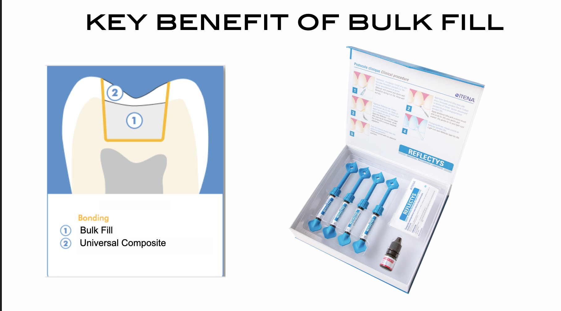
Fig. 4
Key benefits of Reflects flow:
- Enriched with nano-particles
- Light curing
- Contains fluorides
- Low polymerization shrinkage
- Radiopaque
- Ideal for areas with difficult access
- Excellent thixotropy
The direct filling has been performed using REFLECTYS universal restorative composite.
Reflectys is a light-cured micro-hybrid composite reinforced with nano-fillers. The filler content (80%) is embedded in a resin matrix (20%). Reflectys contains particles of different sizes and structure that enhance the material’s mechanical resistance and aesthetic properties.

Fig. 5
The adhesive procedures were carried out with a 2-step self etching bonding system, QuickBond, that:
- Delivers high bonding values to both dentine and enamel
- Disolves the smear layer, penetrating the tubules and peritubular dentine, forming resins tags
- If applied to enamel, the Primer produces a significant pattern with an enhanced surface area, leading to improved enamel bonding
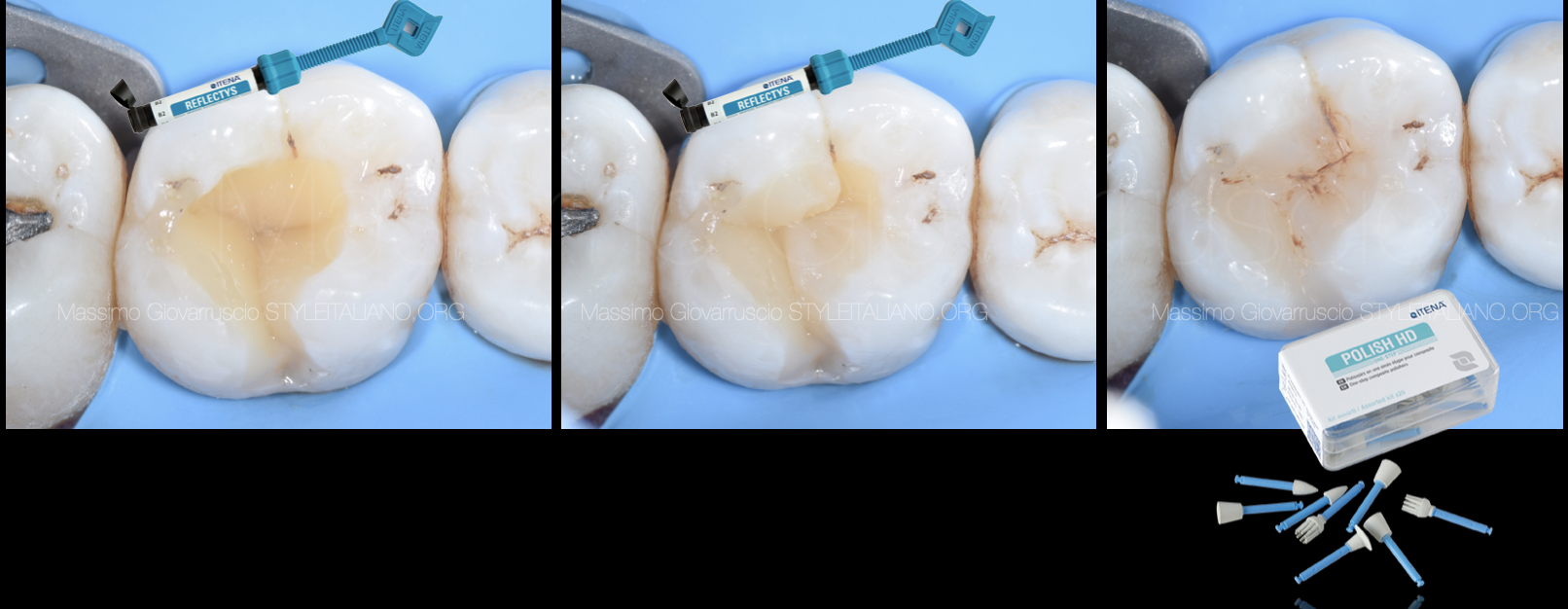
Fig. 6
Here are the main pros of Reflectys:
Exceptional aesthetic
True reflection of natural teeth
Joints cannot be seen
Invisible and naturale restorations
Low polymerization shrinkage: better marginal adaptation
Easy to handle
Easy to polish
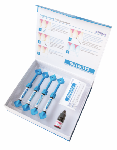
Fig. 7
Reflectys universal composite:
- Exceptional aesthetic
- True reflection of natural teeth
- Joints cannot be seen
- Invisible and naturale restorations
- Low polymerization shrinkage: better marginal adaptation
- Easy to handle
- Easy to polish
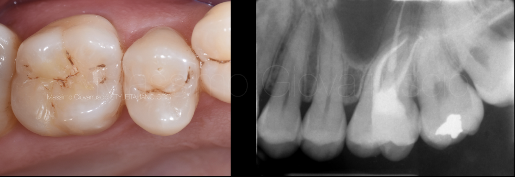
Fig. 8
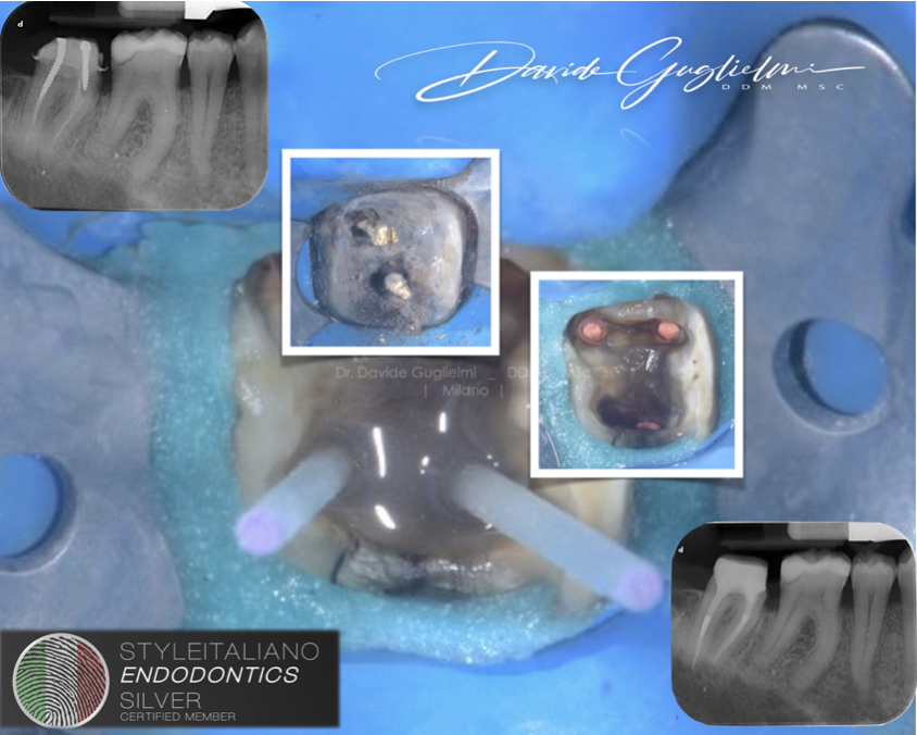
The upper molar has been root canal treated. The access cavity has been restored with a Bulk Filling material. The rest of the cavity has been restored permanently using a Universal composite after applying 2 step adhesive procedures.
Post operative X-Ray shows a conservative root canal treatment respecting the endodontic system. The tooth does not requires any further treatment except a follow-up.
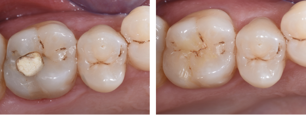
Fig. 9
Pre operative and post operative pictures. The aesthetic result is satisfactory, considering the full respect of the root and dental anatomy.
Conclusions
Based on author knowledge and experience, the Itena products seems to be safe and effective for a direct core Bulk-fill and Universal composite Restoration.
However high-quality clinical trials are needed.
Bibliography
Fuss Z, Lustig J, Katz A, Tamse A. An evaluation of endodontically treated vertical root fractured teeth: impact of operative procedures. J Endod 2001: 27: 46–48
Vire DE. Failure of endodontically treated teeth: classification and evaluation. J Endod 1991: 17: 338–342
Van Ende A, De Munck J, Van Landuyt KL, Poitevin A, Peumans M, Van Meerbeek B. Bulk-filling of high C-factor posterior cavities: effect on adhesion to cavity-bottom dentin.Dent Mater. 2013 Mar;29(3):269-77. doi: 10.1016/j.dental.2012.11.002. Epub 2012 Dec 8
Van Ende A, De Munck J, Lise DP, Van Meerbeek B.Bulk-Fill Composites: A Review of the Current Literature. J Adhes Dent. 2017;19(2):95-109. doi: 10.3290/j.jad.a38141. PMID: 28443833 Review.


