Perforation repair
13/08/2024
Fellow
Warning: Undefined variable $post in /home/styleendo/htdocs/styleitaliano-endodontics.org/wp-content/plugins/oxygen/component-framework/components/classes/code-block.class.php(133) : eval()'d code on line 2
Warning: Attempt to read property "ID" on null in /home/styleendo/htdocs/styleitaliano-endodontics.org/wp-content/plugins/oxygen/component-framework/components/classes/code-block.class.php(133) : eval()'d code on line 2
Perforations are the artificial communications that are formed iatrogenically or pathologically between the tooth and supporting tissues. These perforations can effect the long term prognosis of the root canal therapy.The major complication of a perforation is the secondary infection of periodontal tissues and tooth loss.
Perforations are classified into coronal perforations, furcation perforations, post space perforations, root canal perforations based on the location.
Iatrogenic perforations occurs as a result of
- Inappropriate use of endodontic instruments
- Atypical tooth position in the arch
- Lack of knowledge in dental anatomy
- Calcified pulp chamber
- Endodontic procedure through prosthetic crowns.
The considerations in a successful perforation management include, repair time, level and location,size, access and visibility of the perforation, periodontal status of the tooth, and biocompatibility of perforation repair material.
A definitive diagnosis of a perforation based on symptoms and radiographic findings can enhance the chances of a perforation repair procedure to be effective.
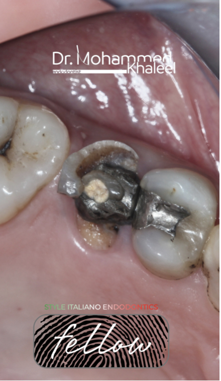
Fig. 1
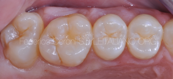
Initial situation
The tooth badly decayed, the patient refused to do extraction.
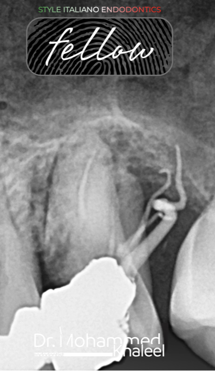
Fig. 2
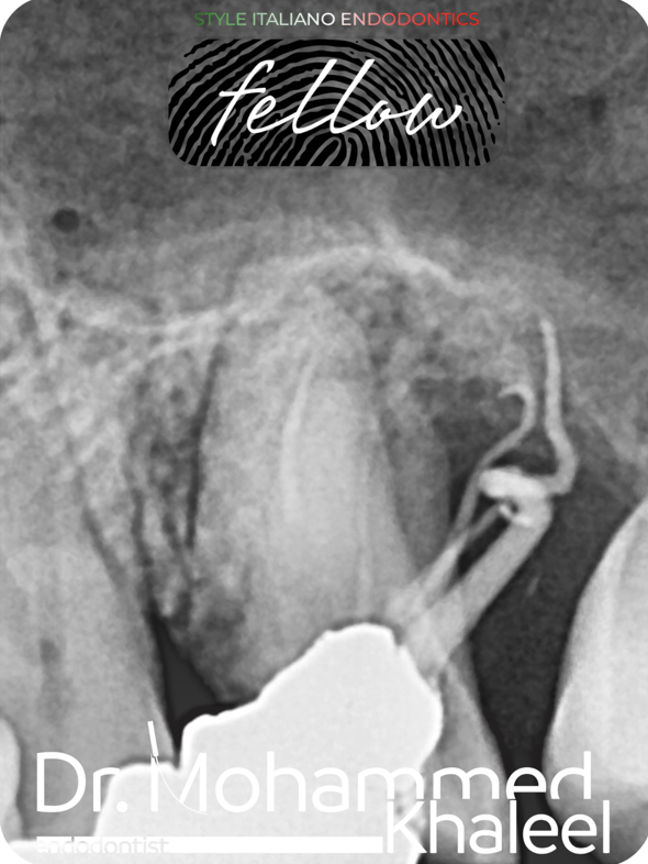
Pre operative x-ray
The tooth had perforation with gutta percha extruded outside.
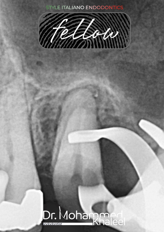
Fig. 3
Intra operative x-ray
Complete removal of extruded gutta percha with small taper 2 file at 1000 rpm.
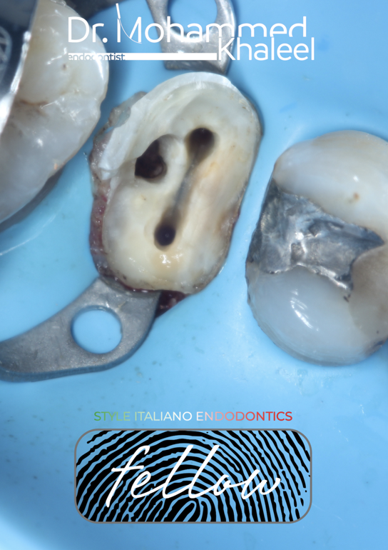
Fig. 4
Cleaning and shaping the canal and perforation site and ready for obturation.
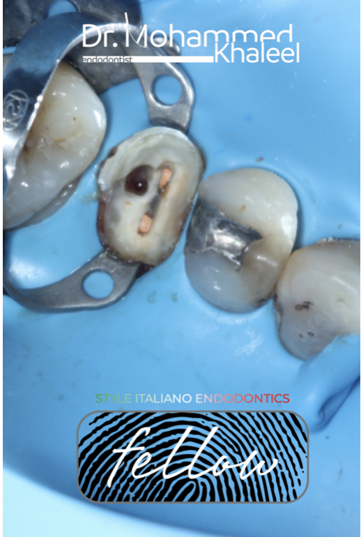
Fig. 5
Obturation of the canals by modified hot technique with bio-ceramic sealer
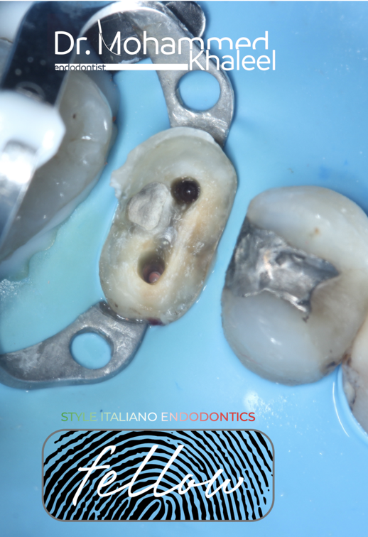
Fig. 6
Repairing perforated site with MTA
& preparing the canal for fiber post
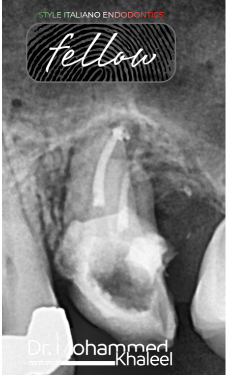
Fig. 7
Post operative x-ray
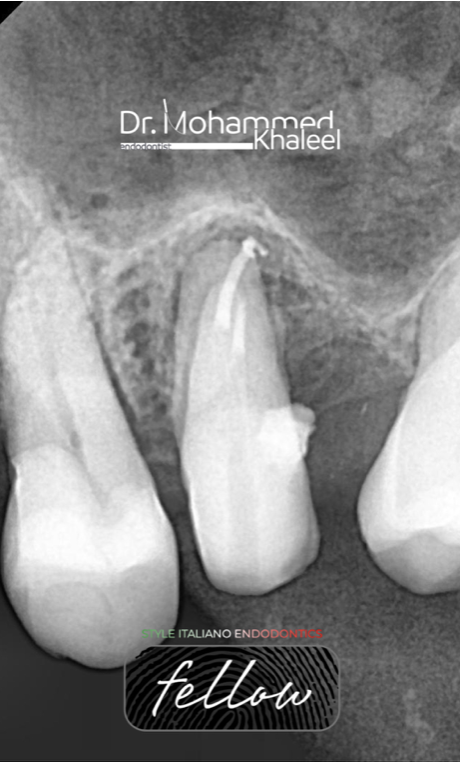
Fig. 8
Follow up x-ray after one year shows great healing!
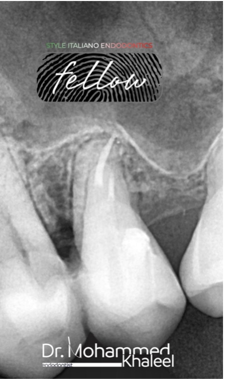
Fig. 9
Follow up 1.5 years

Fig. 10
About the author:
Dr Mohammed Khaleel
B.D.S Hawler medical university /college of dentistry 2011
Key Opinion Leader Denco
Conclusions
All in all, a perforation is an unfortunate mishap during treatment that can happen to the best of us.
Regardless of the approach, surgical or non surgical; there are certain factors that can significantly affect the success of repair.
The clinician should have proper knowledge of tooth morphology, sound clinical judgement and adequate operative skills so as to avoid ending up with a perforation in the first place.
Bibliography
1.Tsesis I, Fuss Z. Diagnosis and treatment of accidental root perforations. Endod Topics. 2006; 13:95-107.
2. Arens DE, Torabinejad M. Repair of furcal perforation with mineral trioxide aggregate : two case reports. Oral Surg Oral Med Oral Pathol Oral Radiol Endod. 1996; 82(1):84-8.
3. Fuss Z, Trope M. Root perforations: classification and treatment choices based on prognostic factors. Dental Traumatology. 1996; 12(6):255-64
4. Breault LG, Fowler EB, Primack CPD. Endodontic Perforation Repair with Resin-Ionomer: A Case Report. J Contmp Dent Prac. 2000; 1(4):1-7.
5. Jeevani E, Jayaprakash T, Bolla N, Vemuri S, Sunil CR. Evaluation of sealing ability of MM-MTA, Endosequence, and biodentine as furcation repair materials: UV spectrophotometric analysis. Journal of Conservative Dentistry. 2014; 17:340-3.


