Partial Necrosis of a Root Canal System
12/12/2021
Marc Habib
Warning: Undefined variable $post in /home/styleendo/htdocs/styleitaliano-endodontics.org/wp-content/plugins/oxygen/component-framework/components/classes/code-block.class.php(133) : eval()'d code on line 2
Warning: Attempt to read property "ID" on null in /home/styleendo/htdocs/styleitaliano-endodontics.org/wp-content/plugins/oxygen/component-framework/components/classes/code-block.class.php(133) : eval()'d code on line 2
A white male patient was referred for a root canal treatment on seemingly necrotic first right lower molar tooth #46.
Patient was exhibiting episodes of pain from time to time that started few days ago.
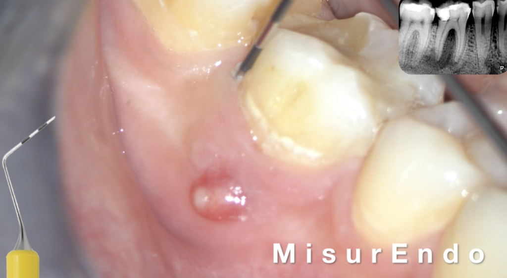
Intra Oral examination showed a sinus tract on the buccal aspect of the gum facing first molar #46.No probing was measured in the furcal area or at any point around the crown.
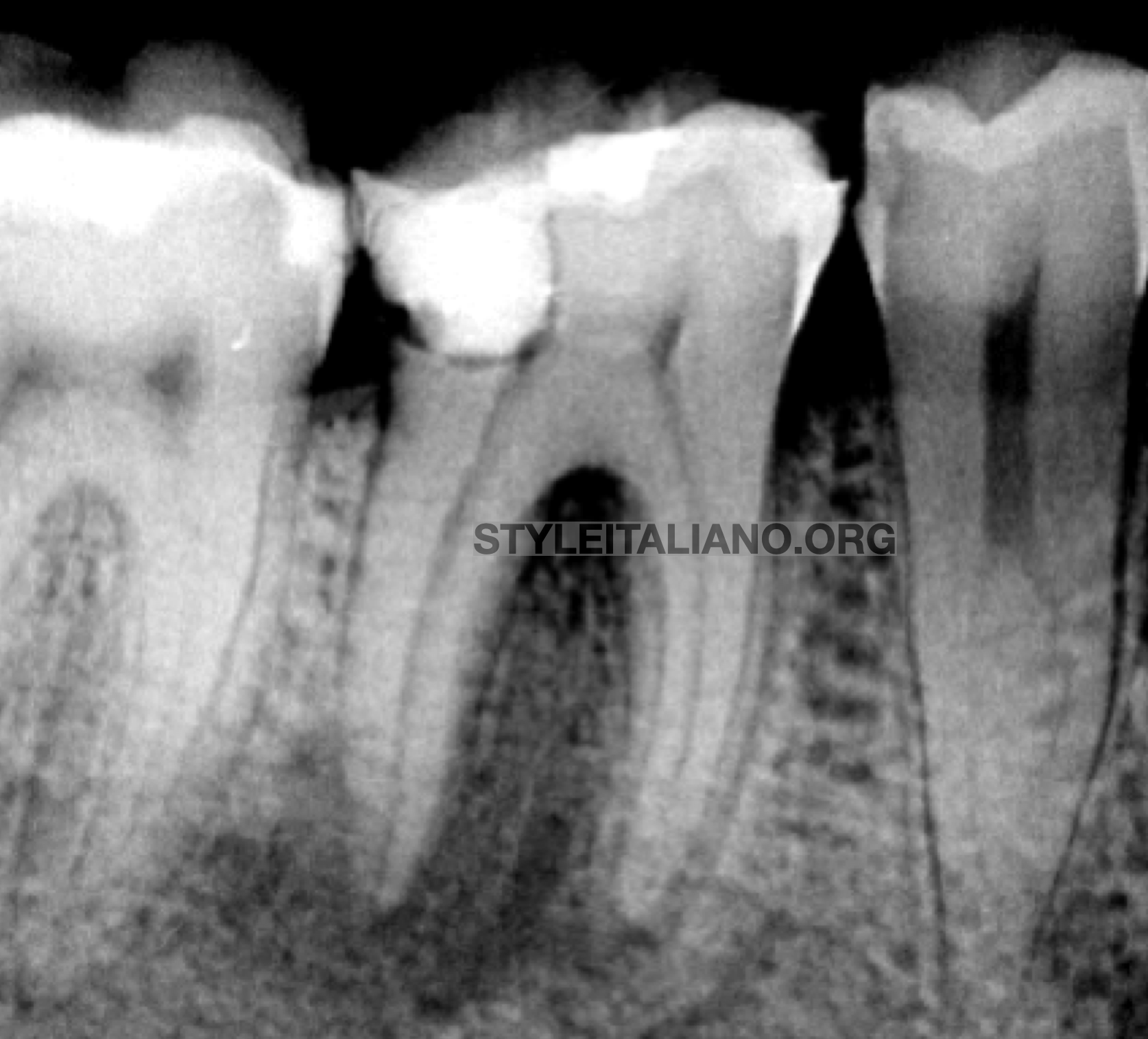
Fig. 1
Pre Op xray showing distal filling in proximity with distal pulp horn and pulp stones in chamber.
Peri apical lesion with bone radiolucency all around distal root
Pulp necrosis is the origin of the radiolucency in most of the time.
Periapical lesion only occurs once the necrotic tissue becomes infected. Necrotic pulp alone does not cause apical periodontitis unless it is infected.
Intra Oral Examination showing Sinus tract on the buccal aspect of the gum facing tooth #46
No pathological pocket probing was measured using MisurEndo
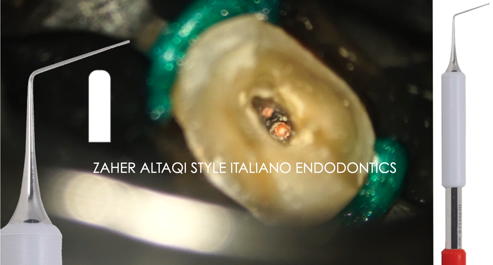
After access cavity, ultrasonics were used under microscope to remove pulp stones separating mesial and distal pulp chamber space.
Canal orifices were located with Explora from the Smart Endo kit.
Vital tissue was found in both mesial canals while distal pulp tissue was necrotic very clear in the pulp chamber microscopic view.
Multiplo connected to the Deppeler mirror is just very ergonomic for file measurement, switching between mirror and Multiplo just saves a lot of time during the shaping process.
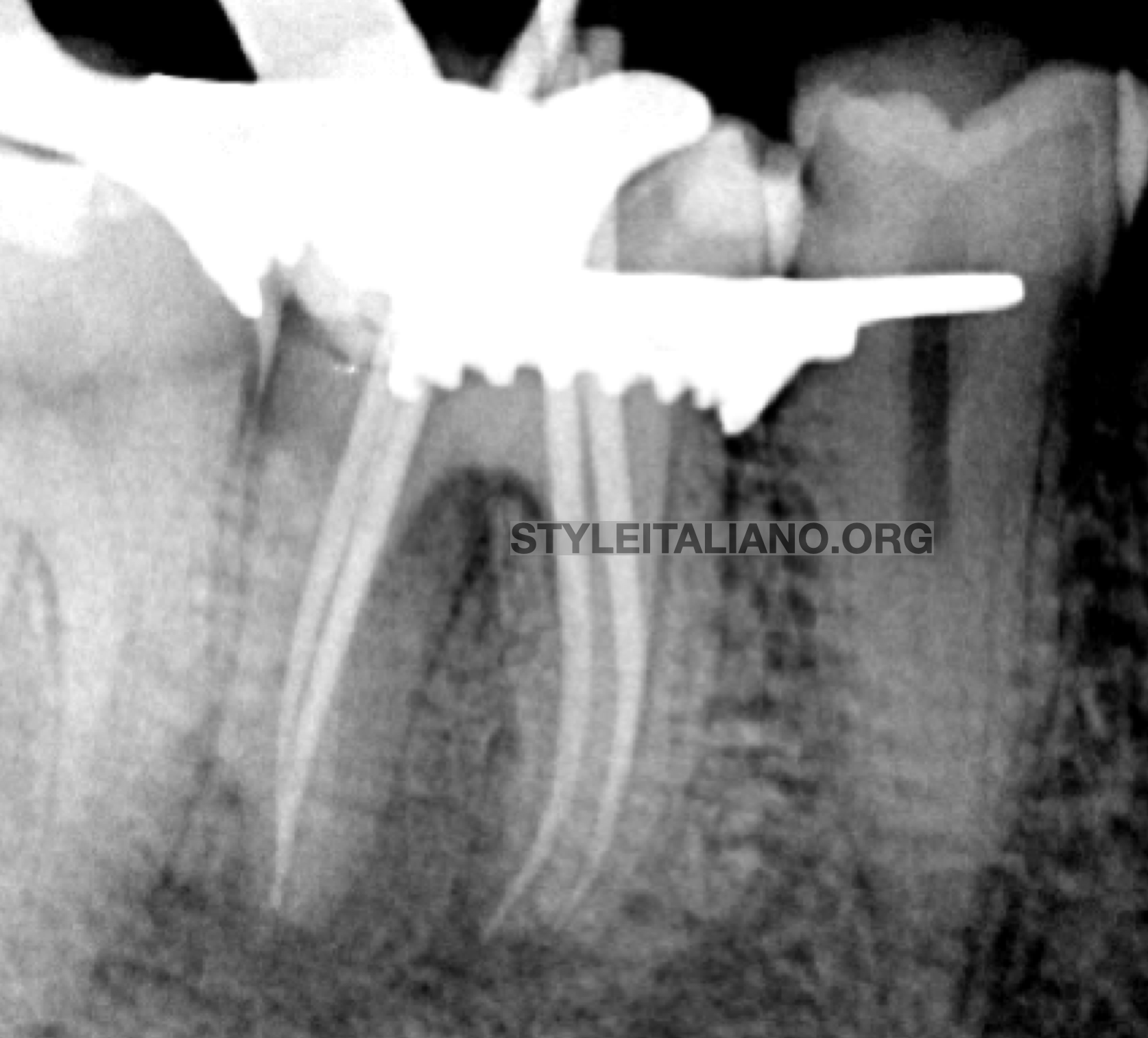
Fig. 2
Cone fit xray
The obturation sequence was managed with warm vertical compaction, multiple waves of heat and compaction of the gutta percha using Prexo from Deppeler.
Prexo is double sided plugger, red side is with reverse taper and rounded edges allowing better visibility under magnification very useful during coronal plug or during perf repair or apical plug with MTA. Gray side is smaller, straight and with rounded edges as well extremely efficient for apical plug during obturation.
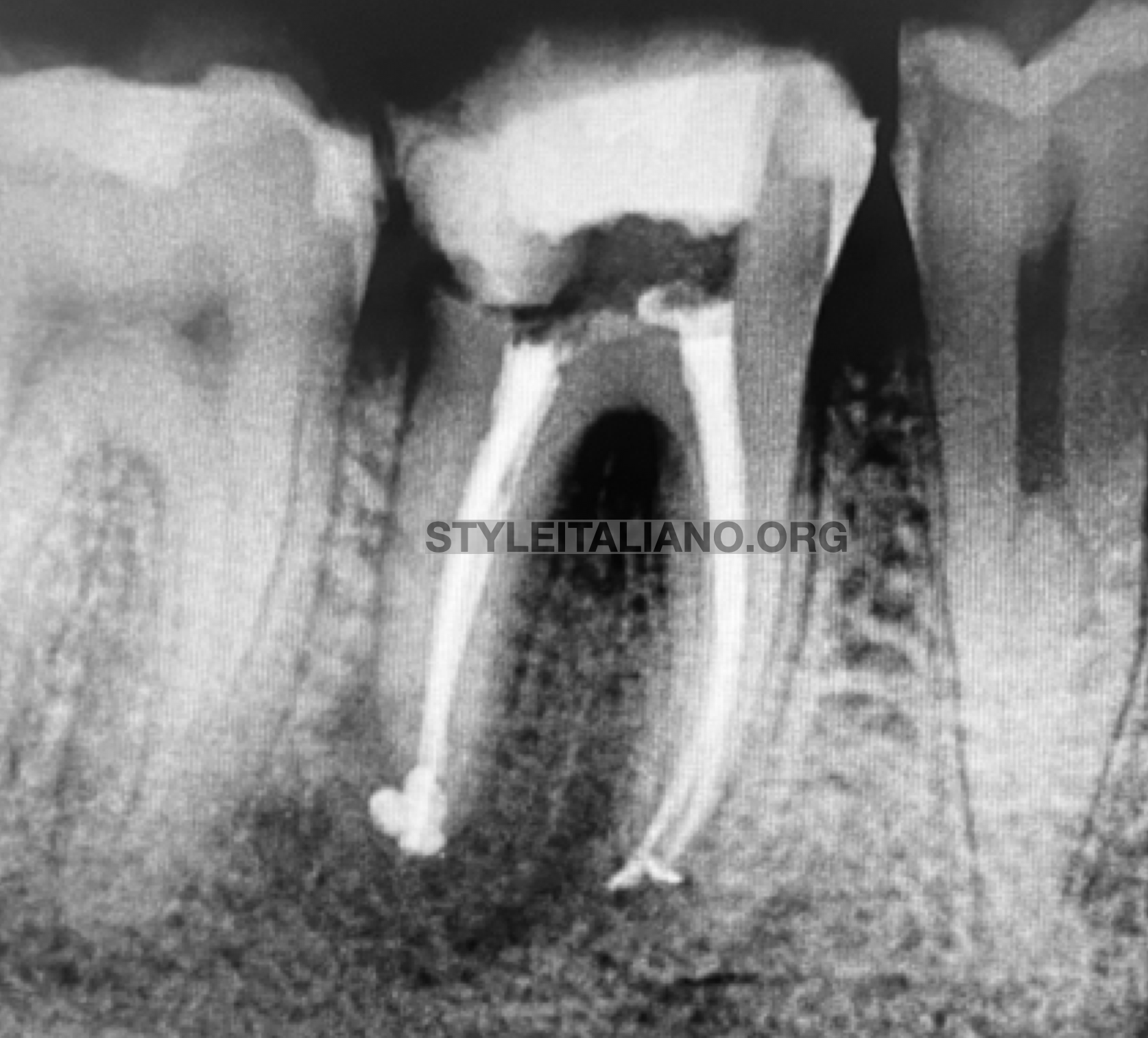
Fig. 3
Final xray.
Conclusions
Pulp tissue might sometimes following exterior irritation and iatrogenic damage get divided due to tertiary dentine created in between like this lower right first molar where the distal root was necrotic and separated from the mesial pulp tissue still vital due to dentine formation creating and tight seal between both parts of the root canal system.
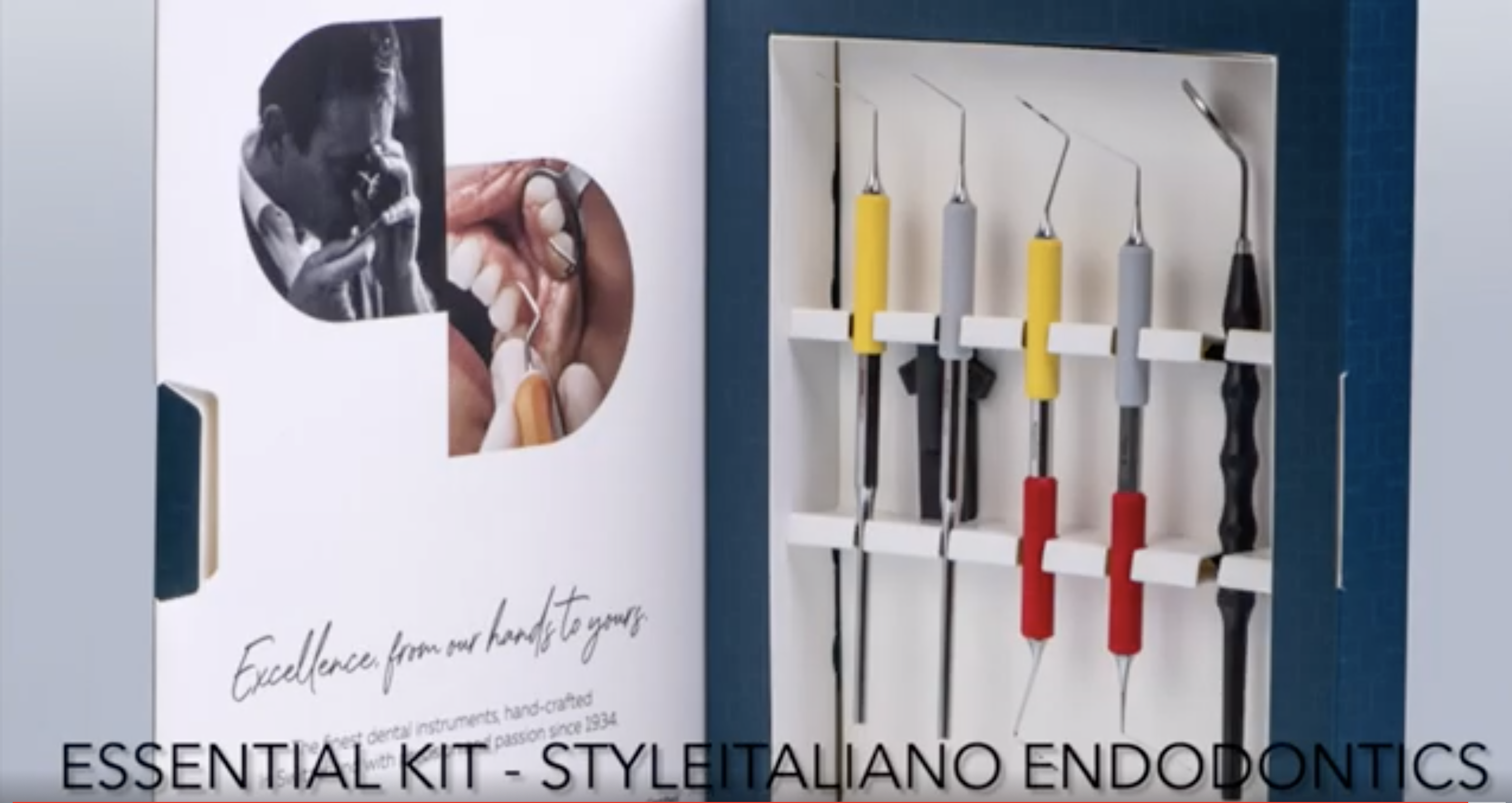
The Deppeler new endodontic instruments Explora, Prexo, Extendo, Misurendo & Multiplo covers all stages of the Endodontic treatment, from diagnosis till obturation. The SmartEndo kit is a mixture of new designs, material and Swiss quality for refined endodontics procedures.
Bibliography
- Abbott PV, Yu C. A clinical classification of the status of the pulp and the root canal system. Aust Dent J. 2007;52(1 Suppl):S17–31.


