Orthodontic Extrusion For proper Endodontic therapy
08/01/2024
Fellow
Warning: Undefined variable $post in /home/styleendo/htdocs/styleitaliano-endodontics.org/wp-content/plugins/oxygen/component-framework/components/classes/code-block.class.php(133) : eval()'d code on line 2
Warning: Attempt to read property "ID" on null in /home/styleendo/htdocs/styleitaliano-endodontics.org/wp-content/plugins/oxygen/component-framework/components/classes/code-block.class.php(133) : eval()'d code on line 2
The major problem with sub-gingival fracture is absence of adequate coronal ferrule and a compromised biological width. This usually complicates the application of the rubber dam during endodontic treatment. The primary objective of tooth extrusion or forced eruption is to provide both a sound tissue margin for ultimate restoration and to create a periodontal environment (biological width) that will be easy for the patient to maintain. Ingber et al. suggested that a minimum distance of 3 mm is required from the restorative margin to the alveolar crest to permit adequate healing and restoration of the tooth that is biologically acceptable.
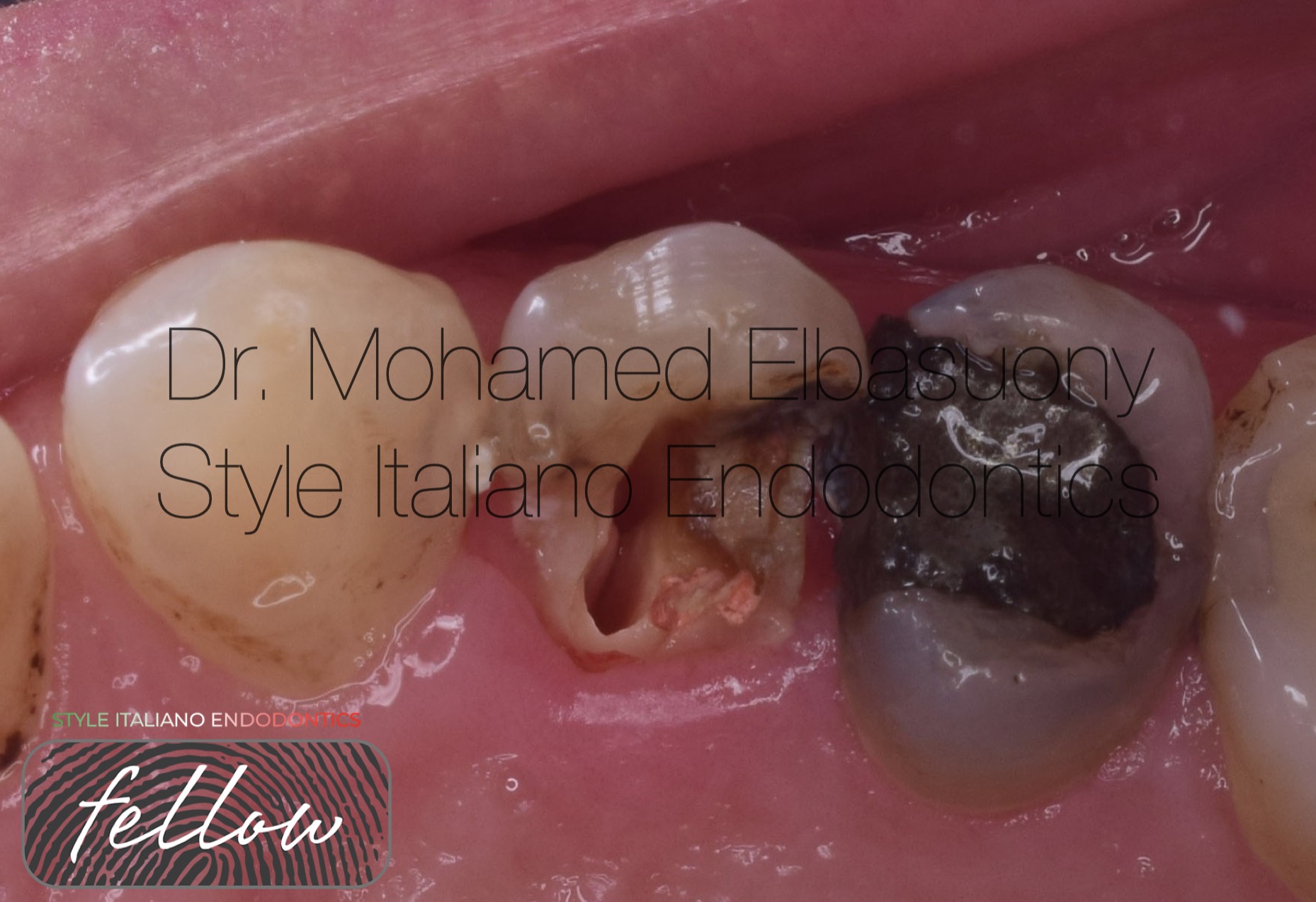
Fig. 1
A 40-years-old female patient presented to my private dental clinic with fracture of palatal cusp of upper right first premolar.
Clinical examination showed fractured line extended sub-gingival on the palatal side making the prosthetic rehabilitation difficult.
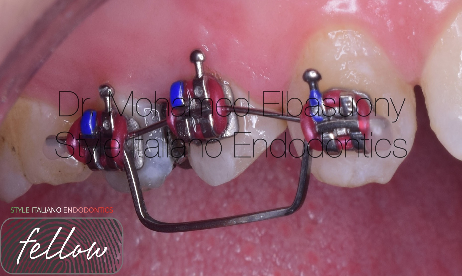
Fig. 2
The main objective of the treatment was to restore the traumatised tooth in respect to all biological, functional and aesthetic principles of all the treatments involved.
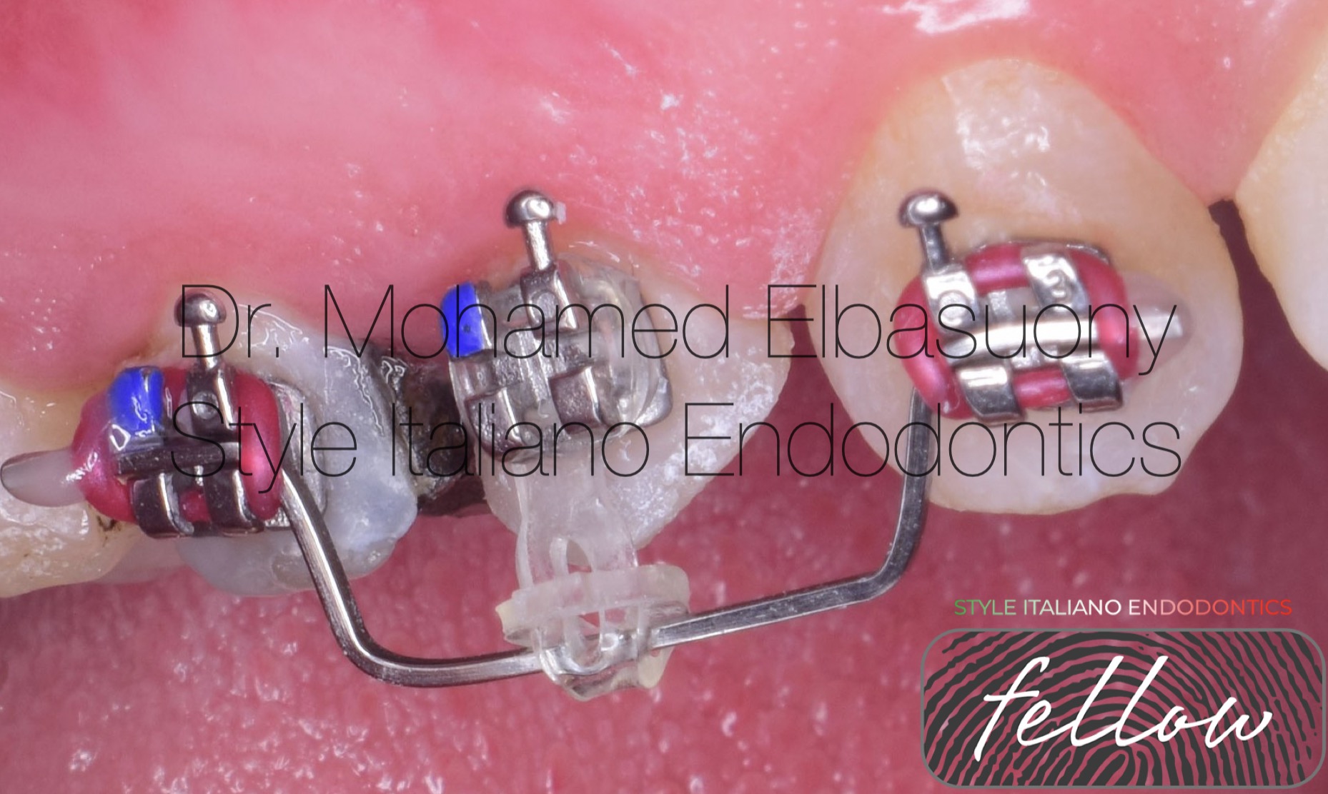
Fig. 3
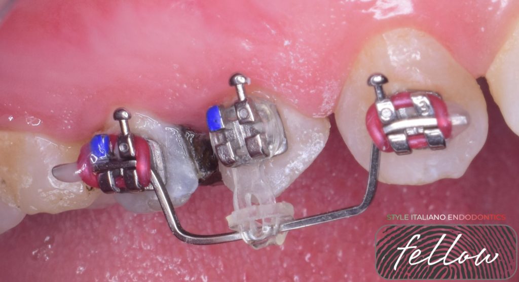
However, as tooth isolation was not possible due to the deep fracture, lengthening of the clinical crown had to be performed. It was decided that orthodontic extrusion was the best and most conservative option. Orthodontic brackets was positioned in its place and more apically on fractured tooth. The time required for wire application is one month and 3 weeks for power chain.
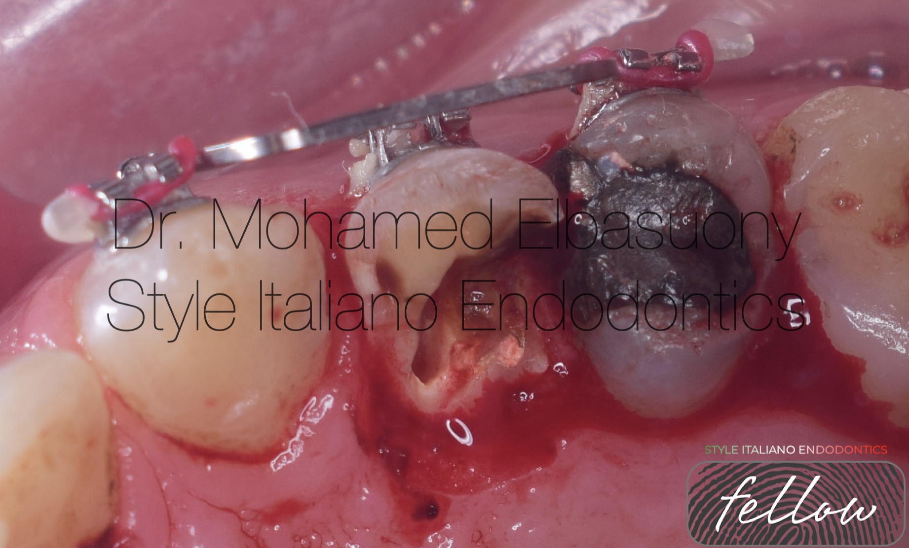
Fig. 4
Minor gingival re-contouring was carried out in order to have a symmetrical gingival margin.
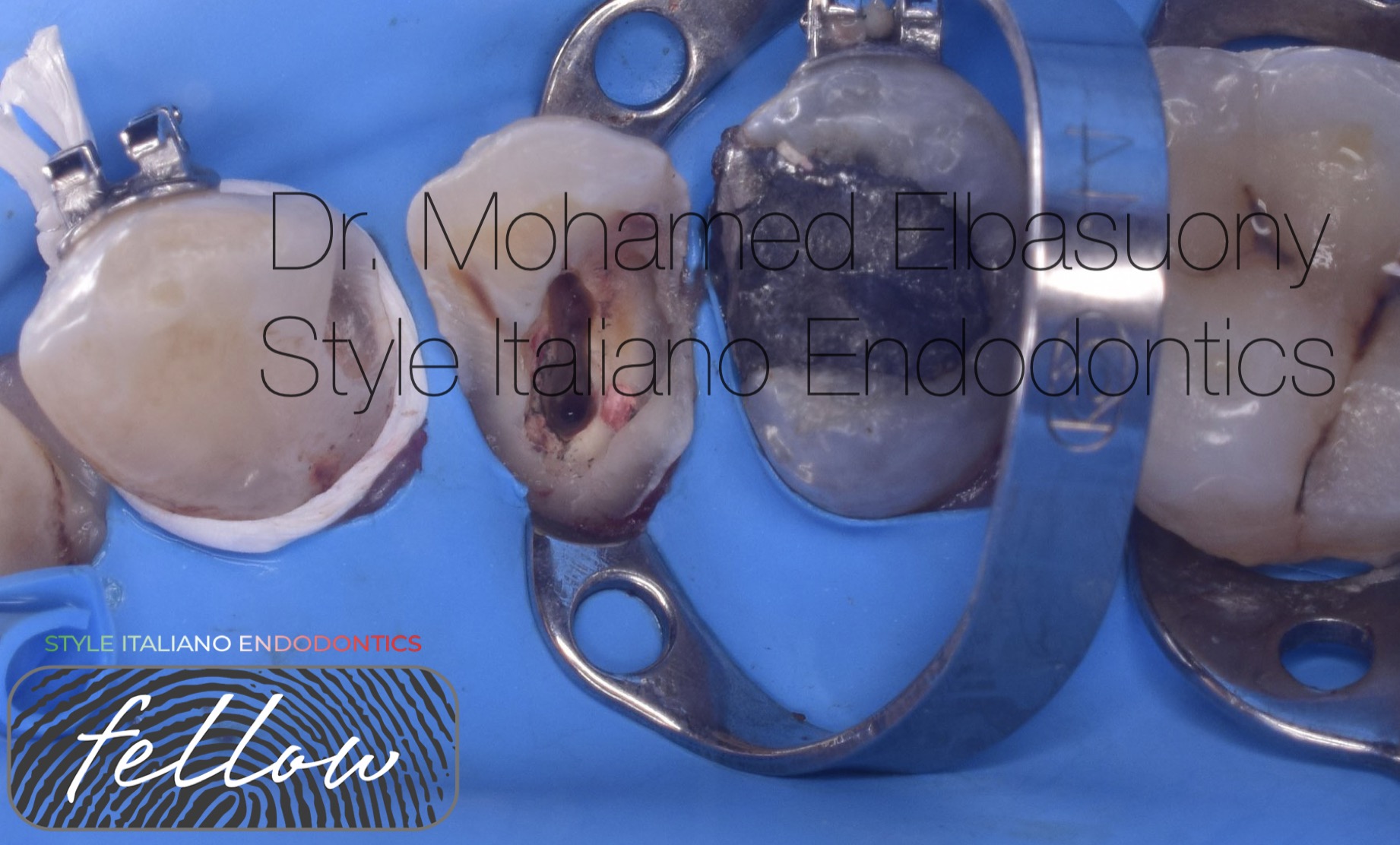
Fig. 5
The coronal extrusion was considered satisfactory as the fractured site had reached the free gingiva level, allowing for a qualitative RDI. Orthodontic wire was removed.
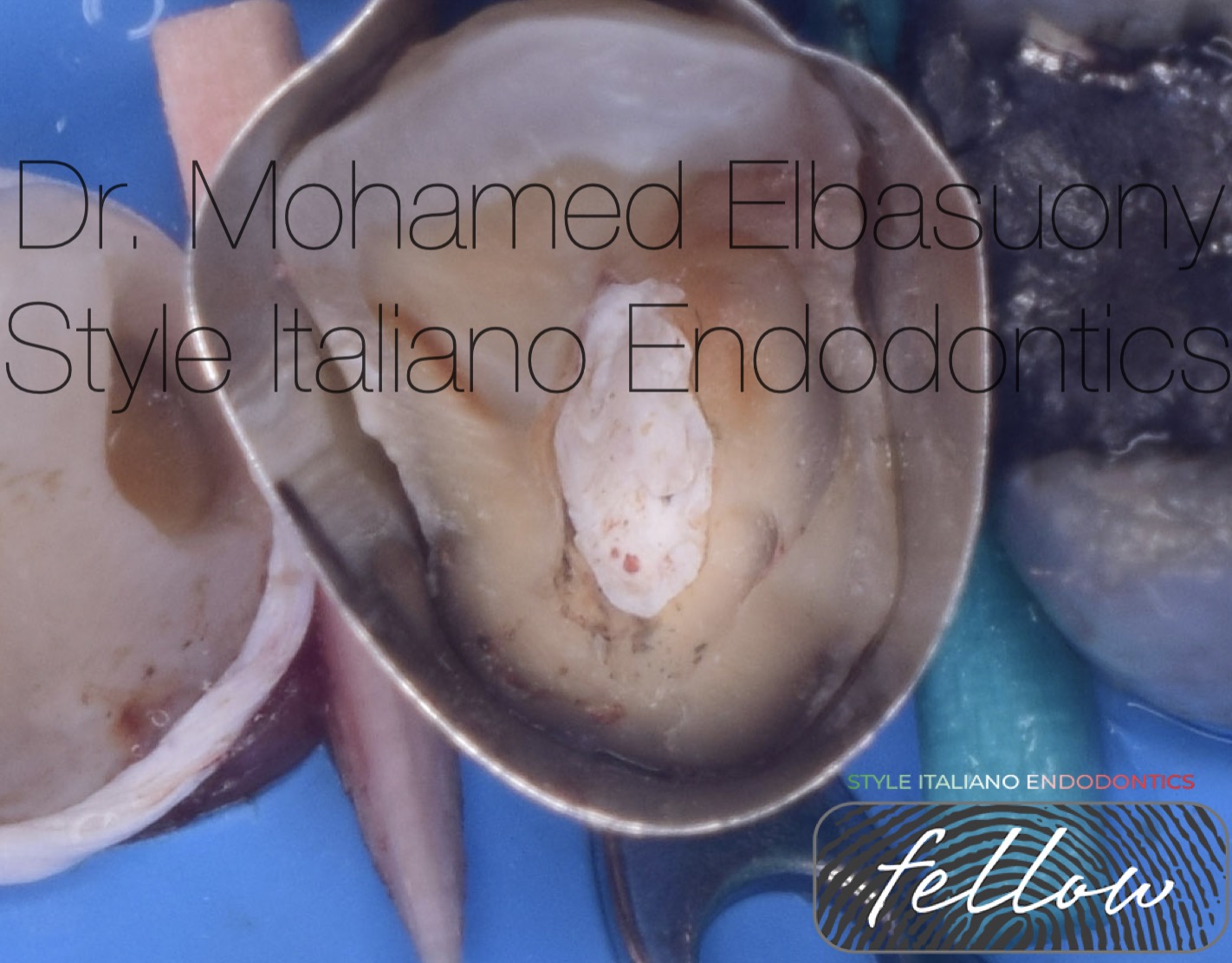
Fig. 6
Band placement.
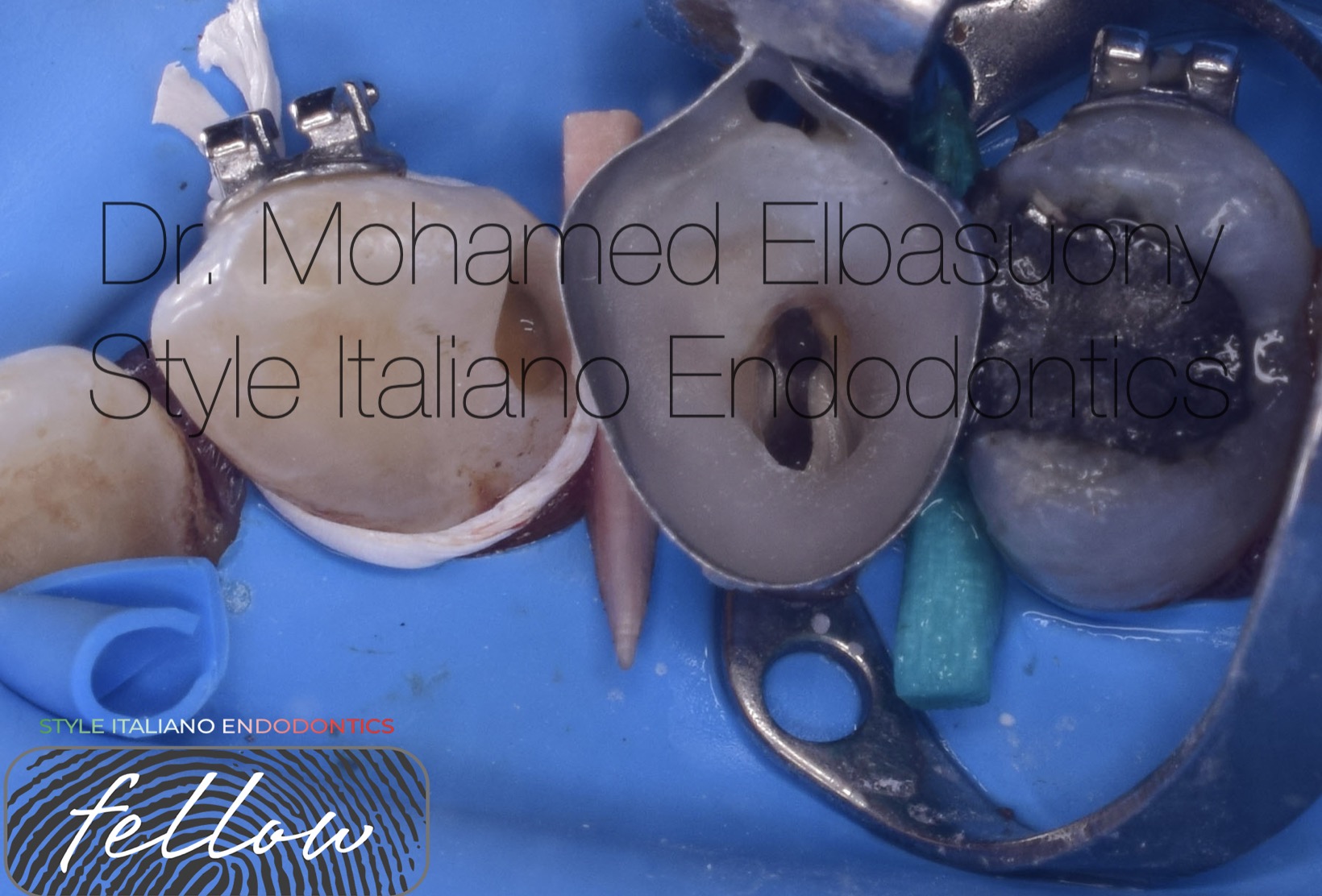
Fig. 7
Composite build up.
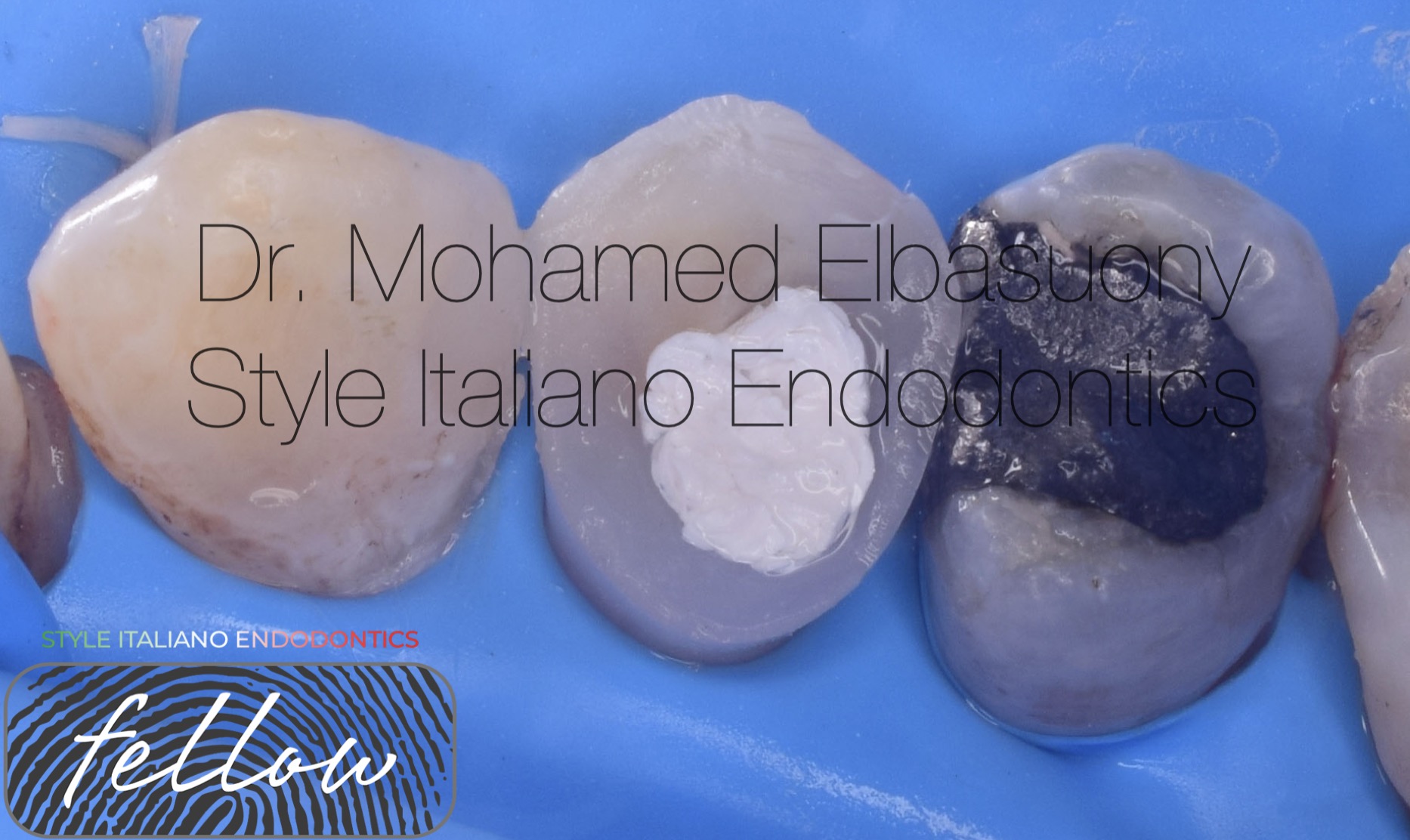
Fig. 8
Composite restoration of adjacent tooth (canine).
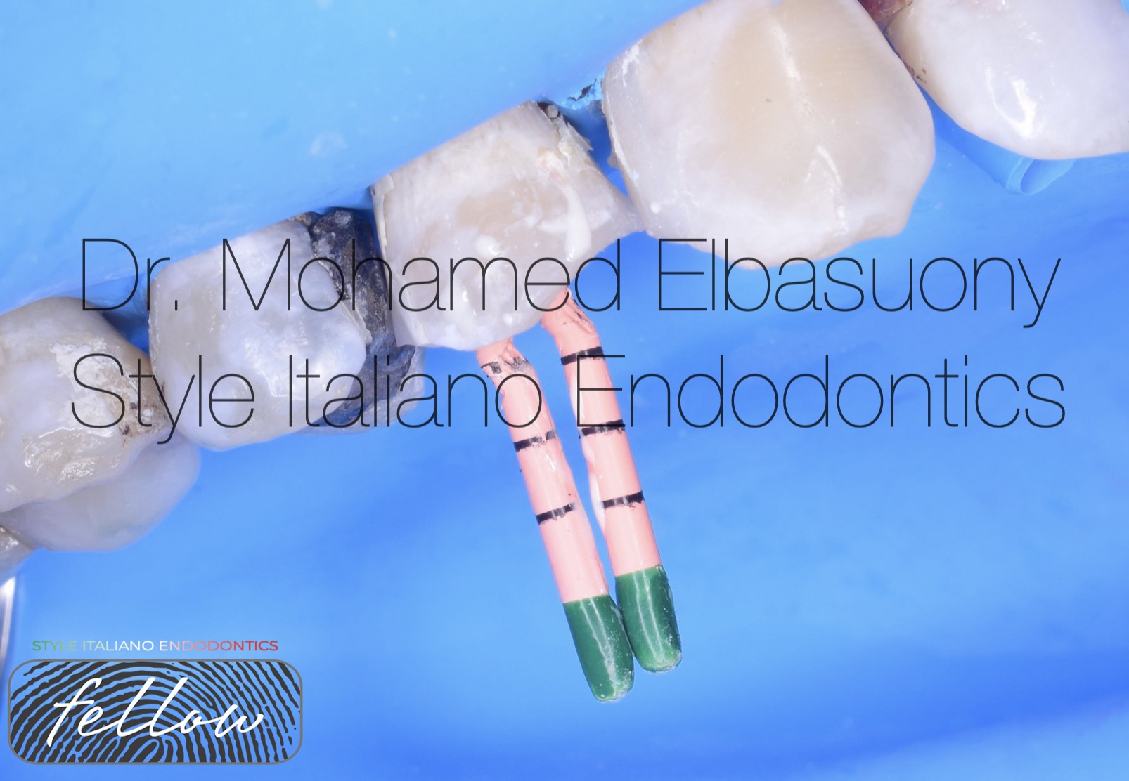
Fig. 9
Master cone in place.
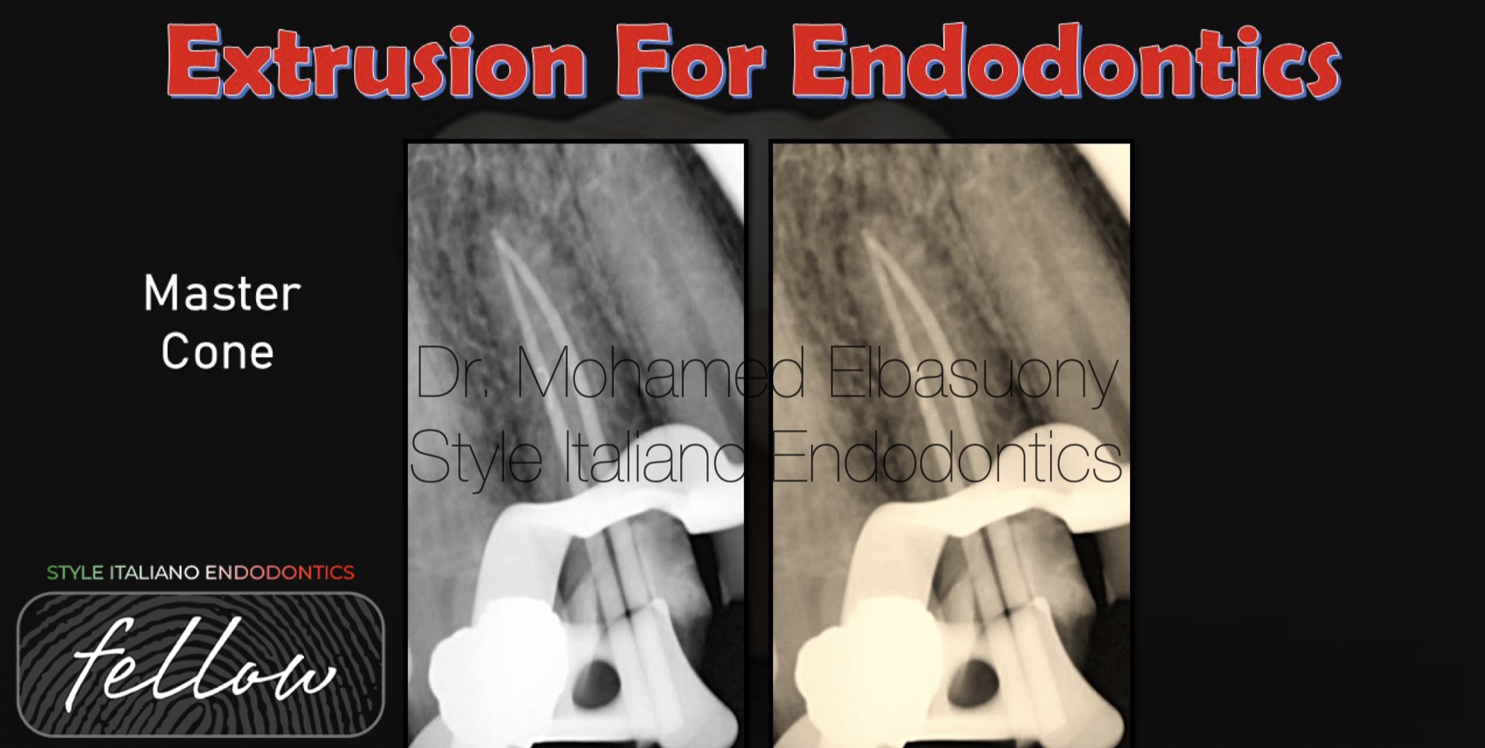
Fig. 10
Master cone radiograph.
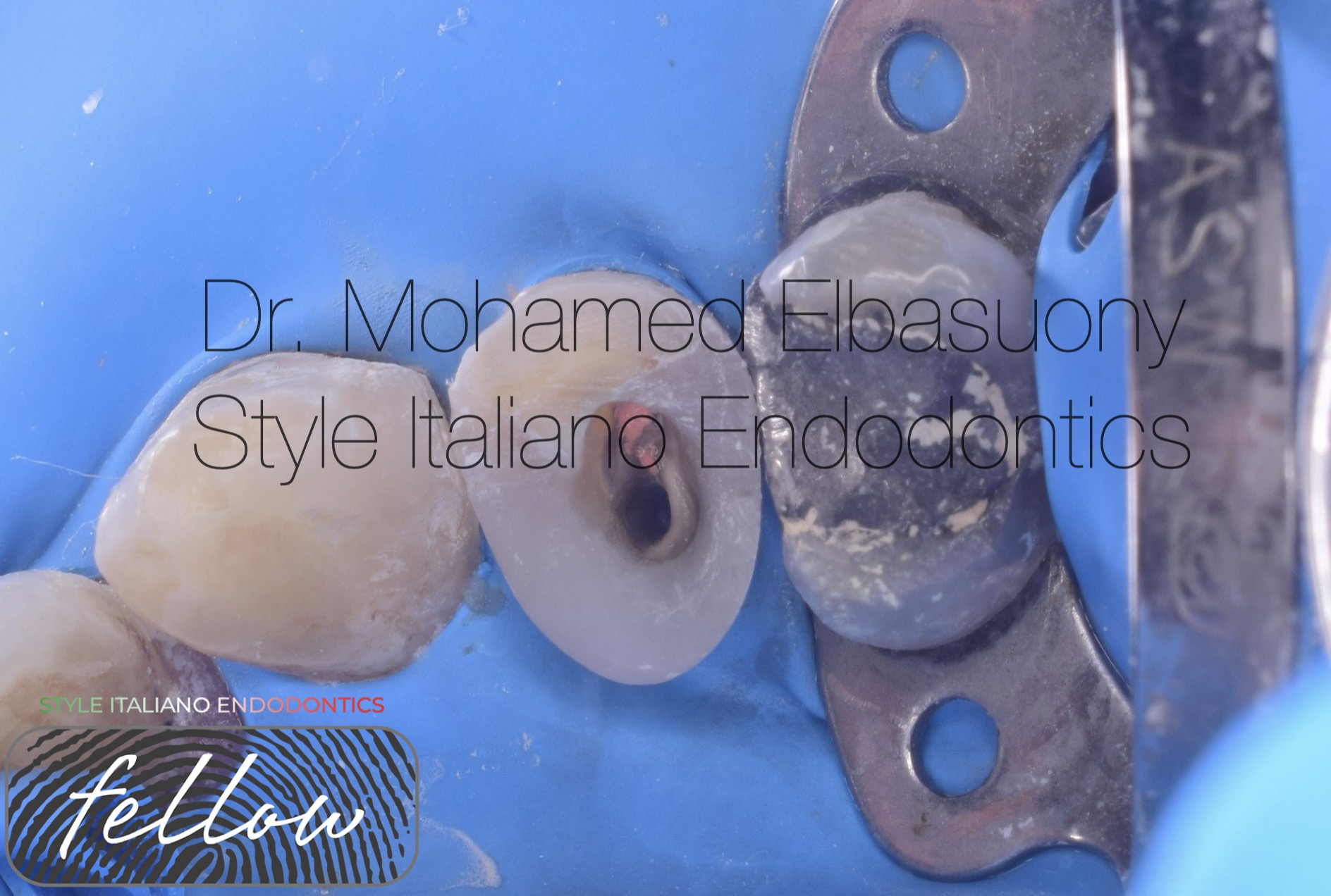
Fig. 11
Post preparation.
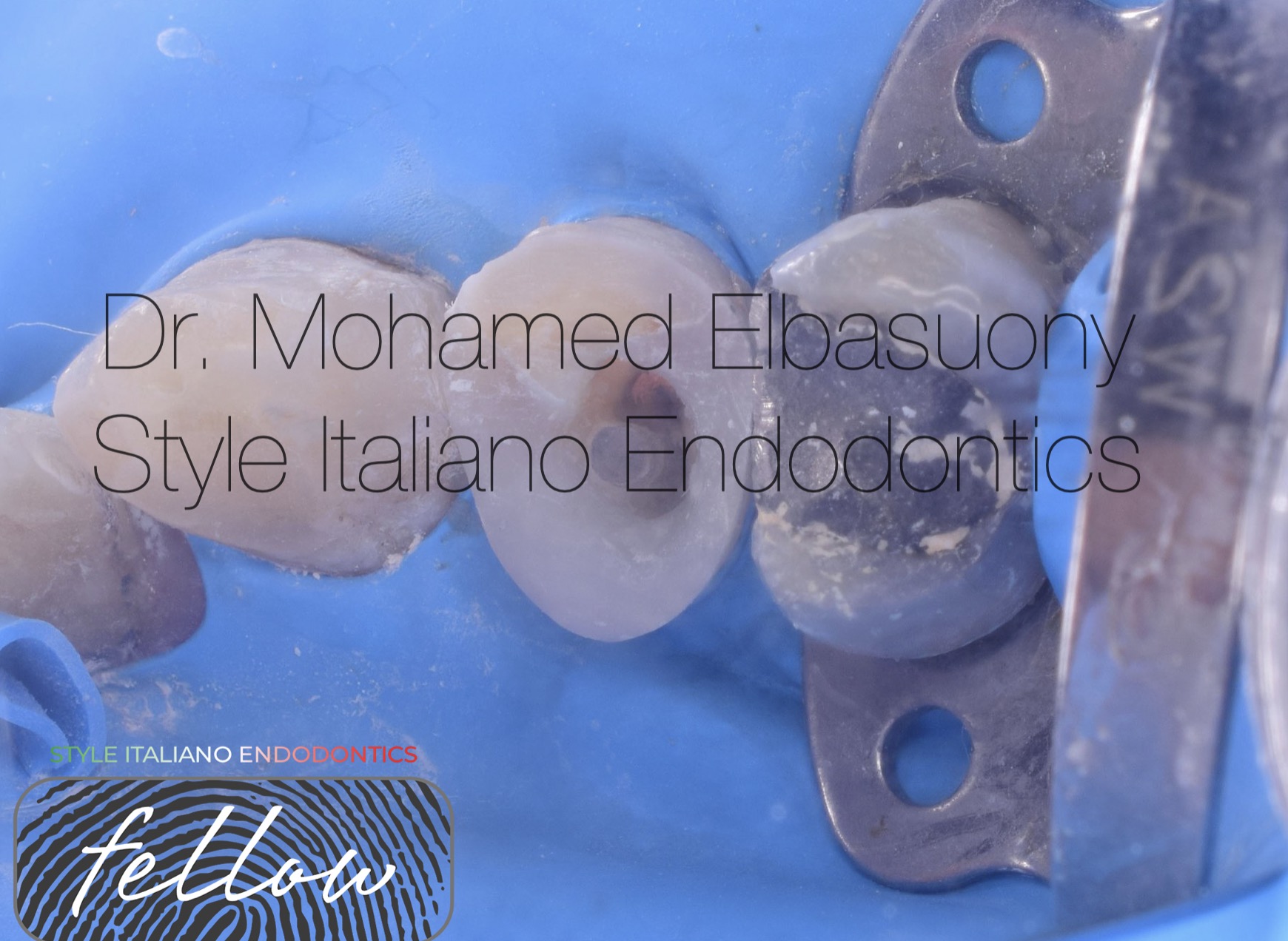
Fig. 12
Insertion of fiber post.
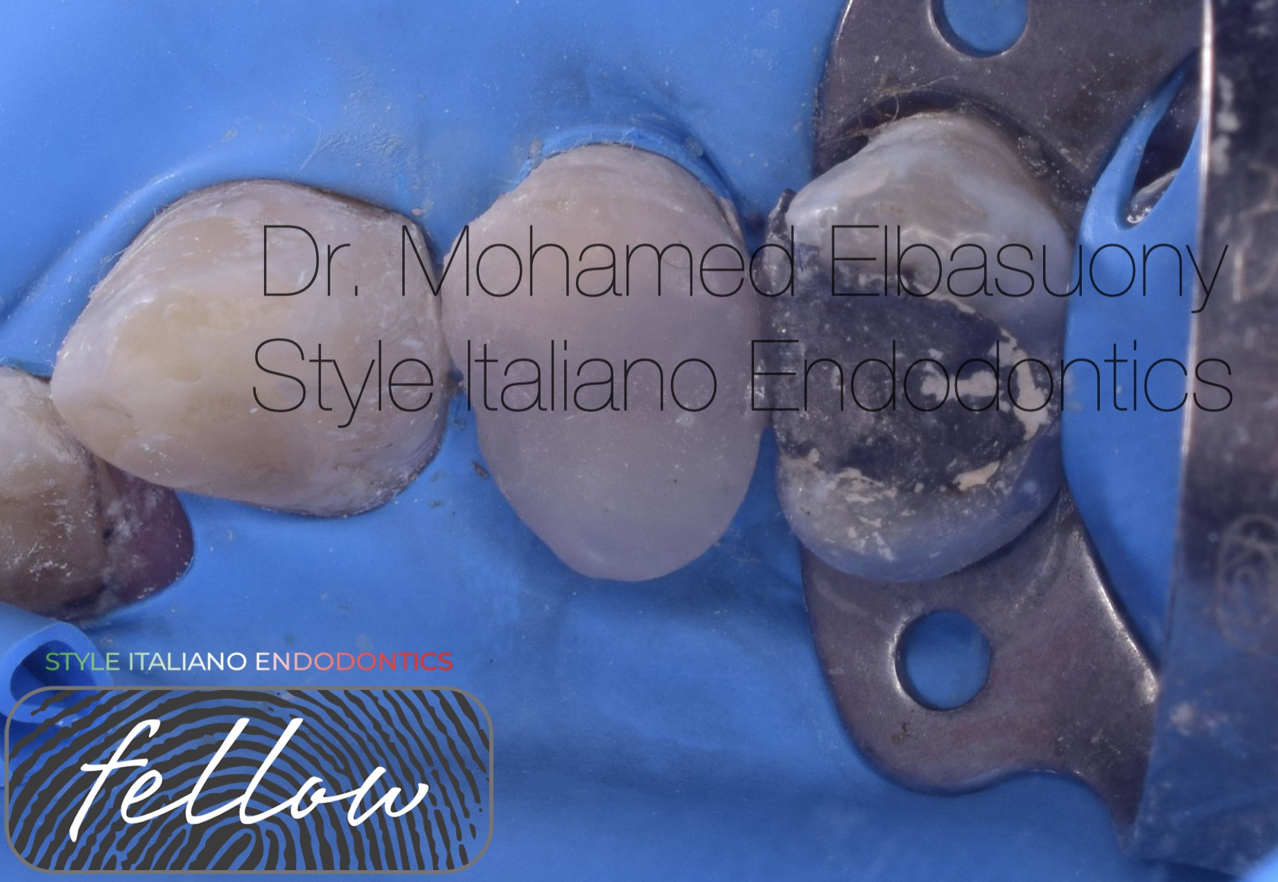
Fig. 13
Composite restoration.
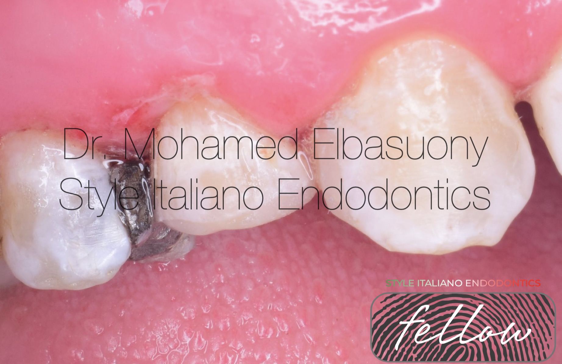
Fig. 14
Facial view.
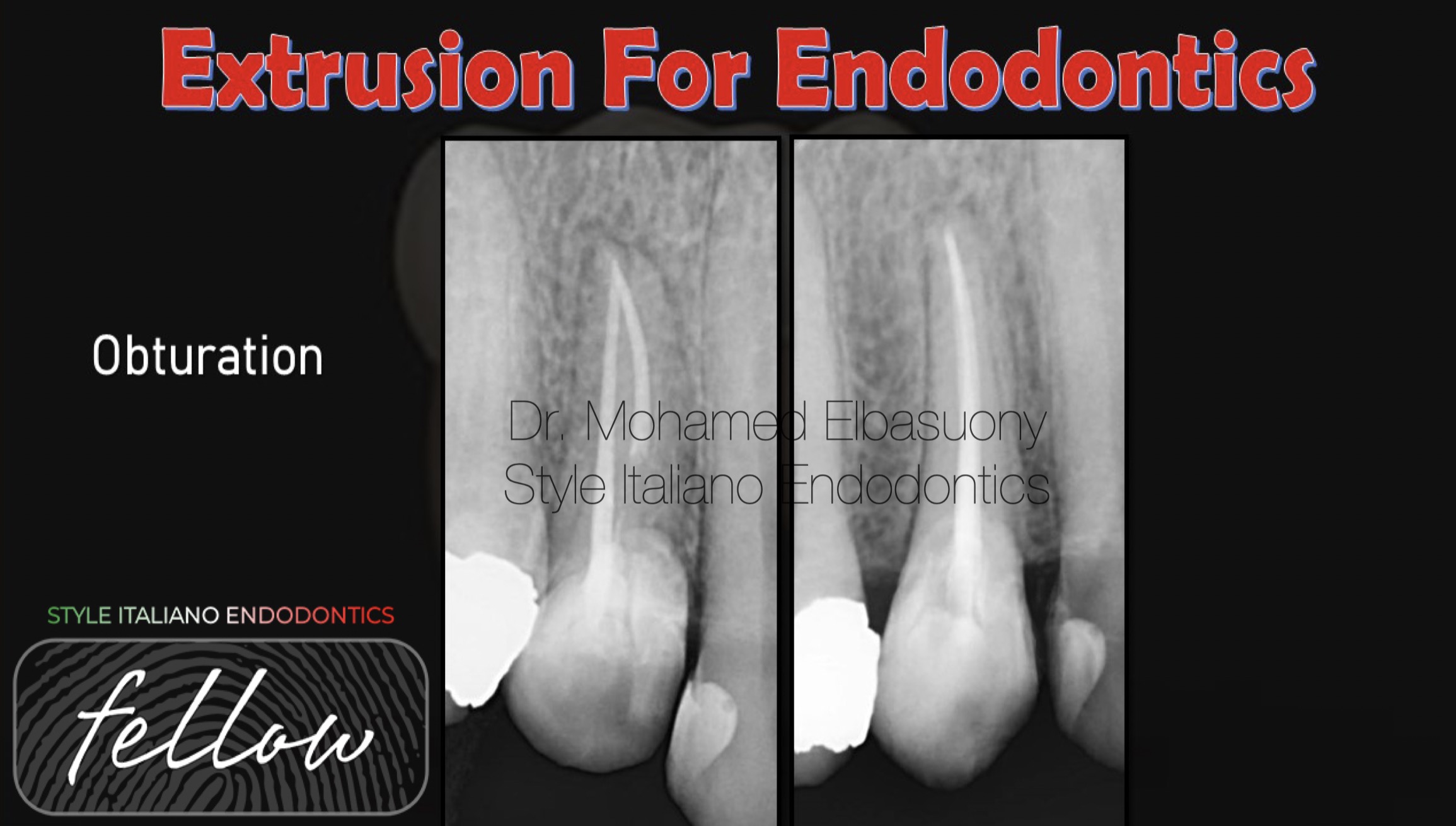
Fig. 15
Obturation radiograph.

Fig. 16
BDS from faculty of dentistry, Mansoura university in 2013.
Diploma in Endodontics at faculty of dentistry, Mansoura university in 2018.
Enrolled in the MSc in Endodontics at faculty of dentistry, Mansoura university in 2020.
StyleItaliano Endodontics Fellow
Endodontic Specialist.
Conclusions
In cases of crown fractures, where endodontic treatment is required and proper field isolation is impossible due to subgingival crown fracture, coronal extrusion of the remaining root via orthodontics, is a viable option before the completion of the root canal and the subsequent coronal restoration under optimal conditions.
Bibliography
- Moghaddam AS, Radafshar G, Taramsari M, Darabi F (2014) Long-term survival rate of teeth receiving multidisciplinary endodontic, periodontal and prosthodontic treatments. J Oral Rehabil 41: 236-242.
- Palomo F, Kopczyk RA. Rationale and methods for crown lengthening. J Am Dent Assoc. 1978;96:257–60.
- Simon JH, Kelly WH, Gordon DG, Ericksen GW (1978) Extrusion of endodontically treated teeth. J Am Dent Assoc 97: 17-23.


