Molars anatomy pt1: upper second molar
27/04/2020
Gabriele Ragucci
Warning: Undefined variable $post in /home/styleendo/htdocs/styleitaliano-endodontics.org/wp-content/plugins/oxygen/component-framework/components/classes/code-block.class.php(133) : eval()'d code on line 2
Warning: Attempt to read property "ID" on null in /home/styleendo/htdocs/styleitaliano-endodontics.org/wp-content/plugins/oxygen/component-framework/components/classes/code-block.class.php(133) : eval()'d code on line 2
The goal of endodontics is to clean, shape and filling 3D the endodontic space, eliminating most of the bacterias presents in the canals. Many of the difficulties found in root canal treatment are due to variations in root canal morphology(1).
Endodontic failure may be associated with the persistence of infection because of a missed canal or incomplete elimination of microorganisms and necrotic pulp remnants during instrumentation. That is why it is very important to know perfectly the anatomy and all its variables.
This short article is intended to be a review of the anatomy of the upper second molar.
The coronal morphology of the upper second molar is very similar to that of the first upper molar, except for the smaller dimensions. Morphologically the three roots are closer and sometimes merged together with two separate canals, or rarely with a single canal. The length is shorter than the first upper molar and the curvatures are less pronounced. According to literature, the variant with three roots, one Palatal and two Vestibular, exists in 91.8% of cases (2-3).
The possible fusion of the mesio-vestibular root with the palatine one can originate a variation with two roots in 15% of cases (4).
In 63% of cases the palatine root is straight, while in 37% it is vestibularly inclined. The disto-vestibular root in 54% of cases is straight; the mesio-vestibular root is straight in 22% of cases or more often inclined distally 54% (5).
The percentage of the second canal (MB2) in the mesio-vestibular root is lower than the first molar (picture 5-6). The position of the pulp chamber is more mesially, it follows that of the first upper molar and is sometimes further emphasized. The finding of the palatine canal, in correspondence with the mesio-palatine cusp, allows orientation in the subsequent opening phase. Extending the chamber opening parallel to the mesial marginal crest up to the mesio-vestibular cusp we localize the entrance of the Mb canal and possibly, on the same line or moved distally, the mesio-distal canal. The presence of three canals along the same line on the floor of the pulp chamber, with the disto-vestibular in the center, has a greater incidence when the coronal perimeter is also compressed in the mesio-distal sense.
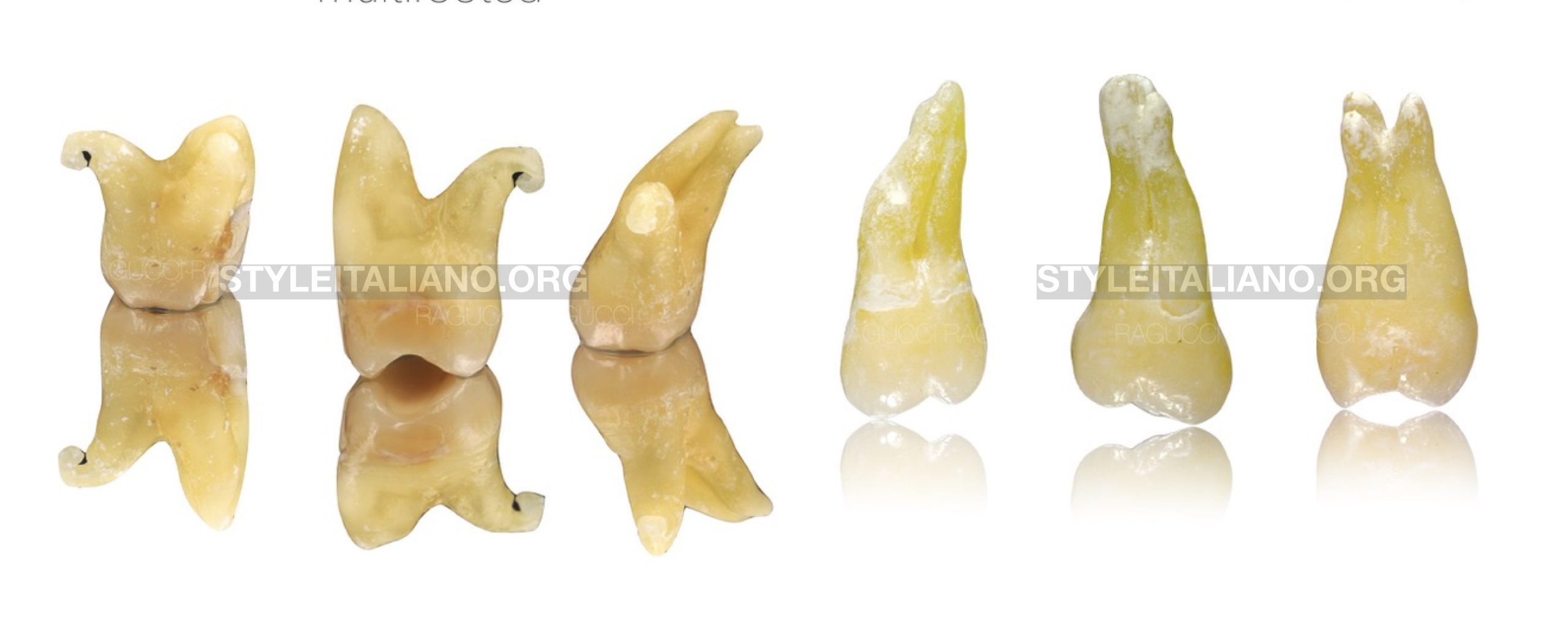
Fig. 1
We can divided Maxillary molars in two big families multirooted and monoradicular or fused.
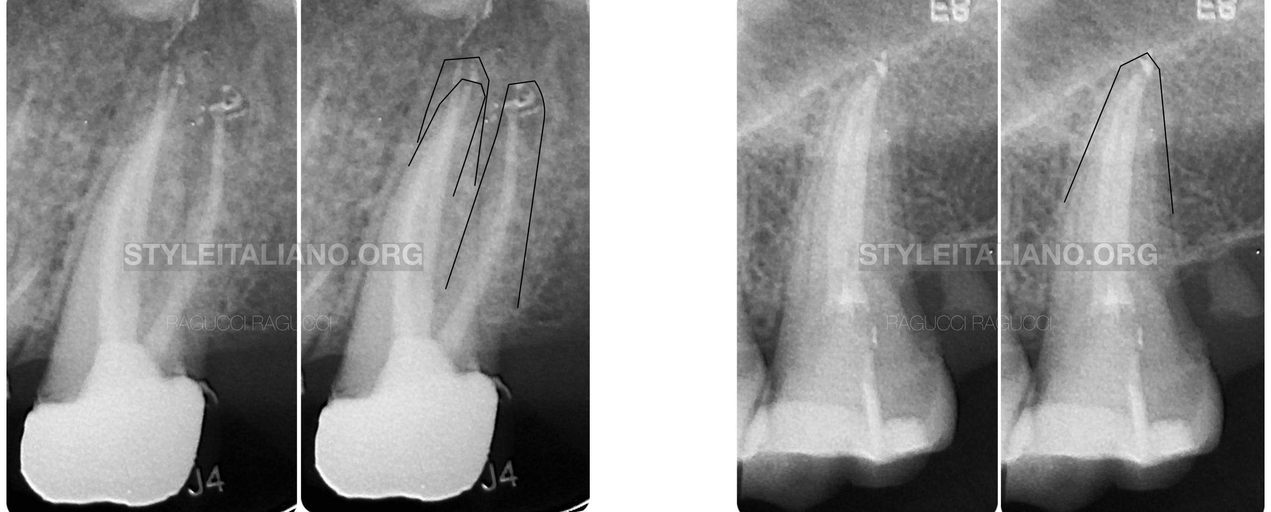
Fig. 2
We can observe in the rx clear differences between multirooted and fused tooth, this will lead us to modify our cavity access ,more mesial in the fused tooth.
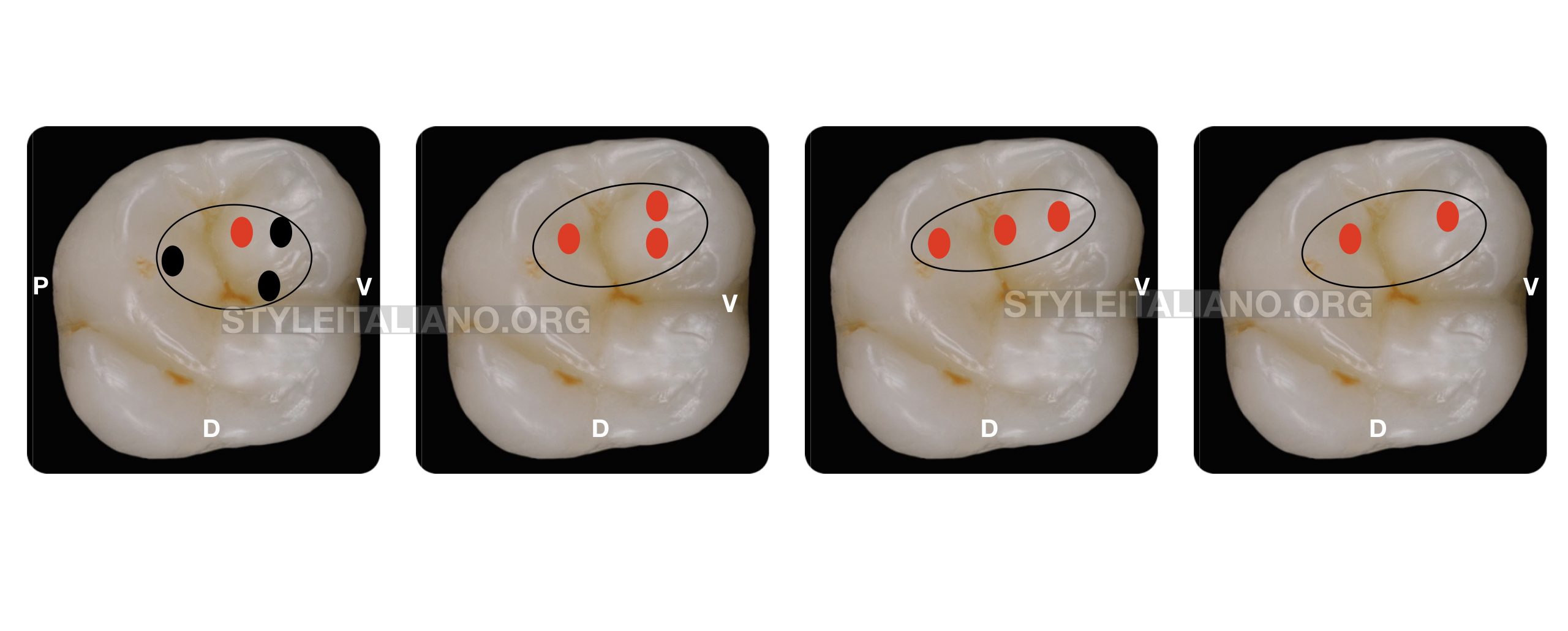
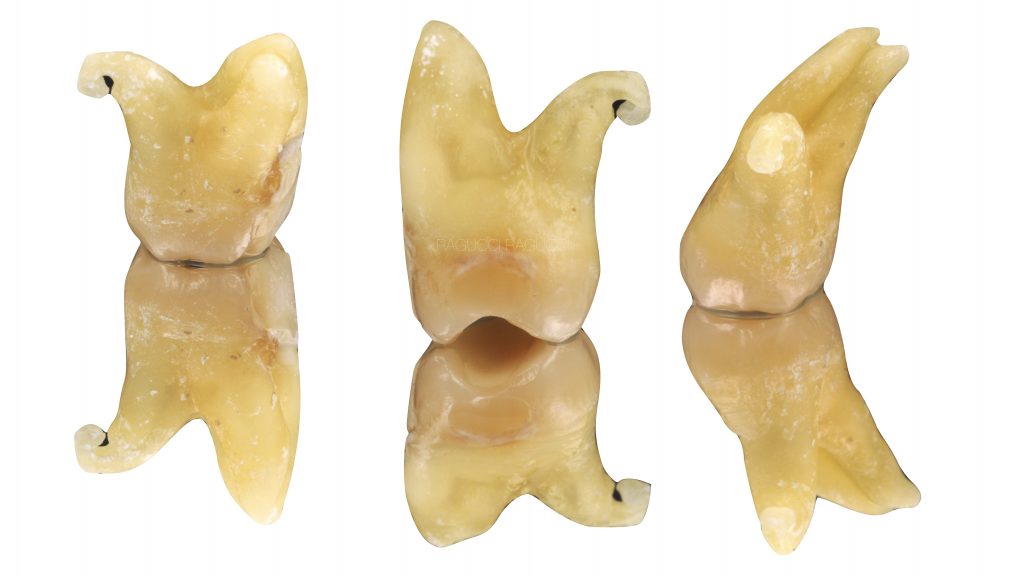
Fig. 3
Shape of the pulp chamber, and location of the root canal orifices.
Multirooted tooth morphology:the pulp chamber is widest in the buccolingual dimension
Fused configuration with two canals: narrow pulp chamber canals in a straight line.
Fused configuration with 3 canals: Mb-Db canals are close
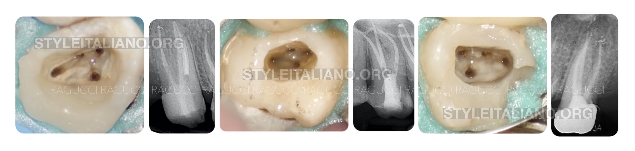
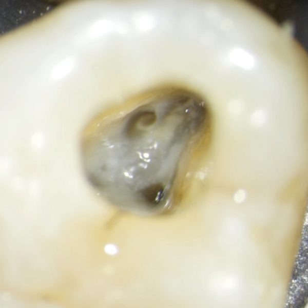
Fig. 4
Multi-rooted upper second molars with mb2 According to literature mb2 is present in 60-70% of the cases. (6-7-9)
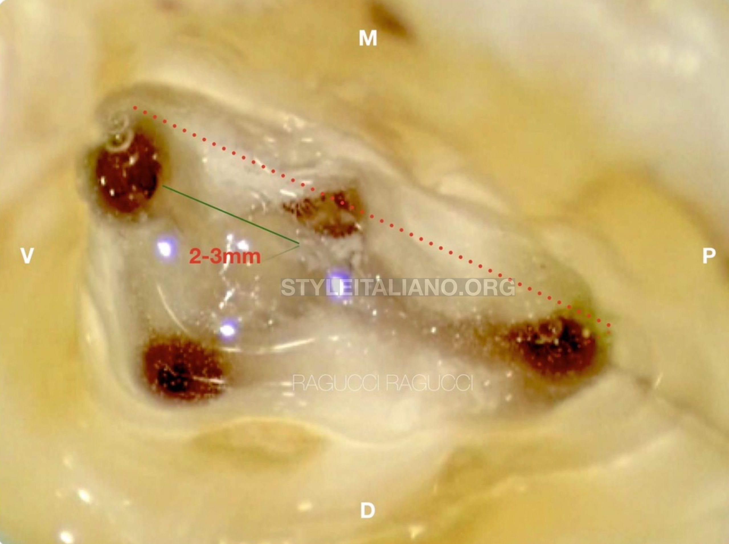
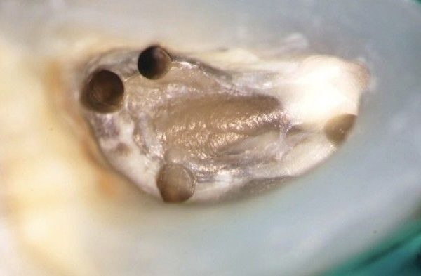
Fig. 5
The mb2 is on a line between mb1 ad palatal canal, it is usually located 2–3 mm from the mesio-buccal canal.
https://endodontics.styleitaliano.org/mb2-in-maxillary-molars-does-it-really-exist/. (suggested article)
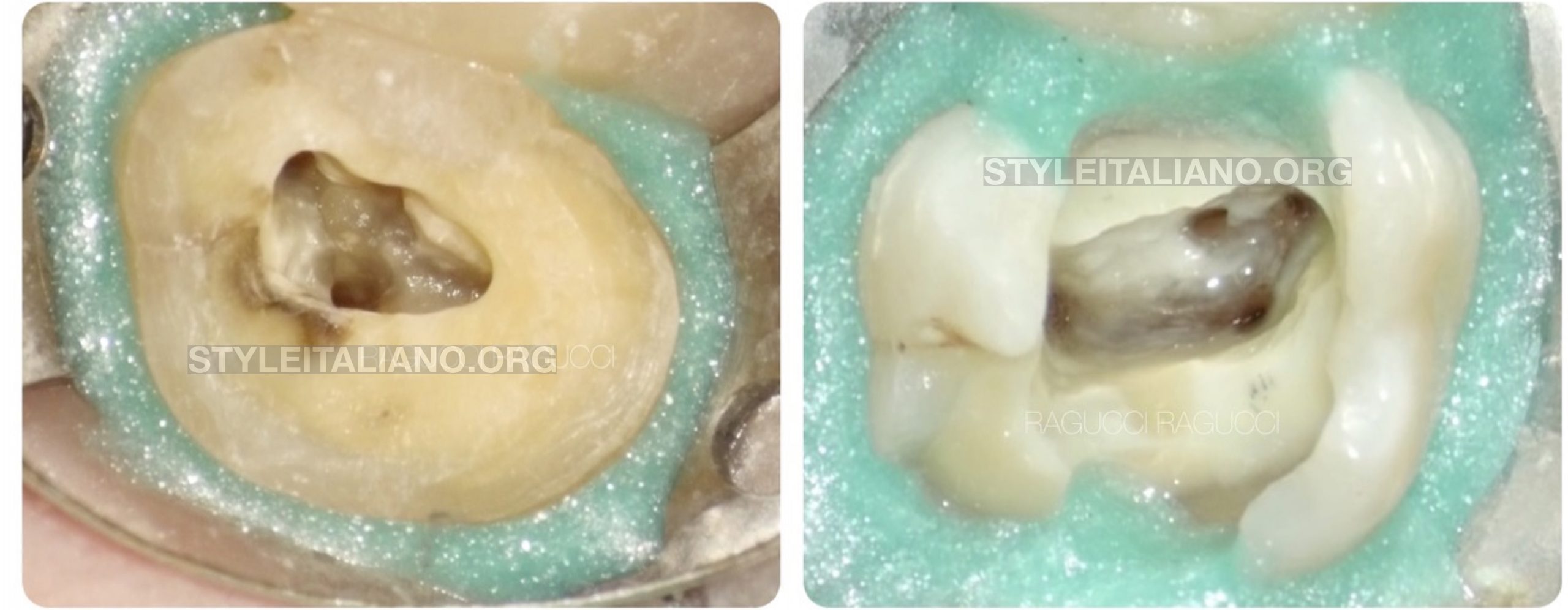
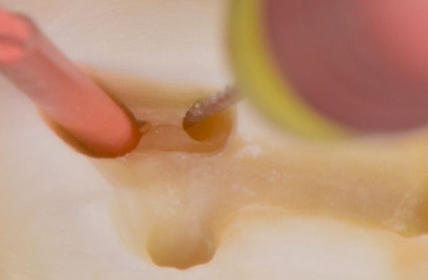
Fig. 6
Color test one of the three laws of pulp chamber anatomy that were published in Journal of endodontic by Krasner et al. 2004 is the law of color change. The developmental root fusion lines or root map are narrow lines on the pulpal floor of furcated teeth and provide a visual trail that has a darker color than the chamber floor. This will lead us to other canal orifices.
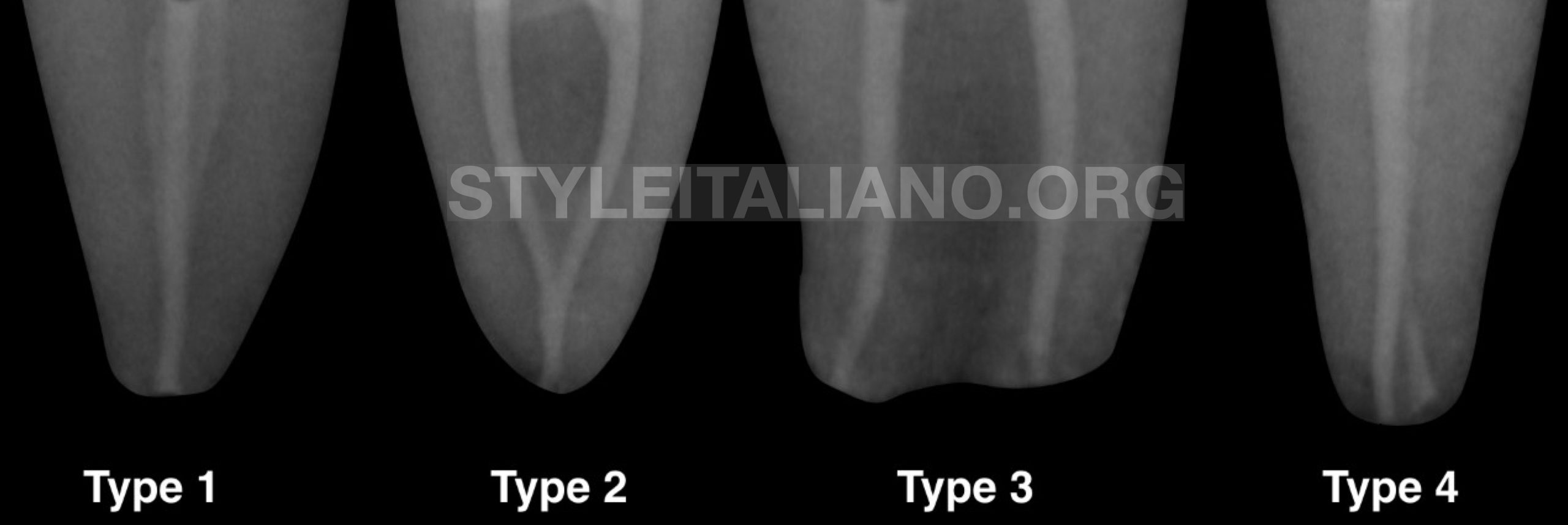
Fig. 7
According to the literature,(7) Weine’s method was used to classify mesiobuccal root canal morphology in upper second molars.
Here is a short video showing canal confluence
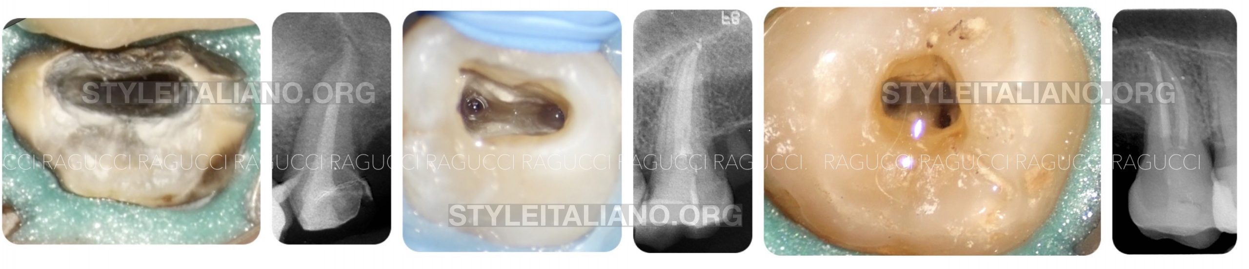
Fig. 8
monoradicular configuration; one tooth out of ten has 1 or 2 canals, while 24% of multirooted and fused has 3 canals.(7-8)
In this clinical case we can see how knowing the correct bases of anatomy and knowing how to correctly interpret the pre-op x-ray can solve many problems and guide us in finding the canals.
Conclusions
The second upper molar is certainly a complex tooth from the anatomical point of view and for the position it has inside the mouth.
Knowledge of anatomy and its variables remains a fundamental step during endodontic treatment.
Everything starts from the correct interpretation of the pre-op RX so as to modulate the treatment according to the anatomy and trying to be as conservative as possible.
Bibliography
- Soares JA, Leonardo RT. Root canal treatment of three-rooted maxillary first and second premolars-a case report. International Endodontic Journal 2003; 36: 705-710
- Eskoz N1, Weine FS.Canal configuration of the mesiobuccal root of the maxillary second molarJ Endod. 1995 Jan;21(1):38-42.
- Ingle JJ, Backland LK Endodontics 5th Edition. Hamilton Ontario Canada 2002
- al Shalabi RM1 Root canal anatomy of maxillary first and second permanent molars.Int Endod J. 2000 Sep;33(5):405-14
- Lautrou A. Anatomia dentaria. Milano . Masson; 1982
- Stropko JJ1Canal morphology of maxillary molars: clinical observations of canal configurations.J Endod. 1999 Jun;25(6):446-50.1
- Naji Kharouf, 2Davide Mancino An In Vivo Study: Location and Instrumentation of the Second Mesiobuccal Canal of the Maxillary Second Molar JCDP 2019
- R. Ordinola-Zapata Morphological evaluation of maxillary second molars with fused roots: a micro-CT study IEJ 2017
- J. Parker, A. Mol, EM. Rivera & P. Tawil CBCT uses in clinical endodontics: The effect of CBCT on the ability to locate MB2 canals in maxillary molars IEJ 2017