MB2: The Most Famous Missed Canal
28/10/2021
Mohamad Zaafrany
Warning: Undefined variable $post in /home/styleendo/htdocs/styleitaliano-endodontics.org/wp-content/plugins/oxygen/component-framework/components/classes/code-block.class.php(133) : eval()'d code on line 2
Warning: Attempt to read property "ID" on null in /home/styleendo/htdocs/styleitaliano-endodontics.org/wp-content/plugins/oxygen/component-framework/components/classes/code-block.class.php(133) : eval()'d code on line 2
Missed Anatomy is one of the main causes of endodontic failure.
The untreated root canal space represent a reservoir for bacteria and necrotic tissue that could be responsible for future failures of endodontic treatment.
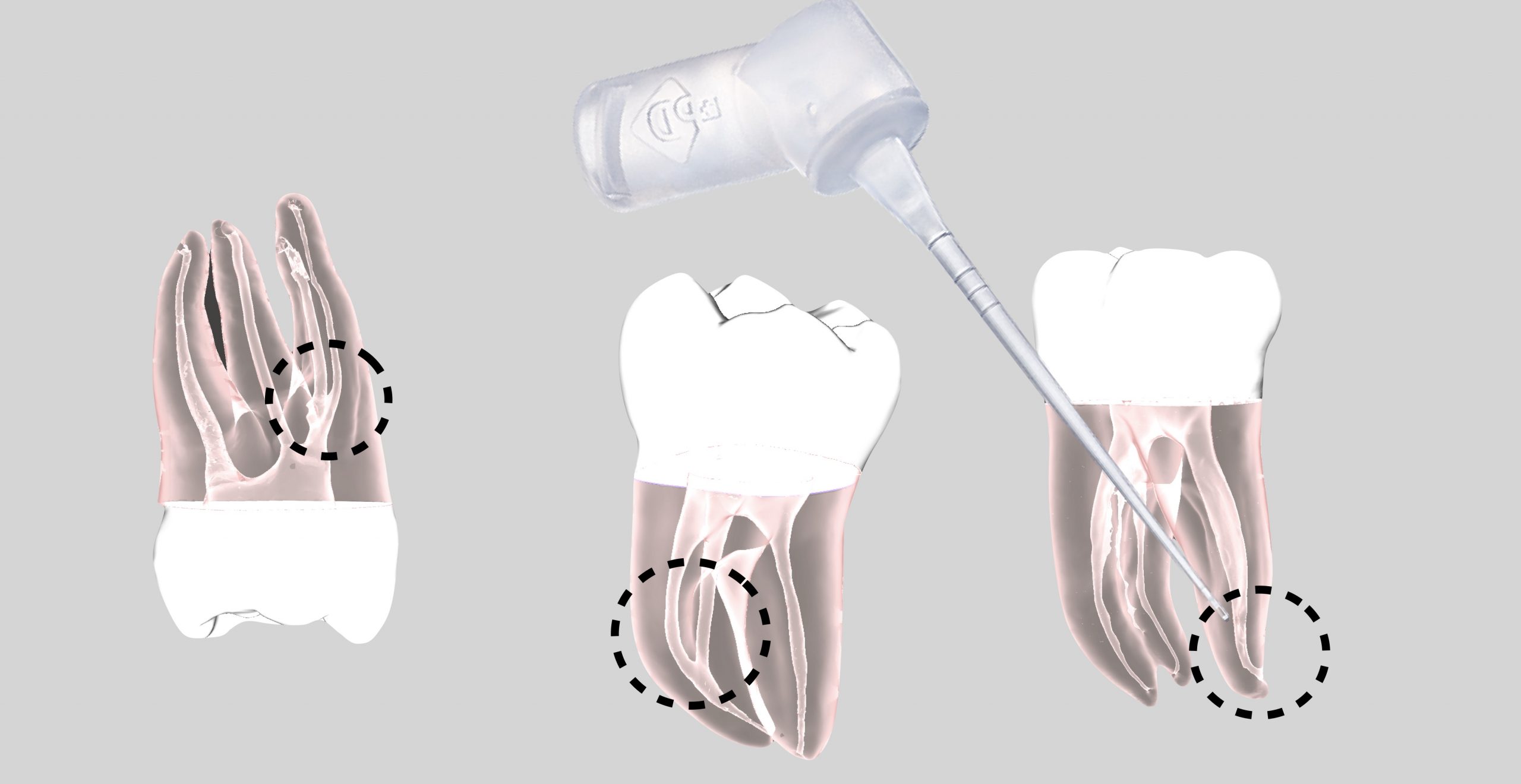
Among maxillary molars MB2 proven to be the most neglected canal during root canal treatment, inability to follow the rules and guides to locate such anatomy will have a big influence on the final treatment outcome.
The results of one study carried out on 5616 molars which were retreated showed that failure to locate the MB2 canal had resulted in a significant decrease in the long-term prognosis of those teeth
The worldwide CBCT assessed MB2 preverlance was 73.8% ranging from 48% in Venezuela to 97% in Belgium
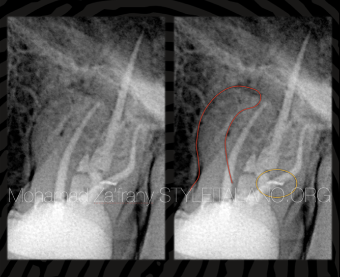
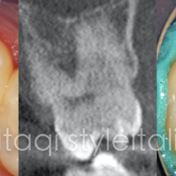
Fig. 1
Preoperative radiographic analysis
Here is a case of endodontic retreatment to Previously treated tooth no.26 with symptomatic apical periodontitis.
From preoperative radiograph it’s shown the obturation of 3 canals MB, D, P with broader MB root outline that indicate the presence of missed anatomy.
A radiopaque object shown superimposed on GP in BD canal that afterwards found to be a separated file in DB canal isthmus.
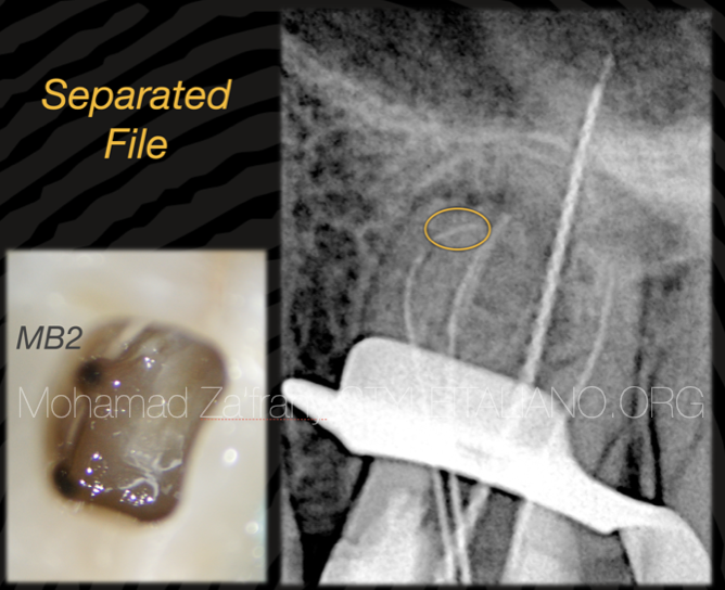
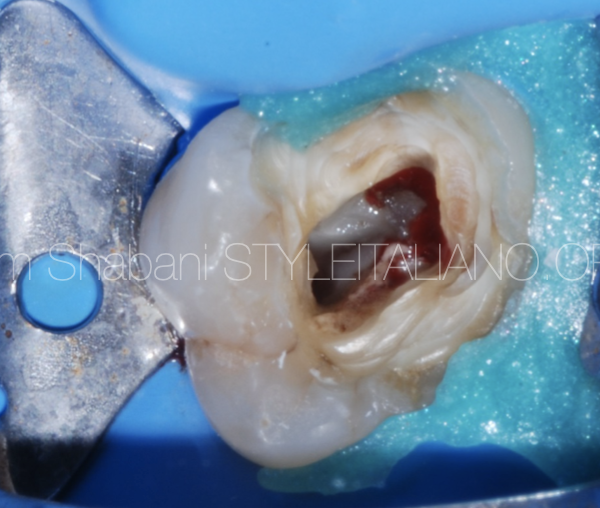
Fig. 2
All old GP removed,
MB2 canal detected quite far from MB canal with the aid of ultrasonics and microscopic magnification.
The separated file in Db canal retrieved.
During manual glide path preparation k_file size 10 got separated in the apical third of MB2 due to the underestimated apical severe curvature.
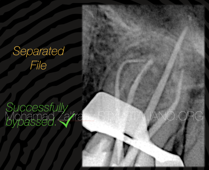
Fig. 3
As the separated file was completely below the curvature and unseen under high magnification so retrieval was not my option and bypass was the most preferred solution as a conservative approach.
By precurving k files size 8,10, separated file was bypassed and canal enlarged to size 20 - 04 not to risk 2nd fracture by enlargement.
Flexible irrigation needle was used for deep delivery of irritant solution.
Master cone fit radiograph showing a very acceptable result.
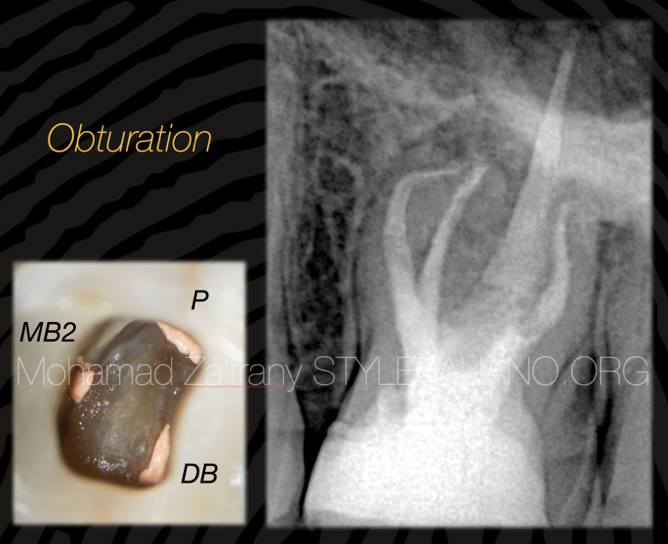
Fig. 4
Obturation done using Modified hot technique with heat friendly BC sealer and warm Gutta Percha.
Clinical video showing the obturation of the 4 canals
Conclusions
- Careful reading and analysis to preoperative radiograph is very important for detection of missed anatomy and identify the reason of treatment failure.
- Clinician should pay attention to detect endodontic anatomy and hidden canals to improve the cleanliness and disinfection for root canal system and improve treatment outcome
- The use of magnification and ultrasonics would facilitate the mission of hidden canals detection
- Unseen separated files under magnification better to be managed by bypass as a more conservative approach.
More details about MB2 in the following link:
https://popdentistry.styleitaliano.org/webinars/the-mb2-canal-a-love-and-hate
Bibliography
Worldwide Analyses of Maxillary First Molar Second Mesiobuccal Prevalence: A Multicenter Cone-beam Computed Tomographic Study. J Endod. 2018 Nov; 44(11):1641-1649.e1. doi: 10.1016/j.joen.2018.07.027. Epub 2018 Sep 19.
Wolcott J, Ishley D, Kennedy W, Johnson S, Minnich S, Meyers J. A 5 yr clinical investigation of second mesiobuccal canals in endodontically treated and retreated maxillary molars. J Endod. 2005;31:262–4.
Hoen MM, Pink FE. Contemporary endodontic retreatments: An analysis based on clinical treatment findings. J Endod. 2002;28:834–6.