Finding the MB2 - part 1
25/10/2020
Fitim Shabani
Warning: Undefined variable $post in /home/styleendo/htdocs/styleitaliano-endodontics.org/wp-content/plugins/oxygen/component-framework/components/classes/code-block.class.php(133) : eval()'d code on line 2
Warning: Attempt to read property "ID" on null in /home/styleendo/htdocs/styleitaliano-endodontics.org/wp-content/plugins/oxygen/component-framework/components/classes/code-block.class.php(133) : eval()'d code on line 2
The famous MB2 canal, which has been studied extensively, has become so popular that it has become a common topic among dentists, especially endodontists. Many studies have shown that not finding and treating this canal has resulted in more endodontic failures.
The methods that help in finding the second mesio buccal canal of maxillary molars are:
- Magnification
- Radiograph
- Access Cavity
- Bubble test
- Blood tracing
- White lines
- Exploring
In this first part of the article i will introduce the Blood tracing method on finding the MB2
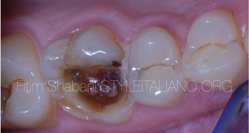
Fig. 1
Initial situation
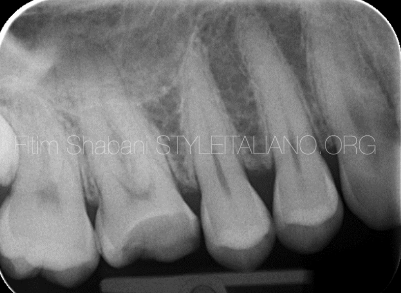
Fig. 2
Pre operative xray
Fig. 3
Isolation and initial preparation
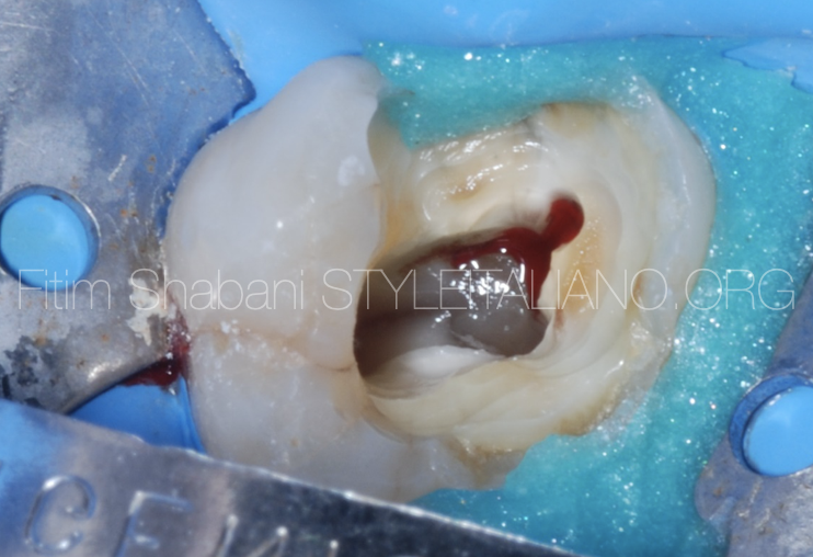
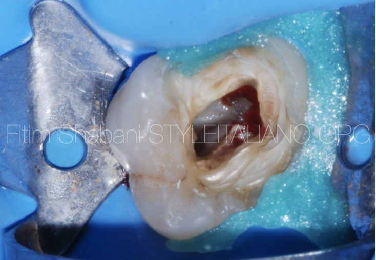
Fig. 4
After mechanical preparation of the P, DB, MB1, we need to search for the MB2
Blood tracing method
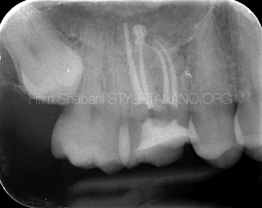
Fig. 5
Post operative xray
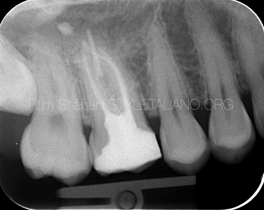
Fig. 6
Post operative xray
Conclusions
Special attention should be paid to the treatment of maxillary molars due to the presence of the MB2 canal.
Before any treatment of these cases we must use all the above methods to verify the presence or absence of this canal. Finding MB2 is in many cases challenging for us, but using a dental microscope makes the job much easier.
Bibliography
-Louis J. Buhrley, DMD, Michael J. Barrows, DDS, MS, Ellen A. BeGole, PhD, and Christopher S. Wenckus, DDS , Effect of Magnification on Locating the MB2 Canal in Maxillary Molars, Journal of Endodontics 2002
-Marcelo Santos Coelho, Mariane Floriano Lopes Santos Lacerda et Al. Locating the second mesiobuccal canal in maxillary molars: Challenges and solutions, Clinical,cosmetic and Investigational Dentistry 2018
-Adham A.Azim BDS, Katharina A.Azim,MA,Med Allan S.Deutsch, DMD, and George T.-J. Huang, DDS, MSD,Dsc Acquisicion of Anatomic Parameters Concerning Molar Pulp Chamber Landmarks Using Cone-beam Computed Tomography, JOE 2014
-Lieutenant Commander Bernard H. Hofmann, DC, USNR and Captain Jeffrey R. Thorpe, DC, USN, Location of the second mesiobuccal canal of maxillary molars in endodontic therapy, Naval Postgraduate Dental SchoolNational Naval Dental Center Bethesda, Maryland December 2002.
-Paul Krasner, DDS, and Henry J. Rankow, DDS Anatomy of the Pulp-Chamber Floor, Journal of Endodontics
-Mitchell H. Davich, DMD, FACD, FICD The MB2 Canal: Following the Map of the Pulpal Floor, Endodontic therapy Vol 5 Nr 2
-Leena Smaldi, BDS, MDentSci, FDSRCS(Ed) and Ameen Khraisat, BDS, PhD, Detection of a second mesiobuccal canal in the mesiobuccal roots of maxillary first molar teeth, University of Jordan