Management of nonsurgical retreatment and final restoration.
06/03/2025
Dennis Quintero Santos
Warning: Undefined variable $post in /home/styleendo/htdocs/styleitaliano-endodontics.org/wp-content/plugins/oxygen/component-framework/components/classes/code-block.class.php(133) : eval()'d code on line 2
Warning: Attempt to read property "ID" on null in /home/styleendo/htdocs/styleitaliano-endodontics.org/wp-content/plugins/oxygen/component-framework/components/classes/code-block.class.php(133) : eval()'d code on line 2
The management of nonsurgical retreatment has proven to be an effective alternative in dentistry for preserving natural teeth. Studies indicate that success rates may decline due to anatomical factors overlooked during the instrumentation phase of primary endodontic treatment, often complicating case resolution. However, despite these challenges, research supports an acceptable success rate for nonsurgical retreatment, reinforcing its viability as a treatment option to help retain teeth in function.
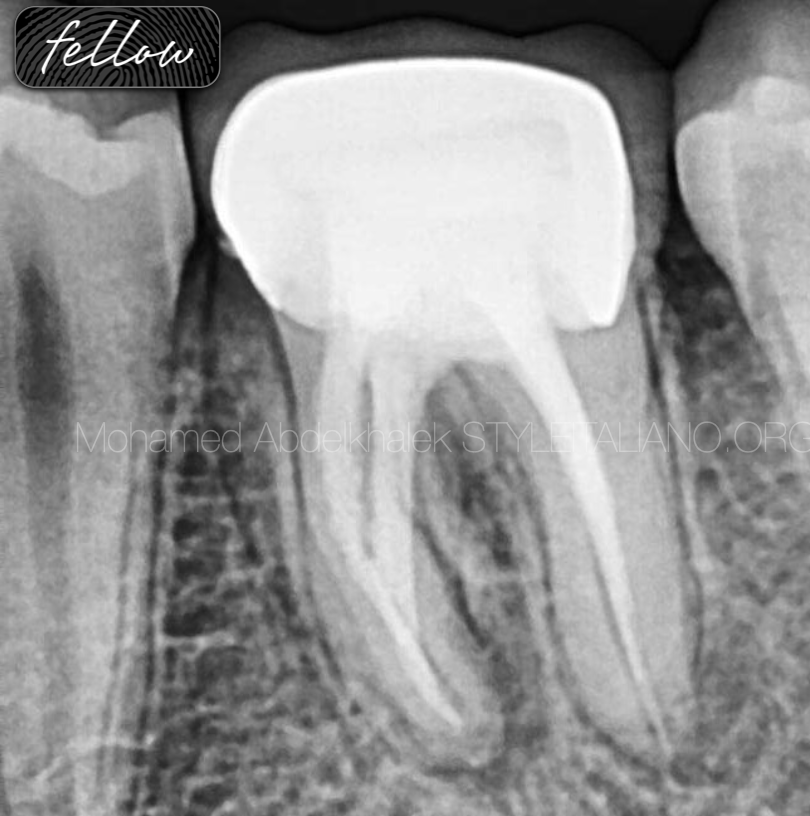
Fig. 1
Female, 35 years old
This patient was referred to me, due to a failed primary endo and also poor final restoration.
Diagnosis:
. Fistula
. Furca lession.
. Extruded gutta-percha in distal root.
. In the mesial root, the anatomy was not respected, resulting in deviation and perforation.
. Very poor final restoration.
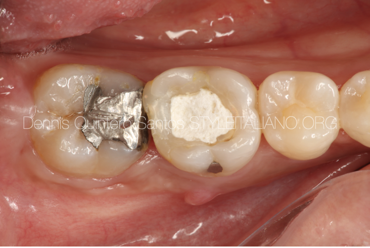
Fig. 2
Initial clinical situation.
. We can observe a very poor restoration.
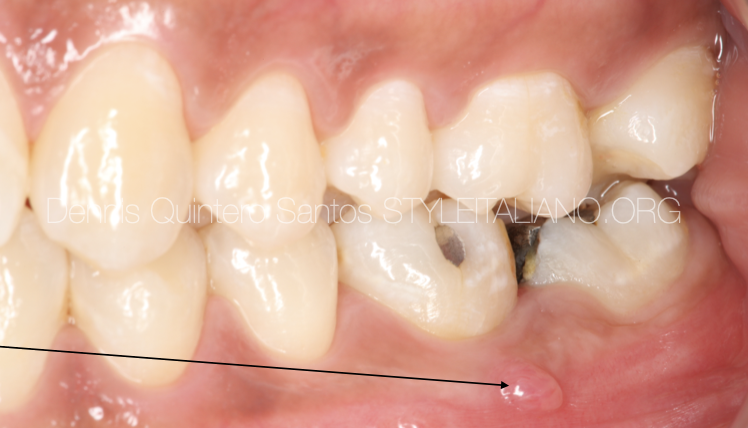
Fig. 3
We can appreciate the occlusion of the patient.
Buccal fistula.
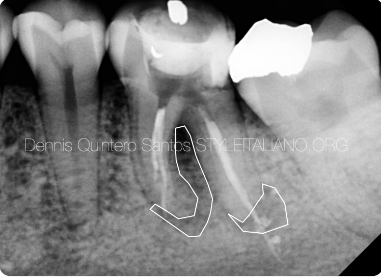
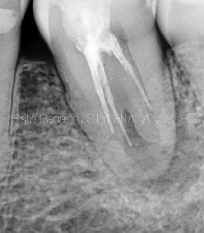
Fig. 4
We can see the size of the periapical lesion.
PROGNOSTIC FACTORS
* Pre-operative periapical lesion
* Unsatisfactory coronal restoration
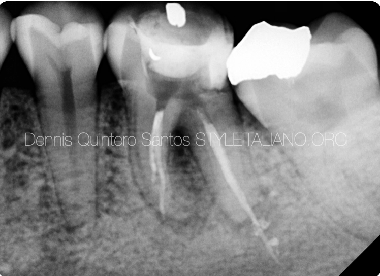
Fig. 5
PROGNOSTIC FACTORS
* Extrusion of filling material during previous treatment
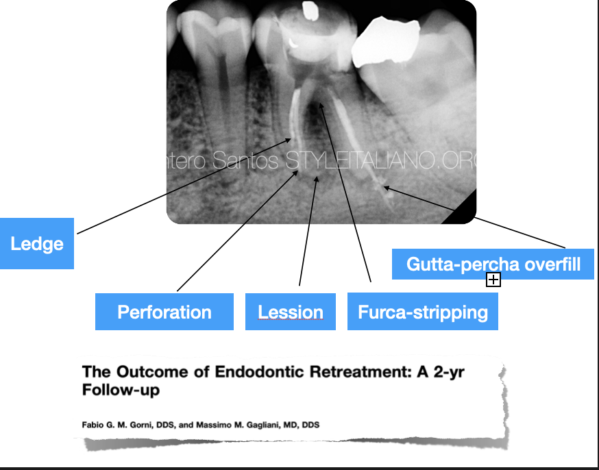
Fig. 6
Internal or external transportation (35.6%)
Apical resorption (71.4%)
Perforation (60.5%)
Stripping (28%)
Internal resorption (71.4%)
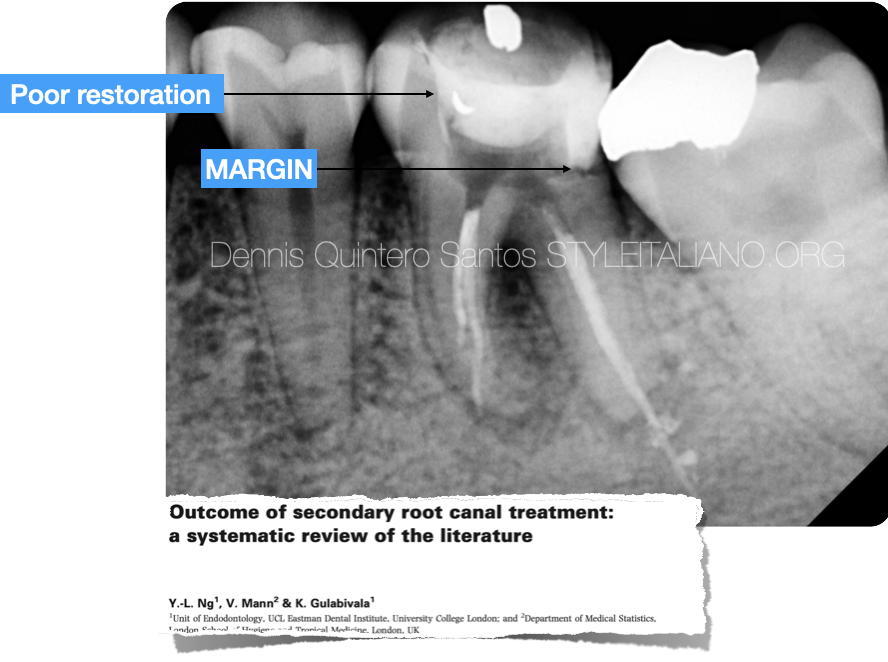
Fig. 7
Teeth that had been restored or permanently restored were associated with significantly higher success rates than their contrary counterpart.
The type of restoration was found to have no significant influence on the outcome 2RCT.
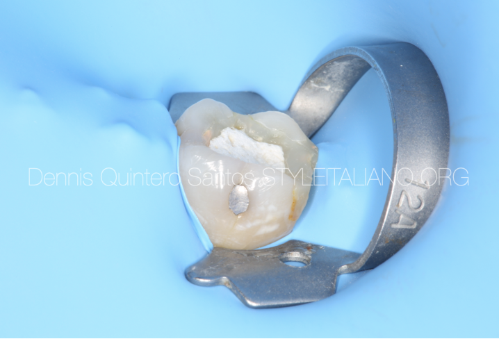
Fig. 8
Isolation with a 12A clamp.
* The use of Rubber dam was associated with significantly higher success rates versus cotton roll isolation.
* The success rates of treatment using rubber dam isolation was 77%, based on strict criteria.
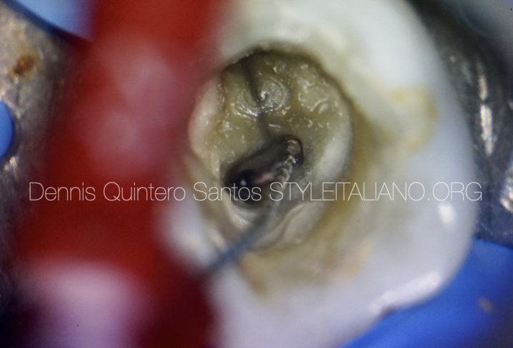
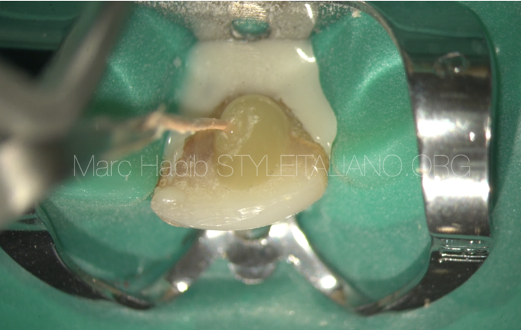
Fig. 9
Removing gutta-percha with a hedstroem file.
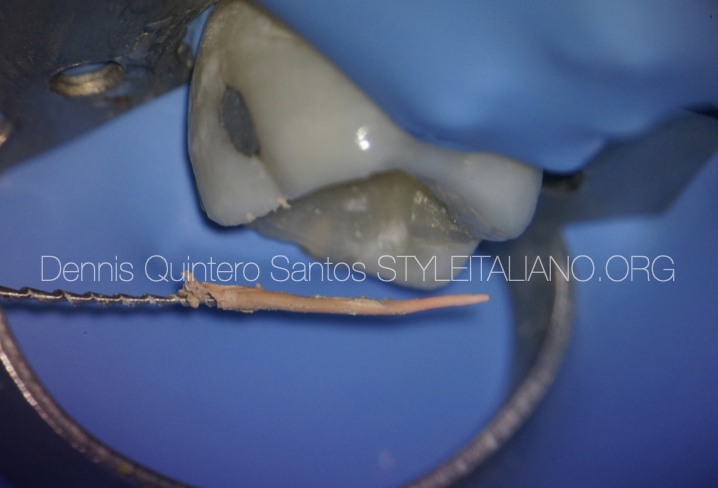
Fig. 10
Guttapercha was removed from distal root, to avoid more extrusion.
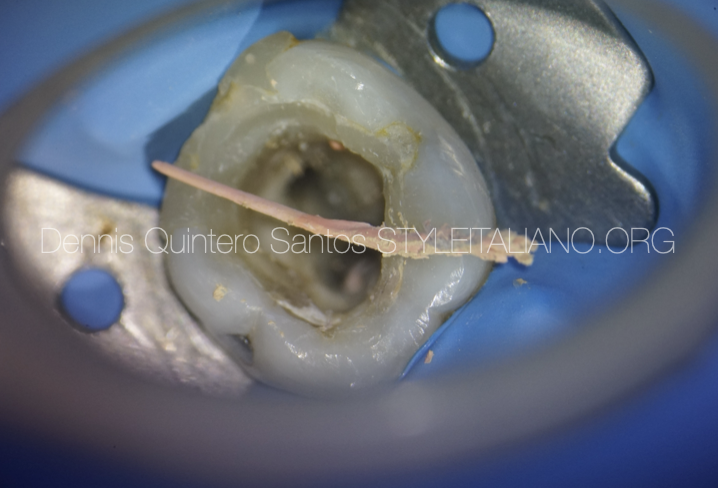
Fig. 11
Single cone was removed.
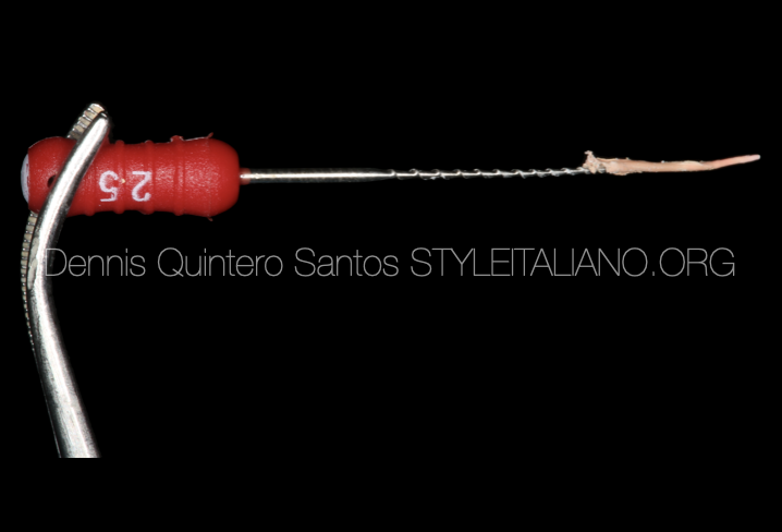
Fig. 12
Hedstrom file.
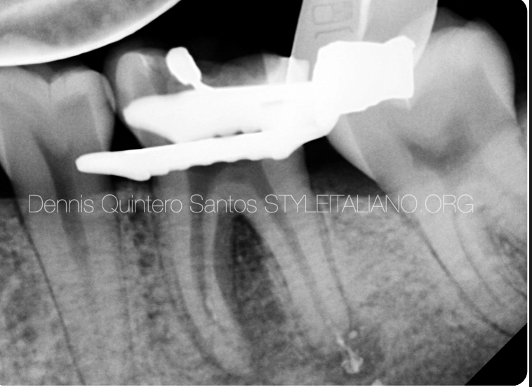
Fig. 13
Guttapercha was removed from all the canals.
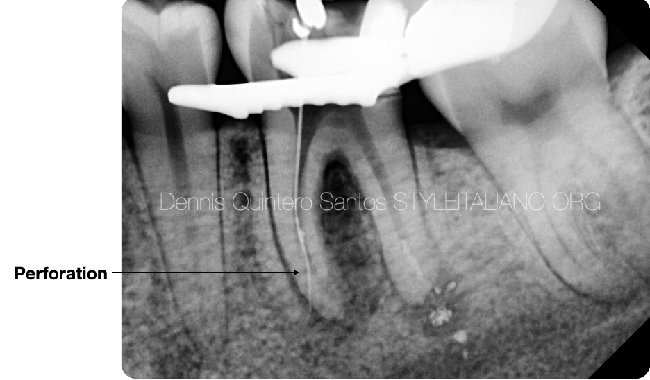
Fig. 14
We found a perforation in mesial root.
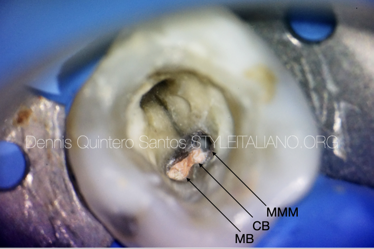
Fig. 15
We found 3 mesial conducts.
. MB, CB, MMM.
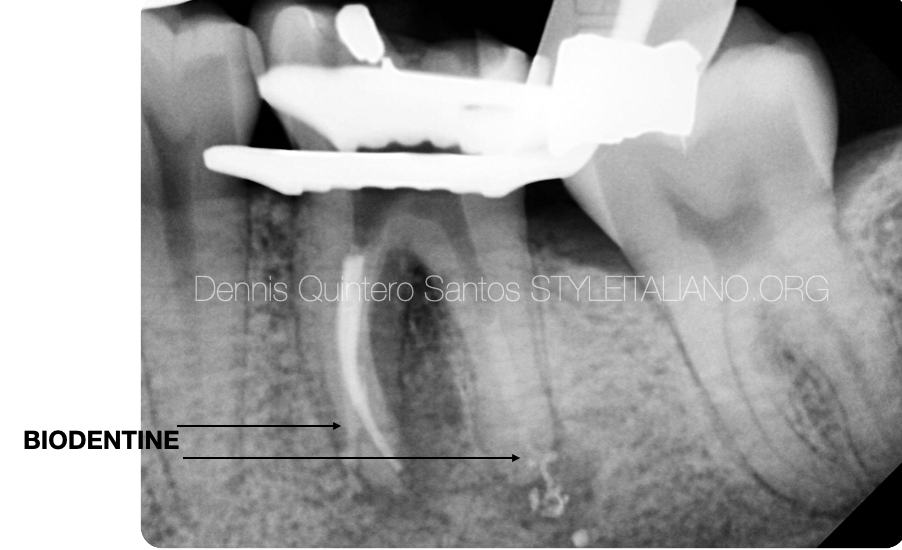
Fig. 16
Biodentine was use to seal the perforation in mesial and distal root.
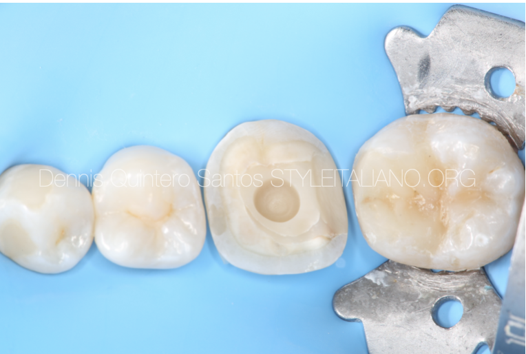
Fig. 17
Final restoration was done for tooth 3.6 and 3.7 for a better long term prognosis.
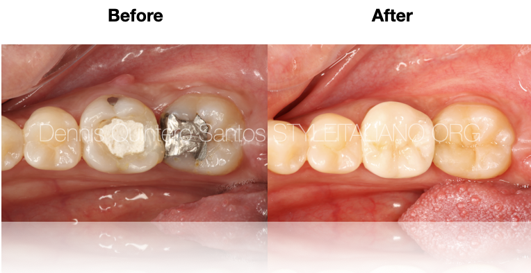
Fig. 18
Final restoration 3.6 indirect emax overlay.
3.7 direct composite restoration.
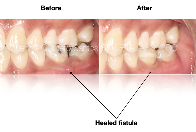
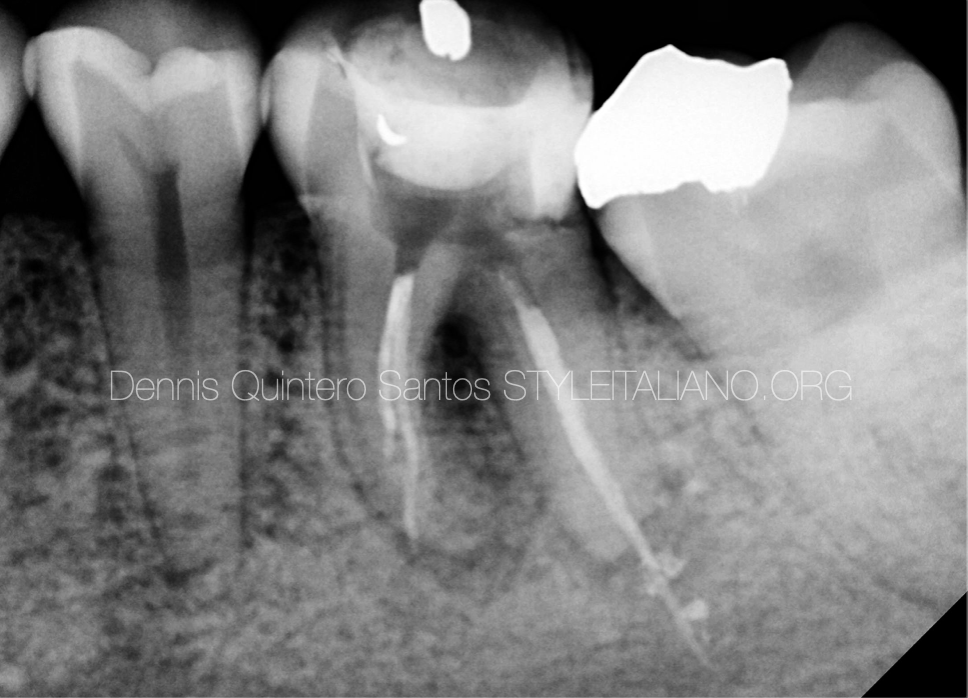
Fig. 19
Conservative overlay as a final restoration and healed fistula.
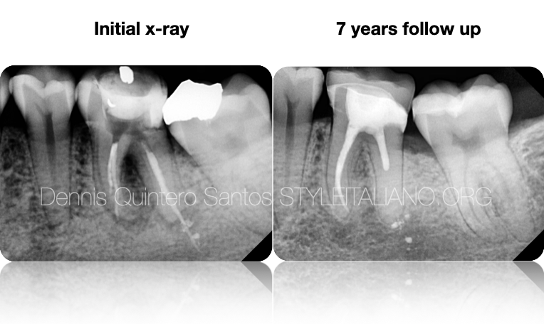
Fig. 20
Before and after with 7 years follow up.
Conclusions
In conclusion, non-surgical retreatment remains a highly effective option for preserving natural teeth. However, each case must be thoroughly evaluated, taking into account both clinical and radiographic findings to select the most appropriate treatment alternative and develop an optimal treatment plan.
Bibliography
Ng YL, Mann V, Gulabivala K. Outcome of secondary root canal treatment: a systematic review of the literature. Int Endod J. 2008;41(12):1026-46.
Kang M, In Jung H, Song M, Kim S, Kim H, Kim E. Outcome of nonsurgical retreatment and endodontic microsurgery: a meta-analysis. Clin Oral Investig. 2015;19(3):569-82.
Imura N, Pinheiro ET, Gomes BP, Zaia AA, Ferraz CC, Souza-Filho FJ. The outcome of endodontic treatment: a retrospective study of 2000 cases performed by a specialist. J Endod. 2007;33(11):1278-82.
Gorni FG, Gagliani MM. The outcome of endodontic retreatment: a 2-yr follow-up. J Endod. 2004;30(1):1-4.