Non surgical retreatment of a lower first molar
15/10/2023
Fellow
Warning: Undefined variable $post in /home/styleendo/htdocs/styleitaliano-endodontics.org/wp-content/plugins/oxygen/component-framework/components/classes/code-block.class.php(133) : eval()'d code on line 2
Warning: Attempt to read property "ID" on null in /home/styleendo/htdocs/styleitaliano-endodontics.org/wp-content/plugins/oxygen/component-framework/components/classes/code-block.class.php(133) : eval()'d code on line 2
Retreatment is one of the most challenging procedures in the endodontic therapy, in this article we will review a case showing the management of a lower first molar within 2 peri-apical lesions related to each root, different Obturation techniques to achieve apical seal and optimum outcome.
In Endodontics we are dealing with a lot of challenges such as anatomy, mishaps, instrument separation, etc. but one of the biggest challenges is to seal the root canal to enhance our prognosis in retreatments.
When facing a retreatment, the clinician must be aware of root anatomy and try to understand the reason of failure to deal with such case in a predictable way, reducing secondary risk of failure.
If we identified the reason we can easily deal with it in the retreatment, hence increasing the success of the endodontic therapy.
Fig. 1
A patient came to my attention complaining about tenderness on chewing on the first lower left molar. To manage this casee 2 pre-operative radiographs with 2 different angulations were taken.
Pre-operative radiograph shows :
Short filling on the mesial system
Poor Obturation of the distal canal and wide apex
Peri-apical lesions related to each root
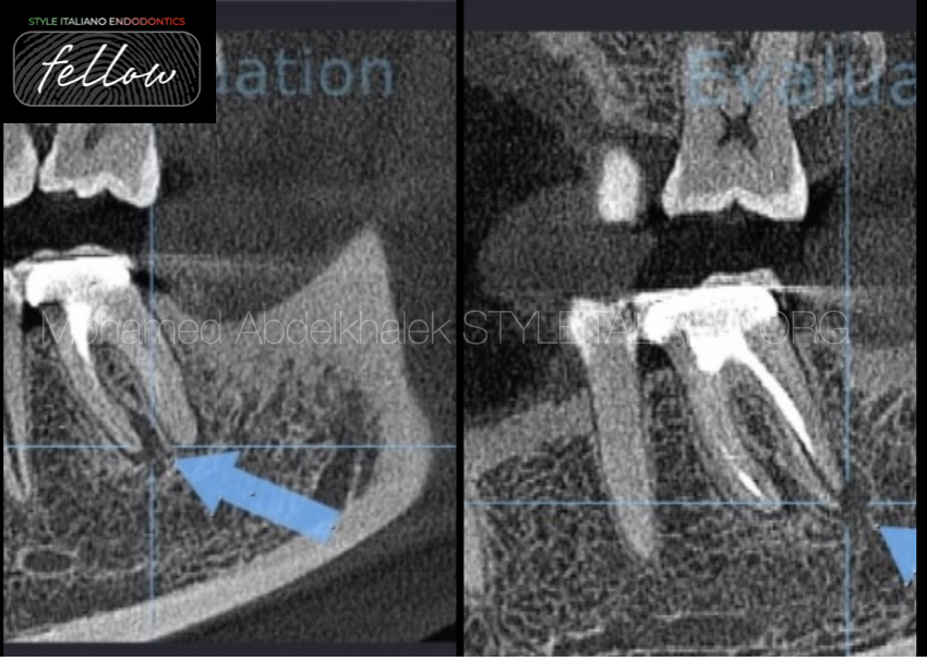
Fig. 2
A CBCT was also taken in order to make a better diagnosis.
Saggital cuts of CBCT scan shows improper seal of wide apex related to distal canals
Poor shaping and Obturation related to mesial system.
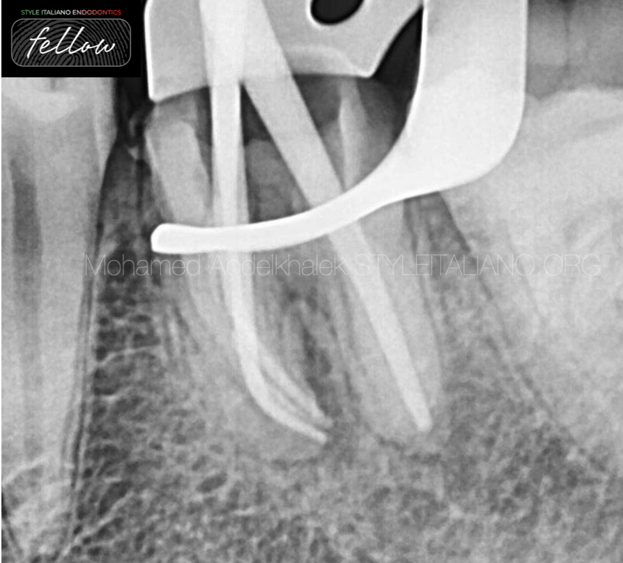
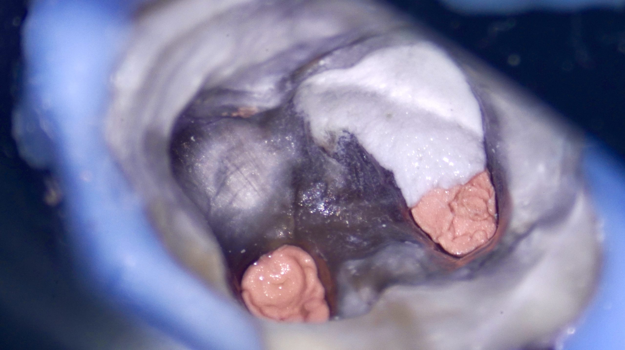
Fig. 3
After the disassembling, the existing filling material was removed and the root canal system was shaped and cleaned.
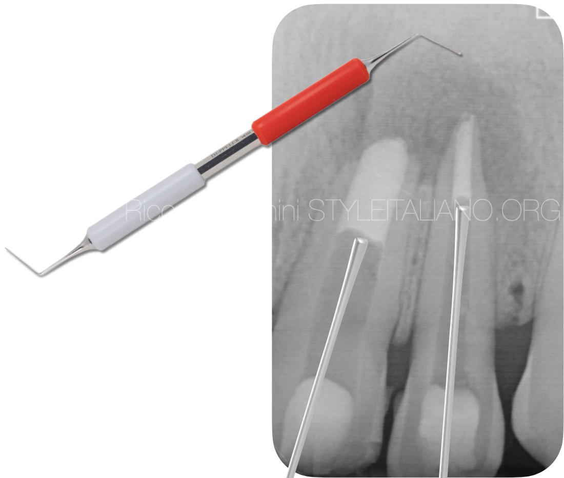
The X-ray shows the master cone fitting.
Mesial system shows well fitted guttapercha to the full working length.
In the distal canal, a customized guttapercha point was cut by 2 mm sort of the apex to preform adequate MTA apical plug.
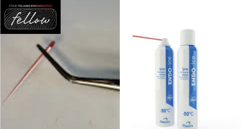
Fig. 4
The apical size of the distal canal, in fact, was larger than ISO .60, which is considered by some authors the cut off above which apical plug with bioceramics or MTA is suggested to achieve apical seal.
Map one system was used as a carrier for increment placement inside the the canal.
Endo ice was applied to the customized guttapercha to harden the guttapercha to pack the MTA at the apical third.
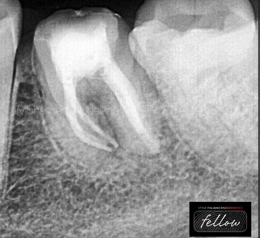
Fig. 5
Post-operative radiograph shows the distal canal filled with a 4 mm MTA apical plug followed by thermo-plastized guttapercha. In the mesial canals, obturation was done using bio-ceramic sealer with hot-modified warm vertical compaction technique.
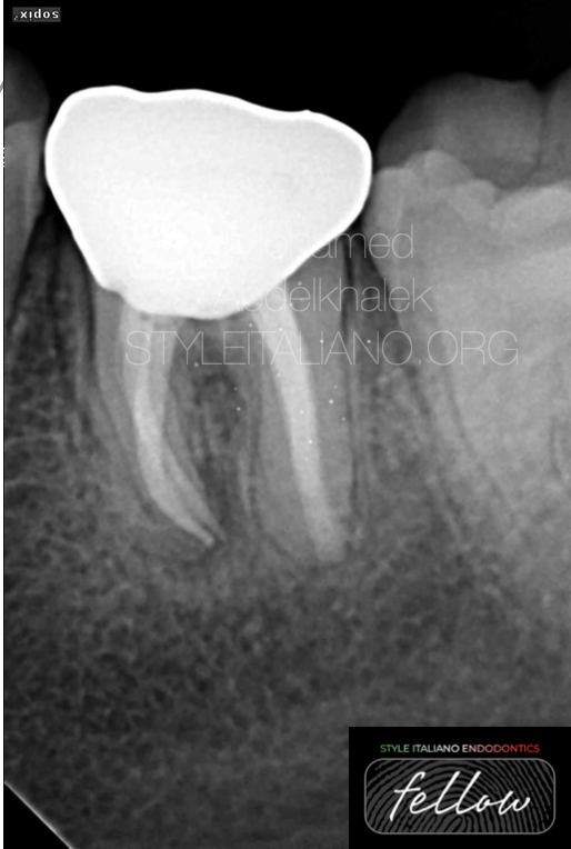
Fig. 6
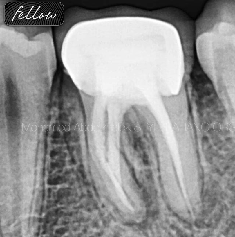
12 months follow up radiograph showing the state of healing of the lesions and new hygienic crown

Fig. 7
About the author
Mohamed Abdelkhalek
I'm a dentist was born and raised in Alexandri, I'm really passionate about endodontics and digital dentistry.
Proudly I'm providing elite services to my patient, also have been sharing knowledge with other colleagues inwebinars and local conference.
EDUCATION
2012- 2017
University of Pharos
WORK EXPERIENCE
Dental derma dreams
Full time conservative dentist related
to micro-scopic and digital dentistry
• Advanced endodontic in Mazen dental clinic
Microscopic dentist
Digital dentistry
ADA certificate in endodontics
Co-founder@dentaldermadreams.eg
Conclusions
A correct pre operative assessment of the case, together with the knowledge of different filling materials, can increase the outcome of endodontic retreatments
Bibliography
Stephen Cohen, Kenneth M. Hargreaves. Pathways of the pulp, St Louis, Missouri, Mosby 2006, 622.
Giuliani V, Baccetti T, Pace R, Pagavino G. The use of MTA in teeth with necrotic pulps and open apices. Dent Traumatol. 2002 Aug;18(4):217-21.
Hachmeister DR, Schindler WG, Walker WA 3rd, Thomas DD.The sealing ability and retention characteristics of mineral trioxide aggregate in a model of apexification. J Endod. 2002 May; 28(5):386-90.
Giovarruscio M, Uccioli U, Malentacca A, Koller G, Foschi F, Mannocci F. A technique for placement of apical MTA plugs using modified Thermafil carriers for the filling of canals with wide apices
Schilder H (1967) Filling canals in three dimensions. Dent Clin North Am, 724-53.
Roggendorf MJ, Ebert J, Petschelt A, Frankenberger R (2007) Influence of moisture on the apical seal of root canal fillings with five different types of sealer. Journal of Endodontics 33(1), 31-33.
Dillon JS, Amita, Gill B. To determine whether the first file to bind at the working length corresponds to the apical diameter in roots with apical curvatures both before and after preflaring. J Conserv Dent. 2012 Oct;15(4):363-6.
Abarca J, Zaror C, Monardes H, Hermosilla V, Muñoz C, Cantin M. Morphology of the Physiological Apical Foramen in Maxillary and Mandibular First Molars. Int J Morphol. 2014 Jun;32(2):671-677.