Management of complex anatomy in the first mandibular premolar, 2 cases in 1
02/01/2025
Fellow
Warning: Undefined variable $post in /home/styleendo/htdocs/styleitaliano-endodontics.org/wp-content/plugins/oxygen/component-framework/components/classes/code-block.class.php(133) : eval()'d code on line 2
Warning: Attempt to read property "ID" on null in /home/styleendo/htdocs/styleitaliano-endodontics.org/wp-content/plugins/oxygen/component-framework/components/classes/code-block.class.php(133) : eval()'d code on line 2
We know that treating a tooth involves different phases or stages, but it is important to know the internal anatomy, in order to approach the case in the best possible way.
I present 2 cases in 1 where we can observe how complex a treatment can be if we cannot approach it correctly, a lower first premolar with aberrant anatomy and a canine with simple anatomy.
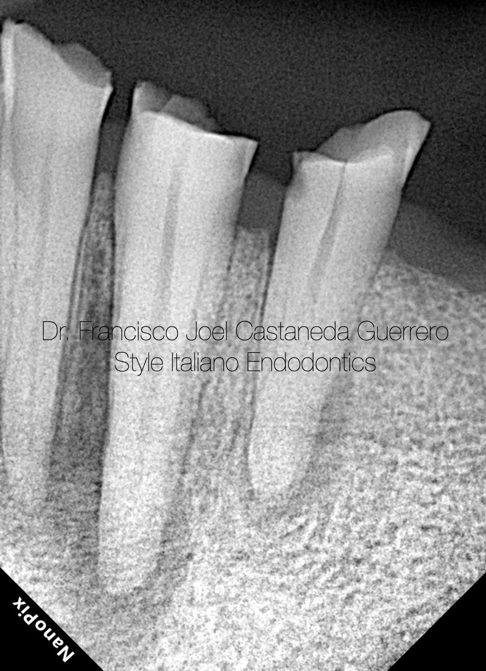
Fig. 1
An 82-year-old man is referred for endodontic treatment.
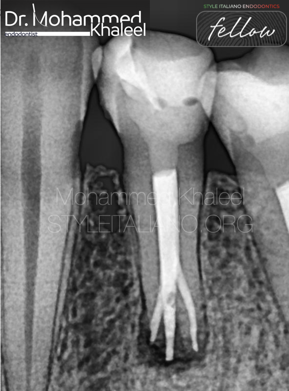
The radiographic examination shows the canine and the lower first premolar with a rarefaction zone, due to pulp necrosis caused by incisal wear of the anterior teeth.
Pulp diagnosis: pulp necrosis
Periapical diagnosis: asymptomatic apical periodontitis
Pulp tests were performed but there was no response in those two teeth.
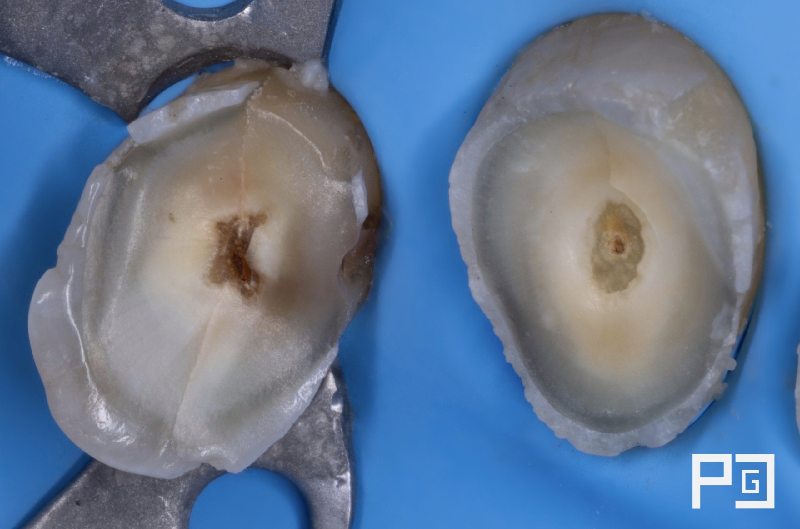
Fig. 2
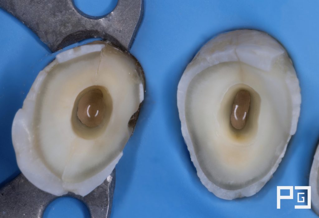
Initial situation after anesthesia and rubber dam placement
As seen in the image, presence of cracks and wear.
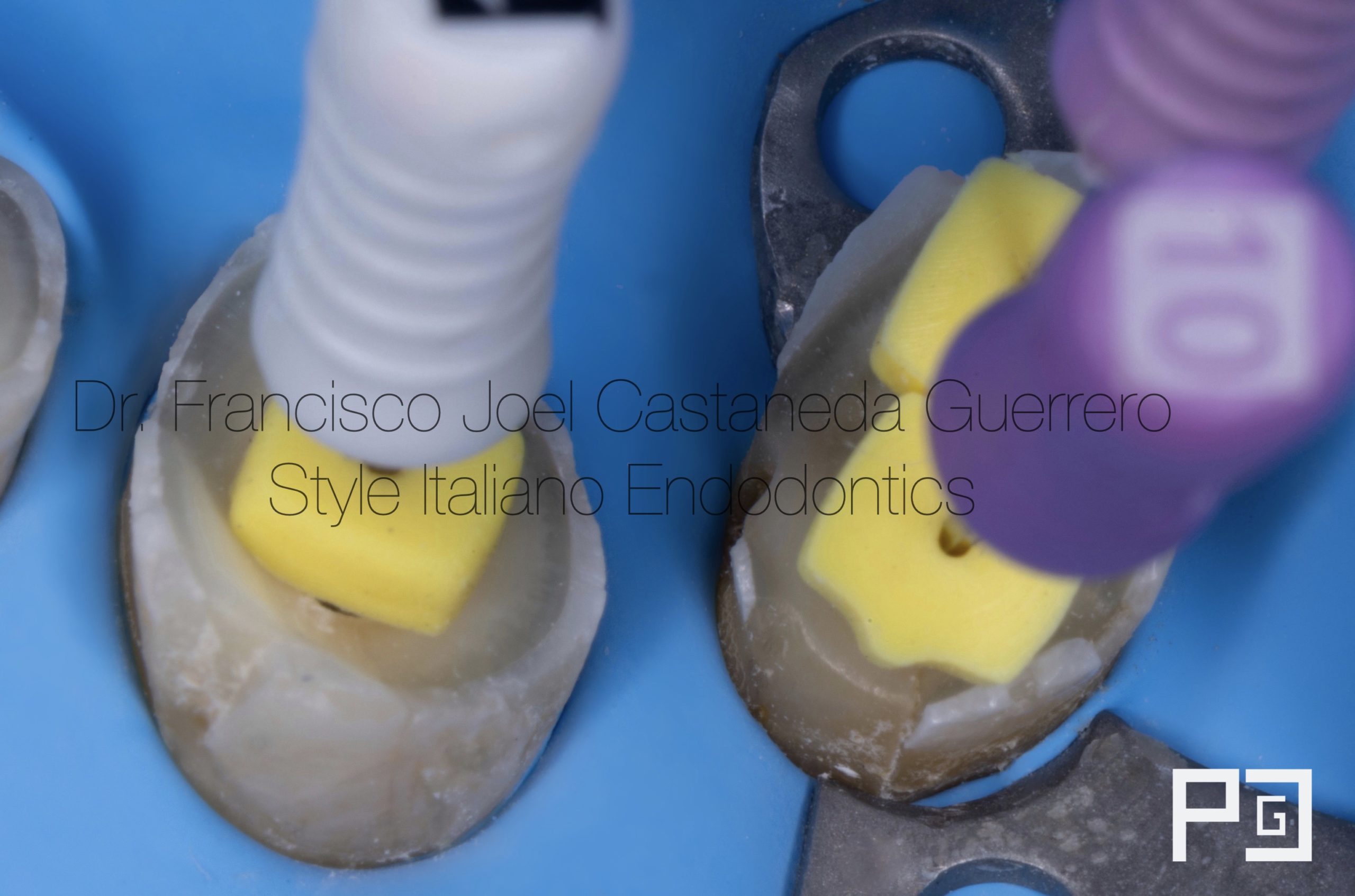
Fig. 3
Access was performed and canals with 10k and 15k files were located.
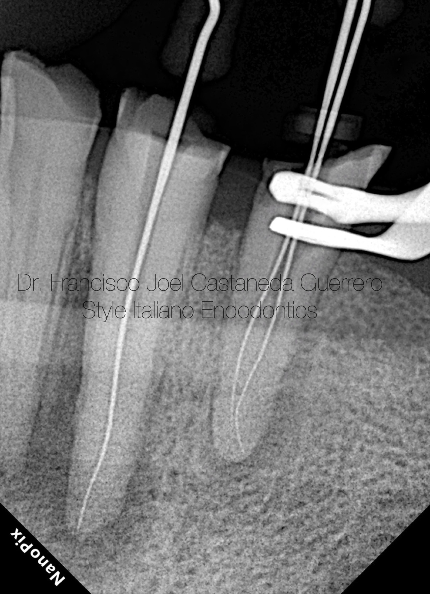
Fig. 4
As seen in the x-ray, the premolar showed a difficult anatomy.
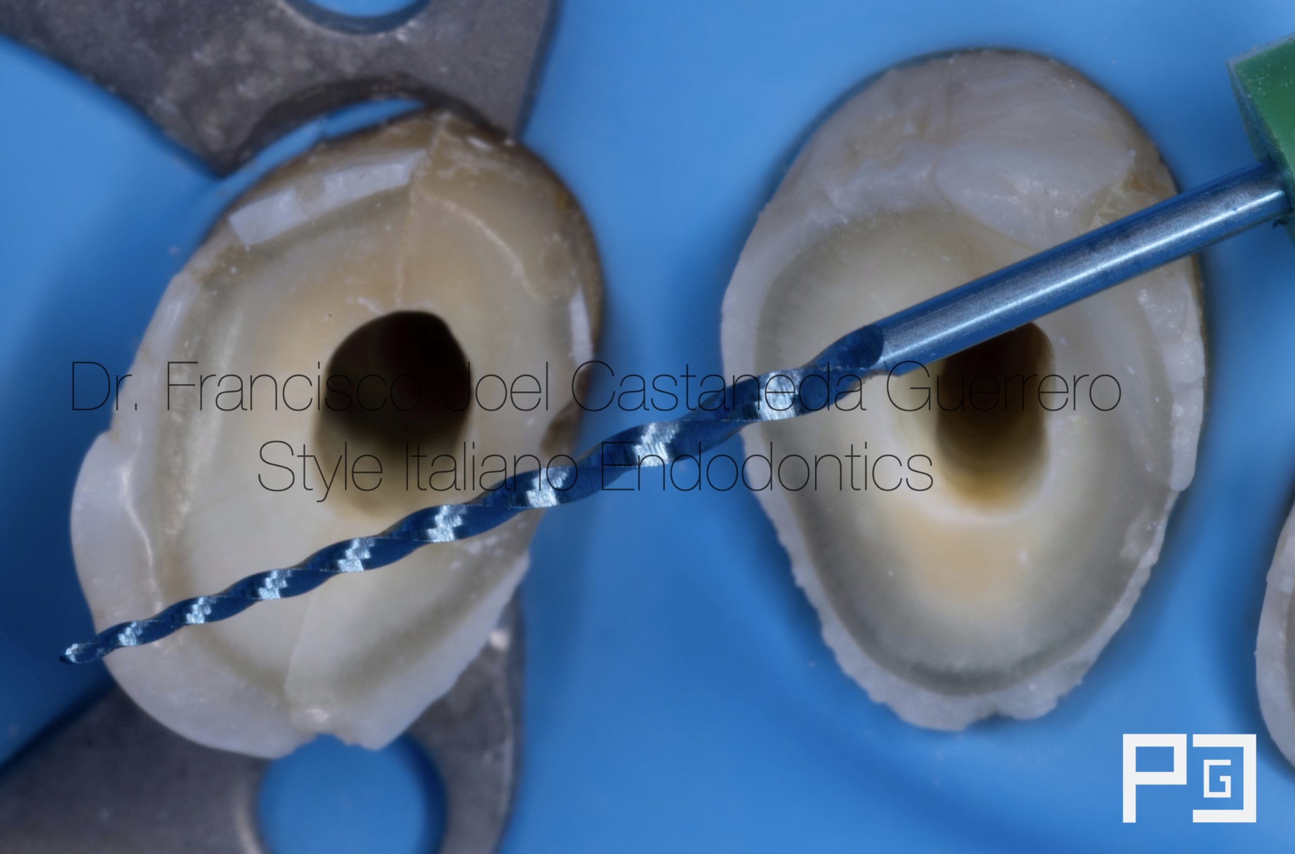
Fig. 5
It was instrumented with martensitic rotary files with a 04 taper due to the anatomy of the canals up to a caliber of 35.04 in the premolar and 40.04 in the lower canine.
Due to the aberrant anatomy and diagnosis, it was decided to focus on disinfection with the help of 5.25% sodium hypochlorite, ultrasonic activation and irrigation with flexible plastic tips with double lateral exit such as irriflex.
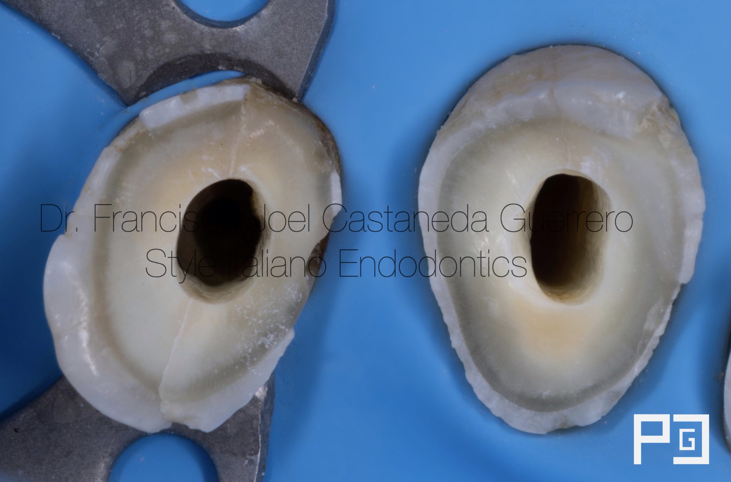
Fig. 6
Instrumented and disinfected canals, ready for filling
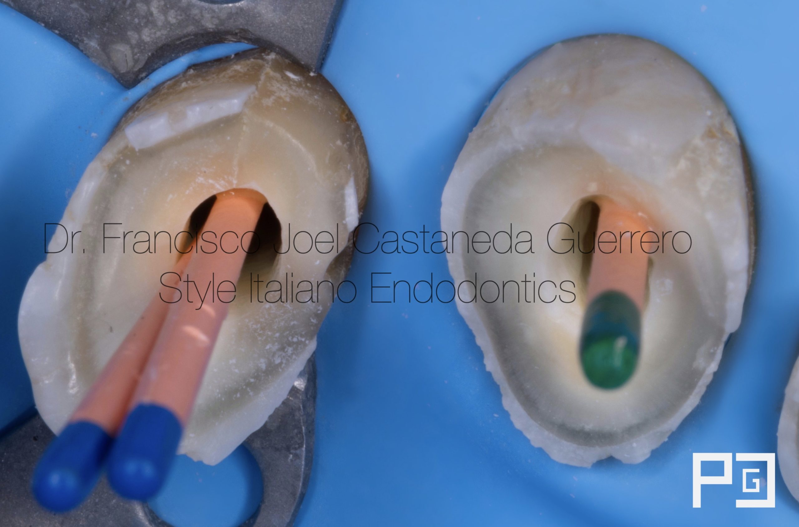
Fig. 7
Cone fit photography
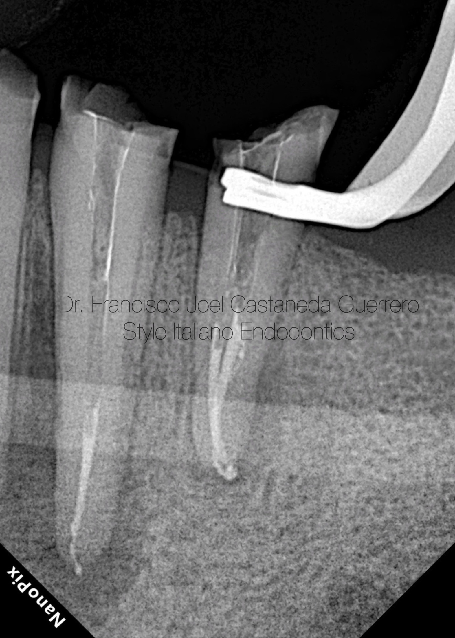
Fig. 8
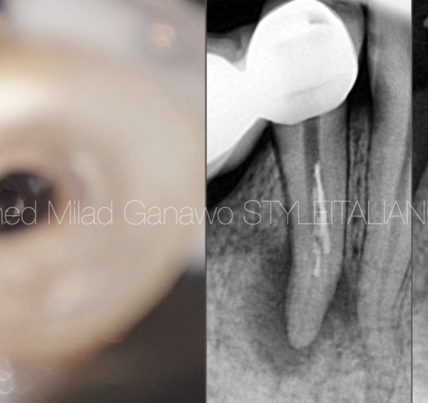
Down pack x-ray, I chose to obturate with continuous wave due to the complex anatomy shown in the initial x-ray
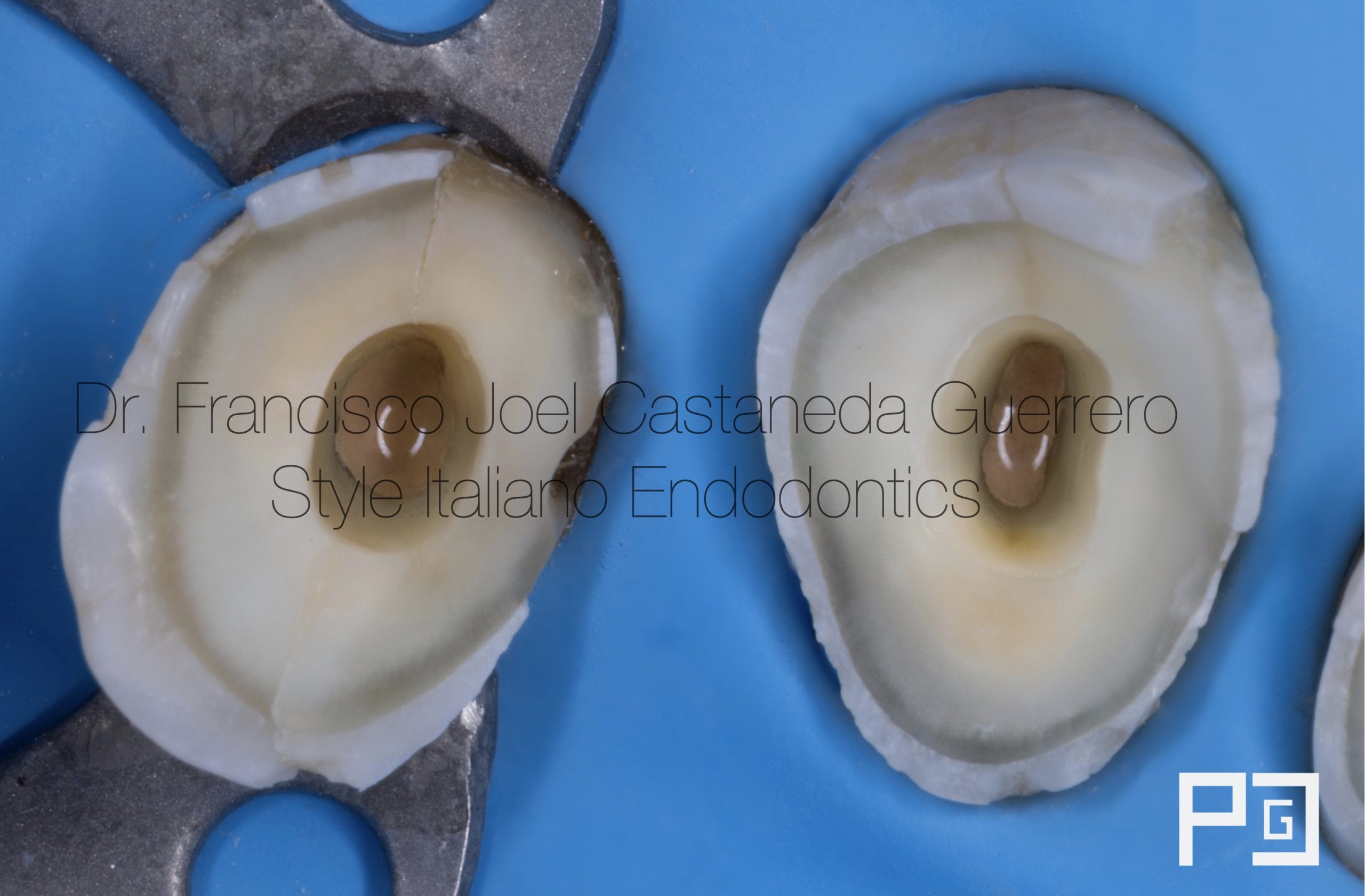
Fig. 9
Once the filling was finished, the pulp chamber was cleaned, removing excess cement and gutta-percha with the help of sandblasting
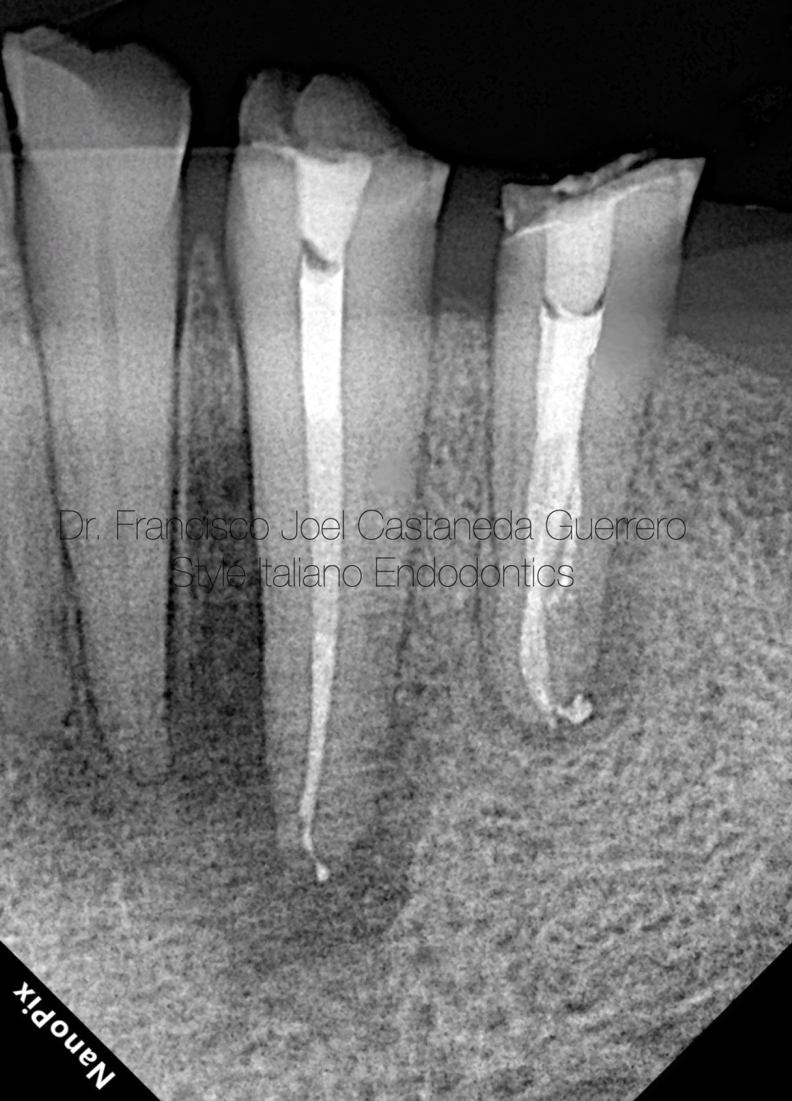
Fig. 10
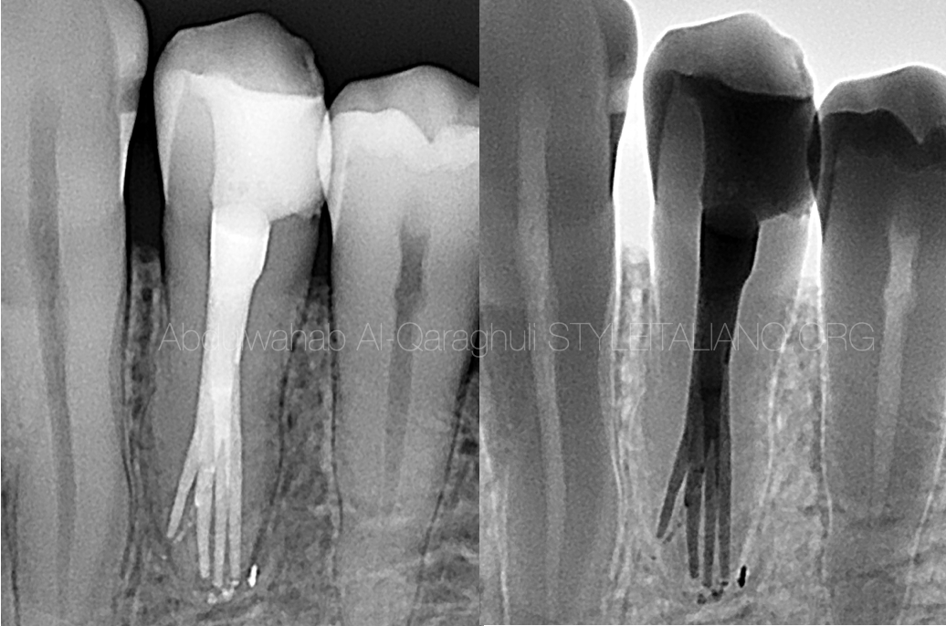
Final X-ray
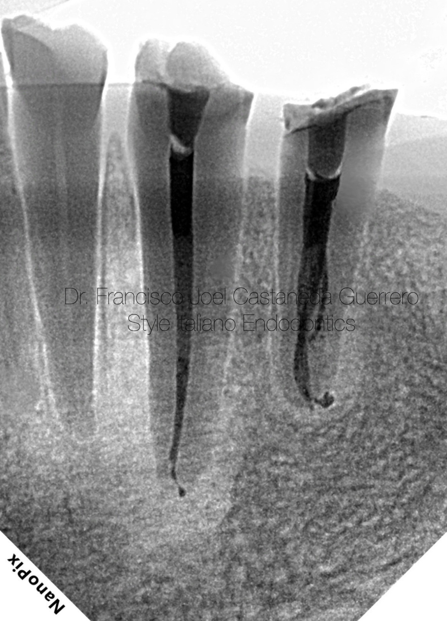
Fig. 11
Inverted Xray
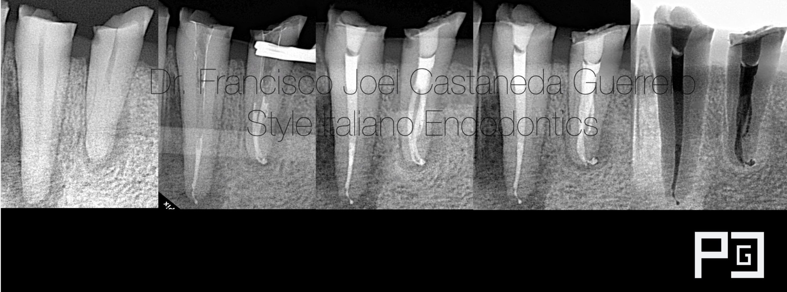
Fig. 12
Radiographic sequence and some angulations

Fig. 13
Francisco Joel Castaneda Guerrero
Guadalajara, Jalisco - Mexico
2014-2018 dental surgeon by UDG (University of Guadalajara)
2021-2023 specialist in endodontics by UAG (University autonomy of Guadalajara)
2021- first place in artistic photography by AME (Mexican Association of Endodontics)
2022- first place in clinical photography, and second place in artistic photography by AME (Mexican Association of Endodontics)
Creator of ENDOGRAPHY
ENDOGRAPHY Courses (Endodontics and photography)
Conclusions
It is very important to have tools and equipment that can simplify the treatment, such as magnification with a microscope, loupes with good light, ultrasound tips and other tools, but the most important thing apart from having these tools is to know the knowledge of internal anatomy, in order to be able to approach the cases in a better way and complement them with the tools for a better job.
Bibliography
- Root anatomy and canal morphology of mandibular first premolars in a Chinese population. Dou L, Li D, Xu T, Tang Y, Yang D. Sci Rep. 2017;7:750.The Effects of Endodontic Access Cavity Preparation Design on the Fracture Strength of Endodontically Treated Teeth: Traditional Versus Conservative Preparation Taha Özyürek, Özlem Ülker, Ebru Özsezer Demiryürek, Fikret Yılmaz
- Prevalence of complex root canal morphology in the mandibular first and second premolars in Thai population: CBCT analysis. Thanaruengrong P, Kulvitit S, Navachinda M, Charoenlarp P. BMC Oral Health. 2021;21:449
- Bürklein S, Heck R, Schäfer E: Evaluation of the root canal anatomy of maxillary and mandibular premolars in a selected German population using cone-beam computed tomographic data. J Endod. 2017, 43:1448-52
- Zhang R, Wang H, Tian YY, et al. Use of cone-beam computed tomography to evaluate root and canal morphology of mandibular molars in Chinese individuals. Int Endod J. 2011;44(11):990–9.
- Zillich R, Dowson J. Root canal morphology of mandibular first and second premolars.Oral surg Oral Med Oral Patho.1973;36:738-44


