Management of a deep bifurcation in a mandibular first premolar, with small instrument fractured in the middle third
17/10/2023
Fellow
Warning: Undefined variable $post in /home/styleendo/htdocs/styleitaliano-endodontics.org/wp-content/plugins/oxygen/component-framework/components/classes/code-block.class.php(133) : eval()'d code on line 2
Warning: Attempt to read property "ID" on null in /home/styleendo/htdocs/styleitaliano-endodontics.org/wp-content/plugins/oxygen/component-framework/components/classes/code-block.class.php(133) : eval()'d code on line 2
It is very important to identify obstacles and plan any strategies from the beginning to avoid problems that could cost us the success of the treatment
Preoperative radiograph identification, root canal access opening, intraoperative identification, location, instrumentation, debridement, disinfection, obturation are improtant for successful endodontic therapy.
The use of the microscope was essential to make the localization of the second canal simple.
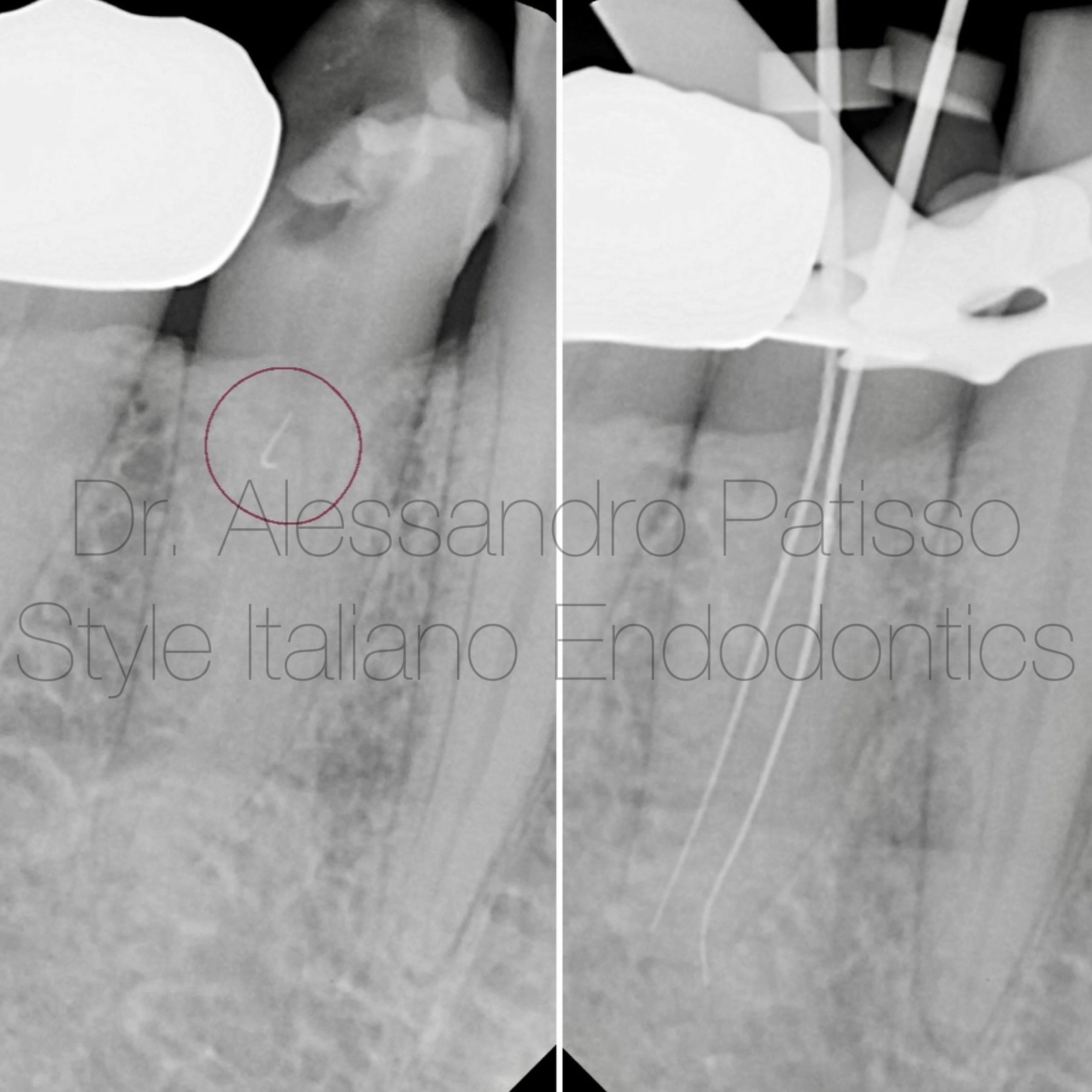
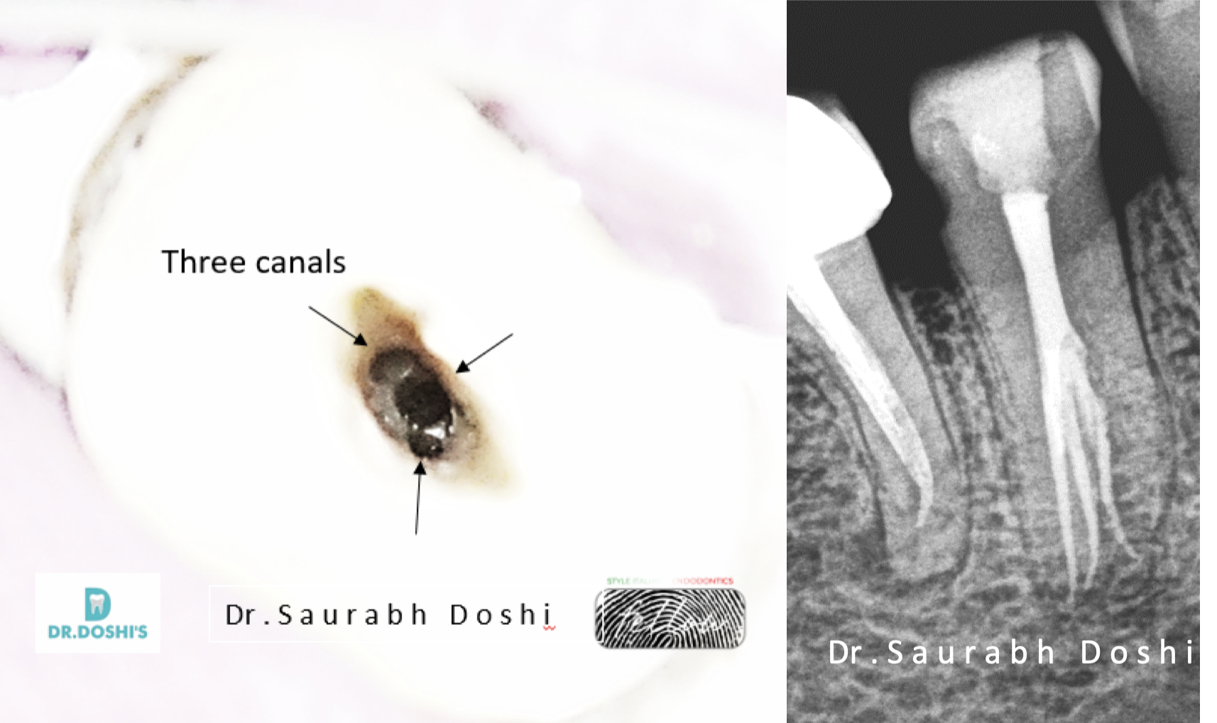
Fig. 1
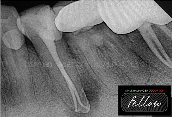
A patient was sent to me with a small manual root canal instrument fractured in the canal entrance. Thanks to the use of ultrasonic tips it was easy to remove the instrument and free the canal.
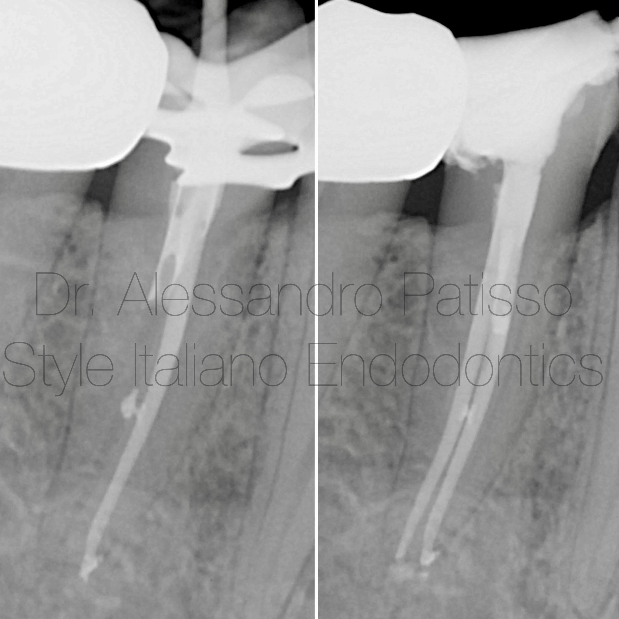
Fig. 2
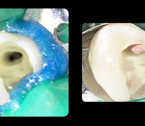
For the obturation of the canals I decided to choose a cold closure with a single cone and bioceramic, due to the close distance of the canal entrances.
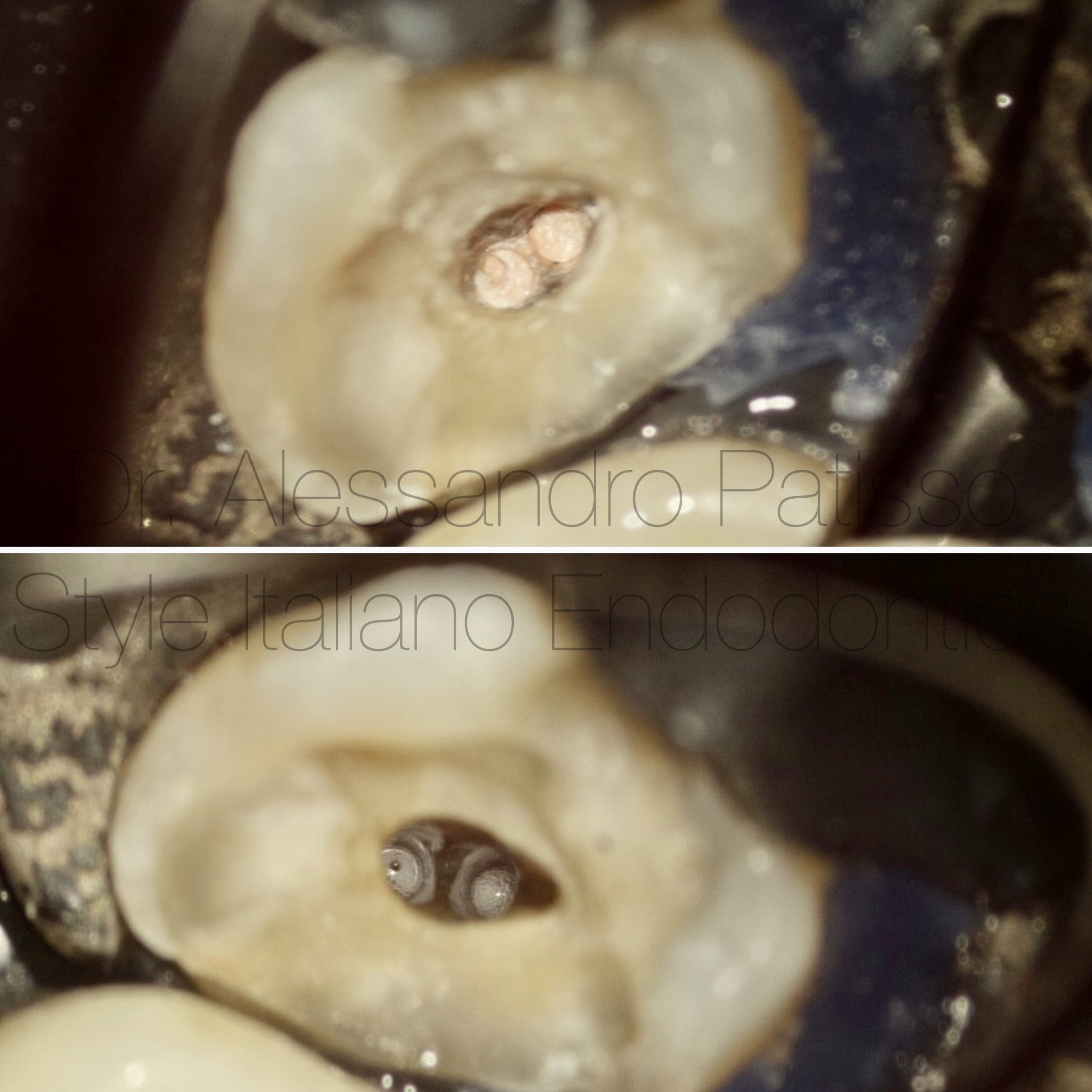
Fig. 3
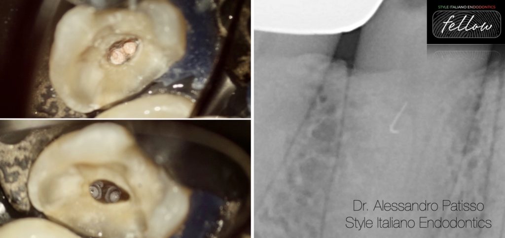
The use of the intraoperative microscope was fundamental to locate the entrance to the canals and preserve the dental tissues.
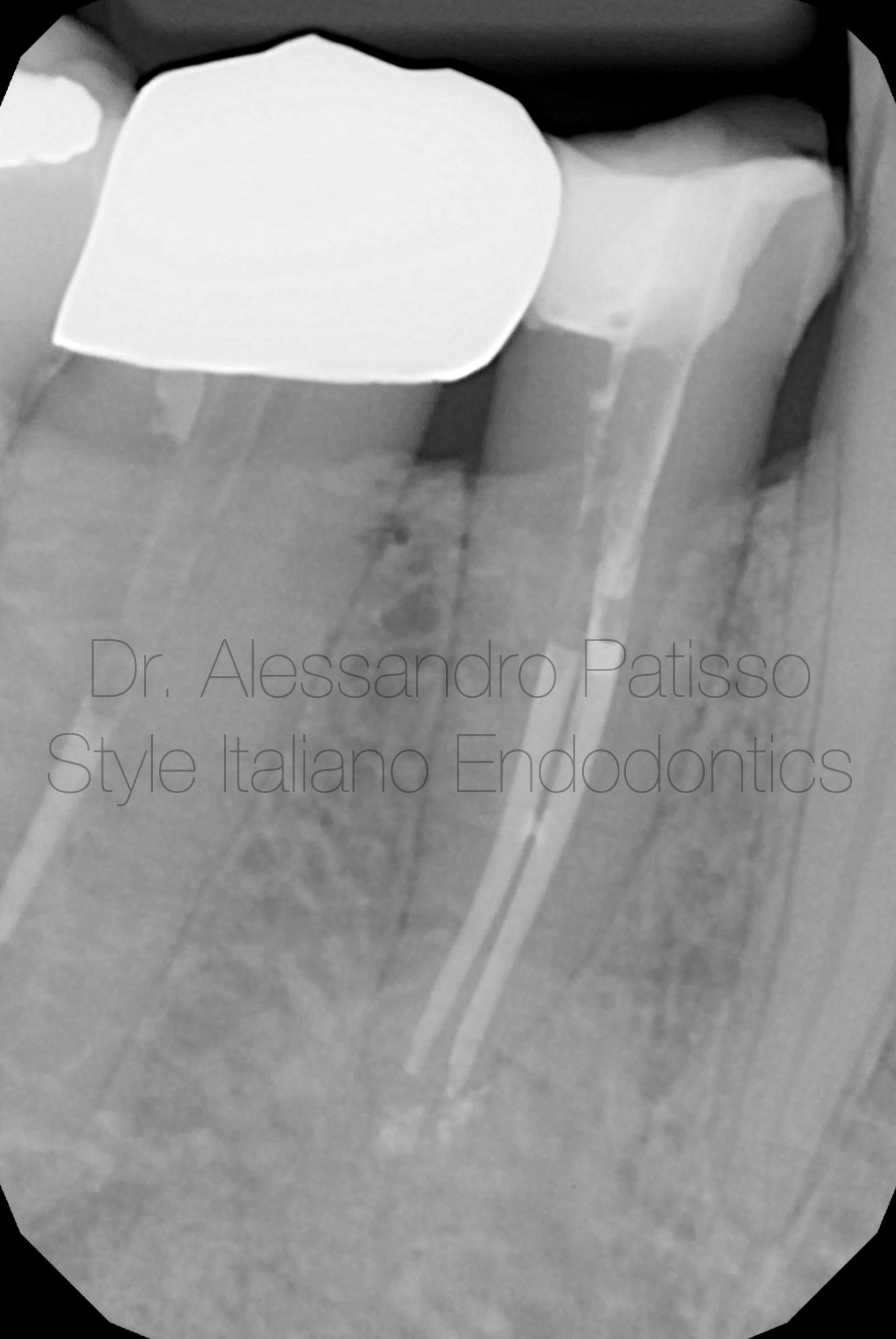
Fig. 4
Follow-up 9 months after reconstruction.

Fig. 5
Graduated in Dental Hygiene at the University of Bari in 2009 and Dentistry and Dental Prosthetics at the Universidad Europea de Madrid in 2015.
From the beginning he has practiced exclusively Clinical Edodontics, perfecting himself through numerous courses around Italy. Among the most recent he attended the courses of Dr Giuseppe Carrieri, finishing first in the course.
Today he works as a freelancer using only the microscope. Tutor in the courses of Dr Giuseppe Carrieri Fellows member Styleitaliano Endodontics.
Conclusions
In cases where the root anatomy is complex, the use of the microscope associated with adequate instruments is essential to achieve a good result without running the risk of making serious errors, therefore we may have to have CBCT to estimate the situation .
Bibliography
- Ree M, Schwartz R. The Endo-Restorative Interface: Current Concepts, Dental Clinics of North America; 54: 345-374, 2010
- Vertucci FJ. Root canal Vertucci FJ. Root canal morphology and its relationship to endodontic procedures. Endod Topics. 2005;10:23–9.
- Association of Endodontists. Glossary of Endodontic Terms, 8th ed. Chicago: American Association of Endodontists; 2012.
- Siqueira JF, Barnett F. Interappointment pain: mechanisms, diagnosis, and treatment. Endodontic Topics 2004, 7, 93109