Inner wall Vs outer wall
25/03/2024
Fellow
Warning: Undefined variable $post in /home/styleendo/htdocs/styleitaliano-endodontics.org/wp-content/plugins/oxygen/component-framework/components/classes/code-block.class.php(133) : eval()'d code on line 2
Warning: Attempt to read property "ID" on null in /home/styleendo/htdocs/styleitaliano-endodontics.org/wp-content/plugins/oxygen/component-framework/components/classes/code-block.class.php(133) : eval()'d code on line 2
The possibility for instrument separation is present during endodontic treatment.
It can be a barrier for the cleaning, shaping and obturation of the root canal system and can therefore be the reason for a future endodontic failure.
I encountered this case of tooth 24 with separated instrument ( rotary fragment) in palatal canal, and it took plenty of time, case took 3 sessions to solve the problem with many manual K files used to achieve the BYPASS
And here's some reasons can lead to file separation :
- Improper use of the instrument
- Inadequate access
- Unusual Root Canal System anatomy
- Manufacturing defects
- Rush to use rotary instruments without creating a proper straight-line access and glide path
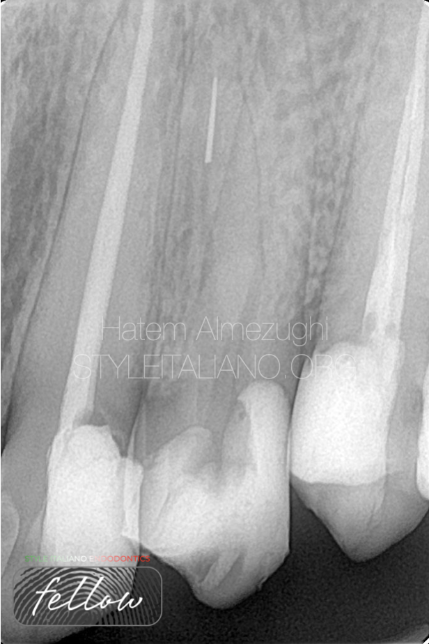
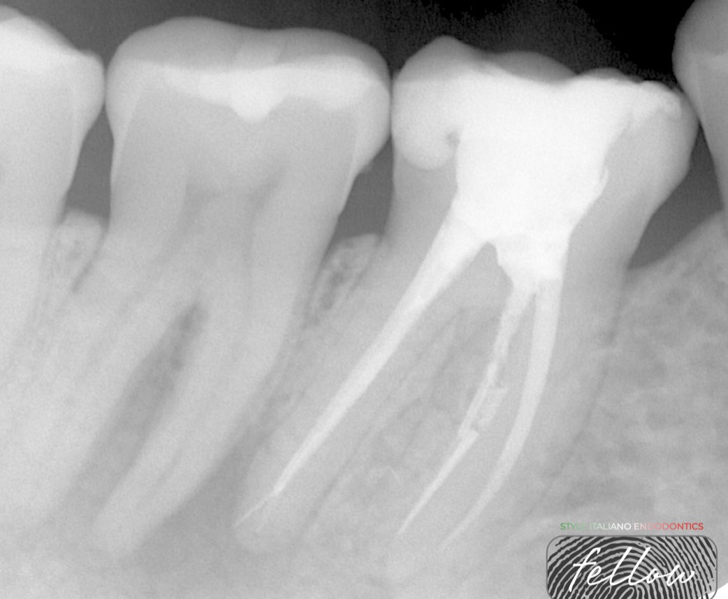
Fig. 1
PRE-OPERATIVE X-RAY
Shows tooth 24 with separated instrument in palatal canal (rotary fragment)
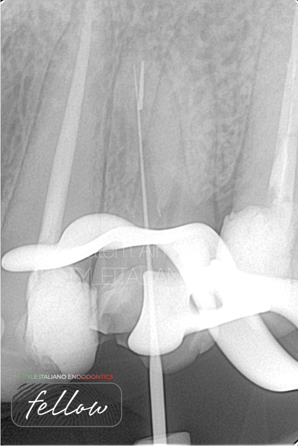
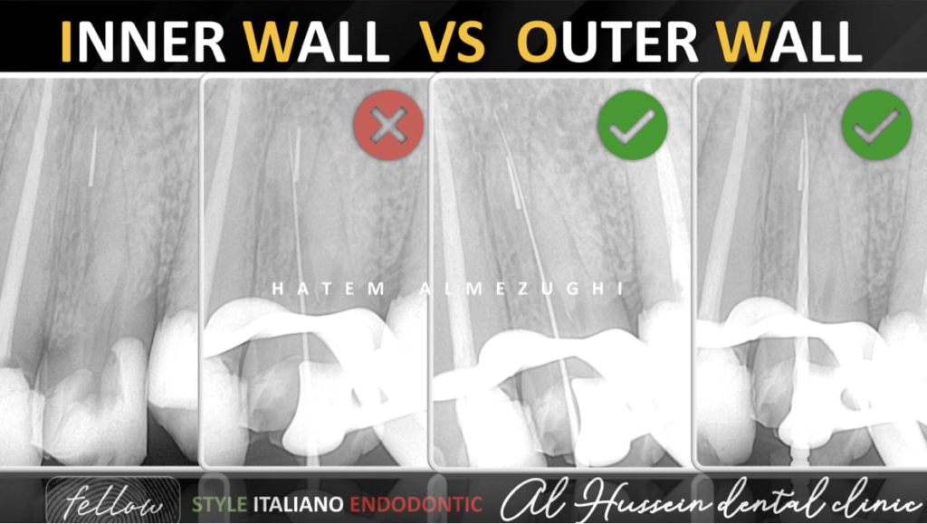
Fig. 2
This x-ray shows an attempt of bypass from outer wall, which is a sign of ledge formation, thus, in such situations bypass from outer wall is not recommended.
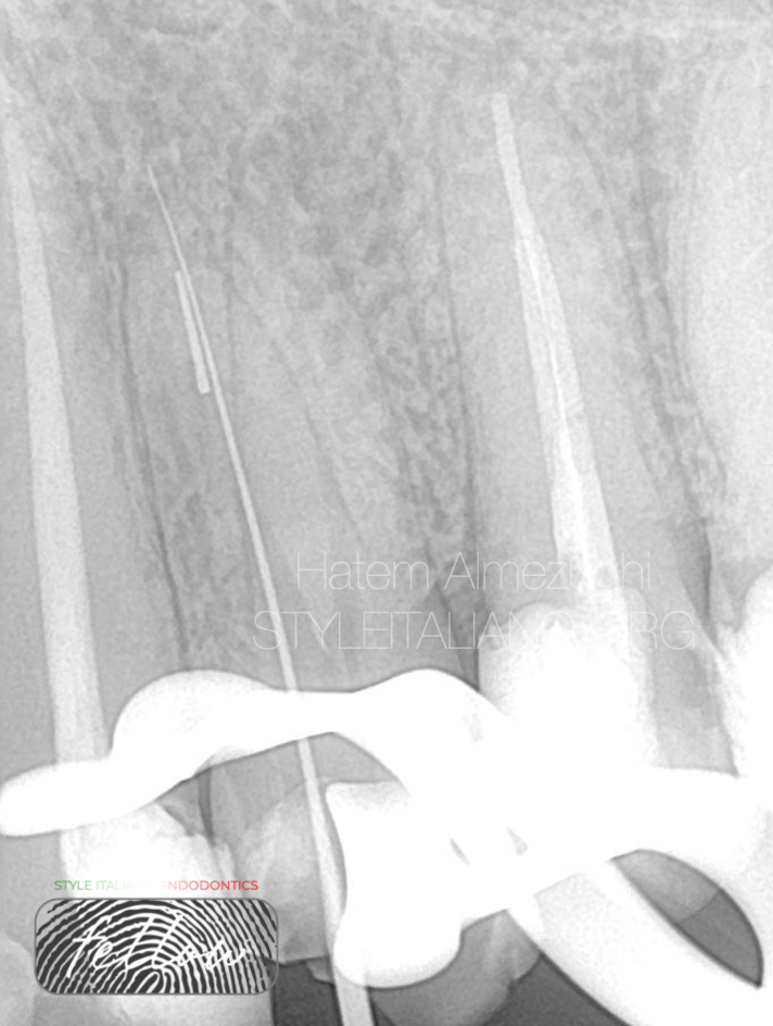
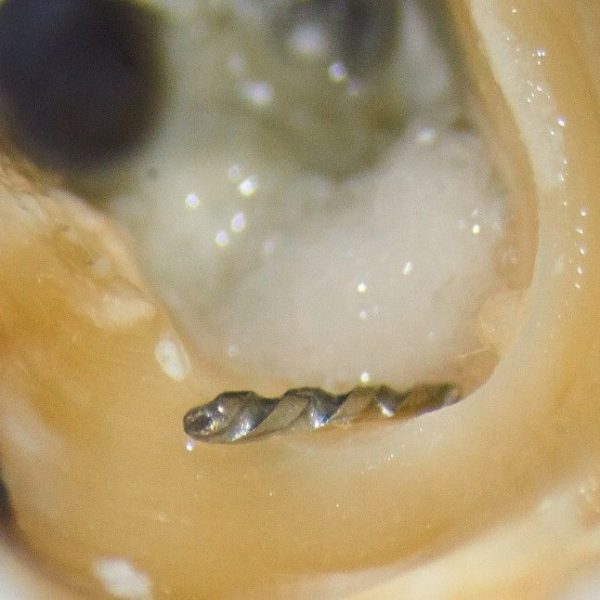
Fig. 3
This x-ray shows bypass from inner wall after many trails to get a catch, now the issue is solved, and will use larger size to bypass then rotary file bypass and so on.
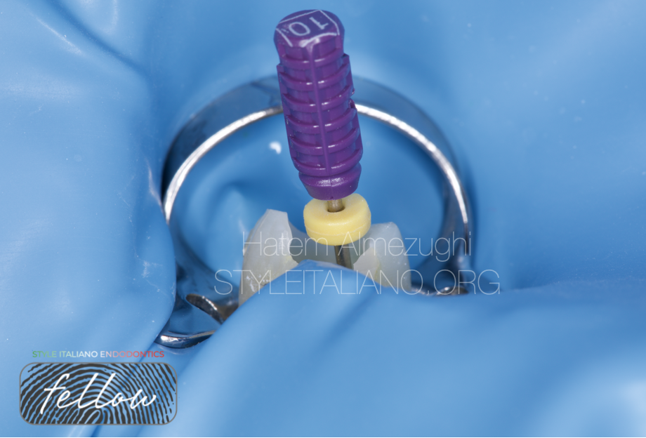
Fig. 4
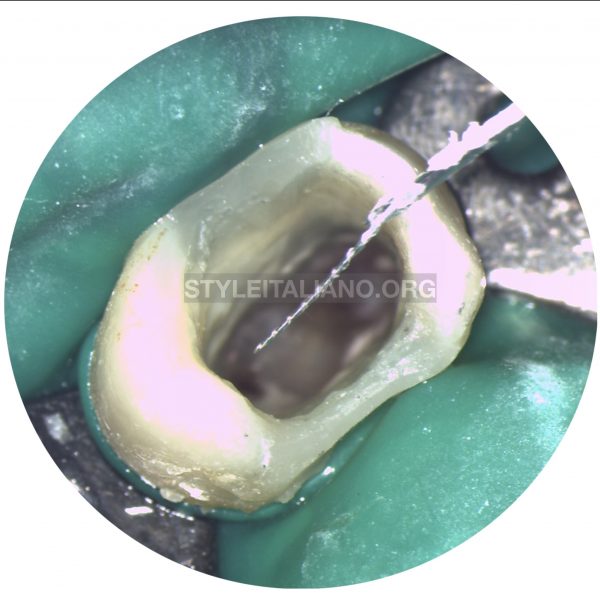
Intra oral shows bypassing SI from inner wall in palatal canal
Fig. 5
Intra oral shows working length by manual files size 8, 10 in both canals after bypass been achieved.
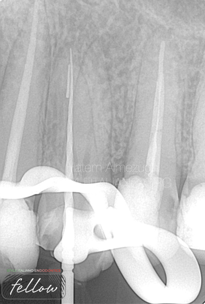
Fig. 6
Here is how bypass by using rotary file and confirming it from inner wall by x-ray to ensure smooth insertion of gutta percha.
Fig. 7
Here is how bypass by using rotary file to ensure smooth insertion of gutta percha
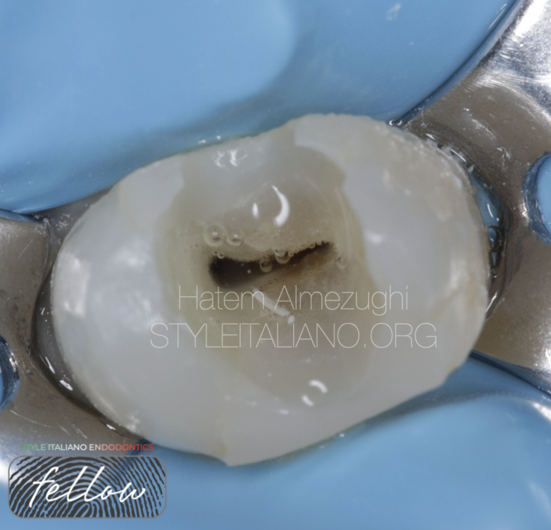
Fig. 8
Intra oral photo shows sodium hypochlorite in action then followed by activation and dryness.
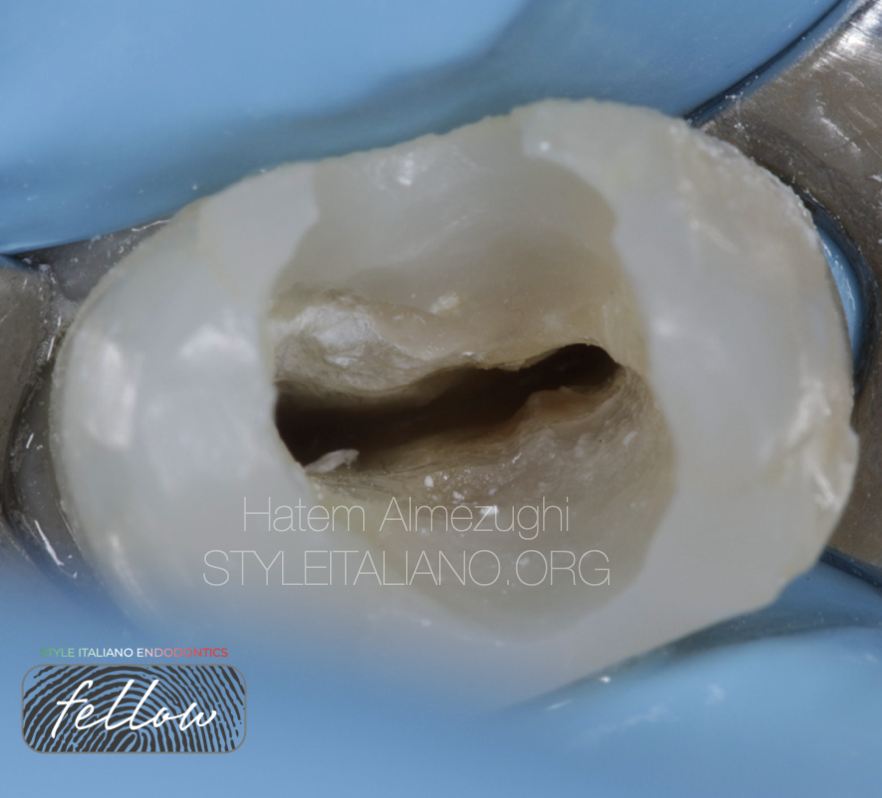
Fig. 9
This intra oral shows pulp chamber ready for inserting GPs after more debris cleaning
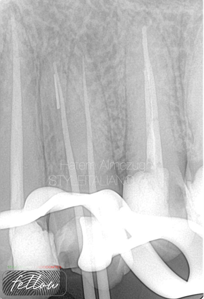
Fig. 10
Cone fit where canals ready for obturation, we can clearly notice gp passing through inner wall
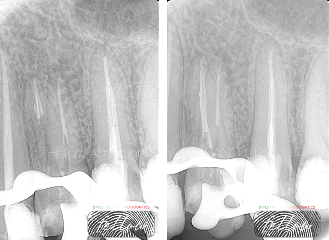
Fig. 11
First stage of down packing done.
Then followed by second stage of down packing.
We can notice the appearance of lateral canal at the apical part of buccal canal.
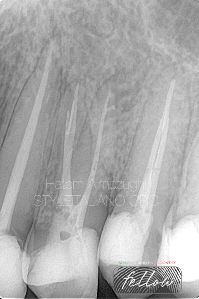
Fig. 12
Obturation done, just a bit more of hydraulic pressure on buccal to reveal lateral canal clearly.

Fig. 13
About the author:
Hatem Almezughi
Was graduated from Tripoli university, Libya.
Local and international speaker in different conferences and forums
Instructor in many endodontic courses.
Dental practice limited to endodontics and restorative .
Fellow member in style Italiano endodontics
Conclusions
Instrument separation can occur under any circumstances. The decision for conservative management to bypass or to completely remove the fractured instrument should be made after careful evaluation and the use of magnification would be handy in such situations.
Root canal system anatomy and morphology, exact location of the fragment, play a major role in determining either the easiness or difficulty in managing such an issue.
In the case I deal with in this article, I reach to a conclusion that if the separated instrument is able to be bypassed, and bypass from inner wall is not a permanent rule to go with, there's always dispensation where some broken files bypassed from outer wall, and it is more conservative to bypass than retrieving.
Bibliography
Tsvetelina Borisova-Papancheva, Silvia Stankova, Slavena Georgieva. Conservative Management Of Intracanal Separated Endodontic Instruments: Treatment Decisions And Related Factors. Scripta Scientifica Medicinae Dentalis, Vol. 3, No 1, 2017, Pp. 23-31.
Rambabu T. Management Of Fractured Endodontic Instruments In Root Canal: A Review. Journal Of Scientific Dentistry 2014;4(2):40-48
Dr Rafaël Michiels,. Bypassing A Fractured Instrument. Case Report. Roots1_2011.
Sultana Parveen, Mozammal Hossain And Md. Farid Uddin. Management Of Broken Instrument By File Bypass Technique. Bsmmu J 2017; 10: 41-43
Dr. Praveen John, Dr. Ramesh Kumar M., Dr. Jayasree S. Bypassing Separated Instruments In The Root Canal – Two Case Reports. IOSR Journal Of Dental And Medical Sciences (Iosr-Jdms) Volume 15, Issue 6 Ver. Xi (June. 2016), Pp 08-13.
M. B. Mcguigan, C. Louca And H. F. Duncan. Clinical Decision-Making After Endodontic Instrument Fracture. British Dental Journal Volume 214 No. 8 Apr 27 2013.