Alternative method for removing fractured instrument: a case report
04/06/2020
João Meirinhos
Warning: Undefined variable $post in /home/styleendo/htdocs/styleitaliano-endodontics.org/wp-content/plugins/oxygen/component-framework/components/classes/code-block.class.php(133) : eval()'d code on line 2
Warning: Attempt to read property "ID" on null in /home/styleendo/htdocs/styleitaliano-endodontics.org/wp-content/plugins/oxygen/component-framework/components/classes/code-block.class.php(133) : eval()'d code on line 2
An alternative method, involving the use of an irrigation needle and cyanoacrylate adhesive, allowed the removal of the fractured instrument from inside of the canal system.
During the mechanical shaping of the root canal system, the possibility of fracture of an instrument is always present. When it occurs, the first step is dealing with our emotions and avoid panicking. But the most important step in avoiding file separation during our procedures is avoiding it, firstly by understanding the causes of this complication (torsion fracture / cyclic fatigue fracture; access errors), and, ultimately, preventing it from happening. On the other hand, one must be prepared to overcome this obstacle and devise a way to still be able to promote the complete disinfection of the root canal system.
The best way to handle with the situation is with honesty, integraty and a plan.
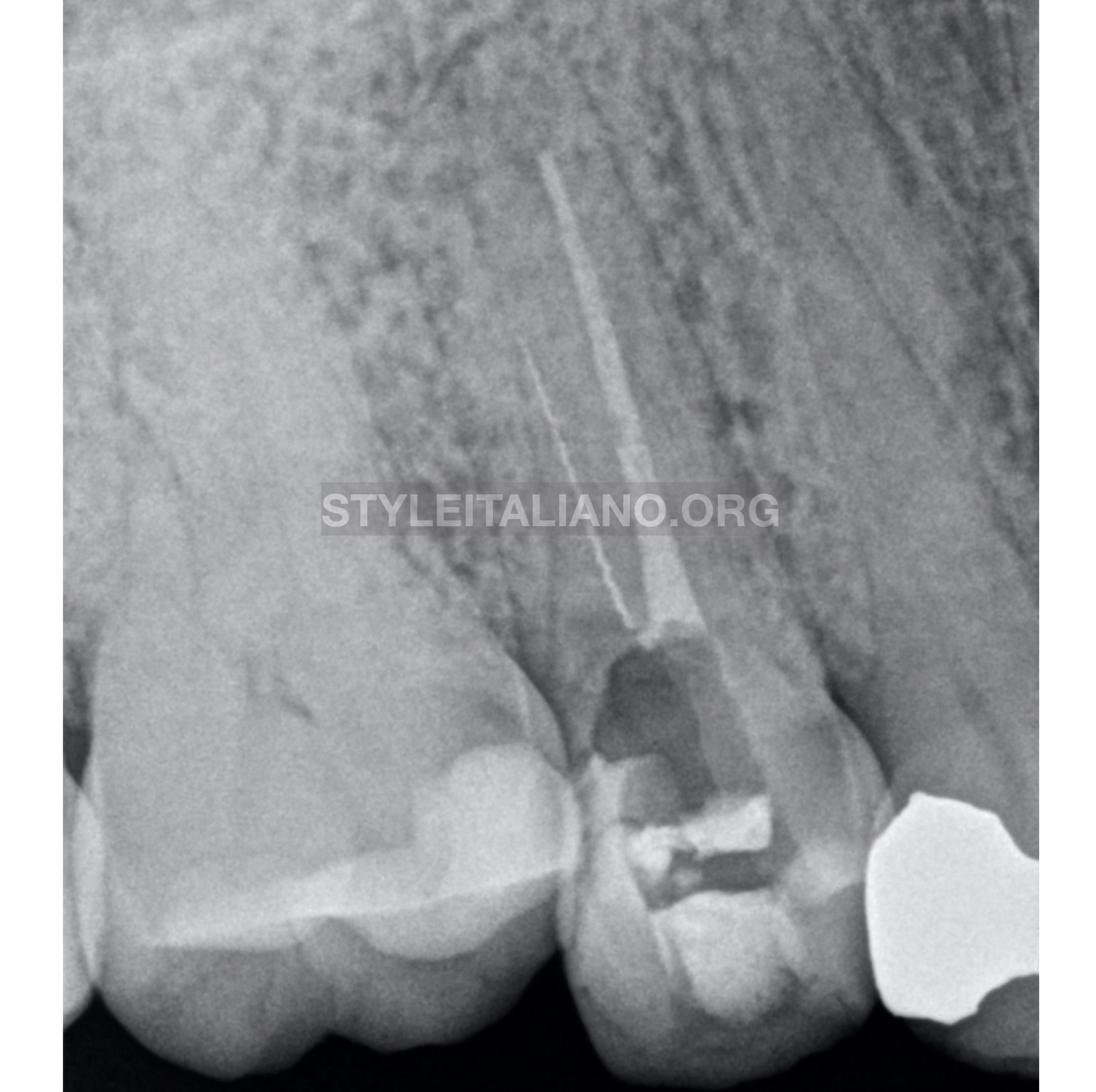
Fig. 1
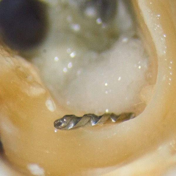
A 58-year-old female patient was referred by a colleague. In the radiographic examination, the second right upper pre-molar presented a fractured instrument located in the middle third of the buccal canal. As a treatment plan, non-surgical endodontic treatment was suggested.
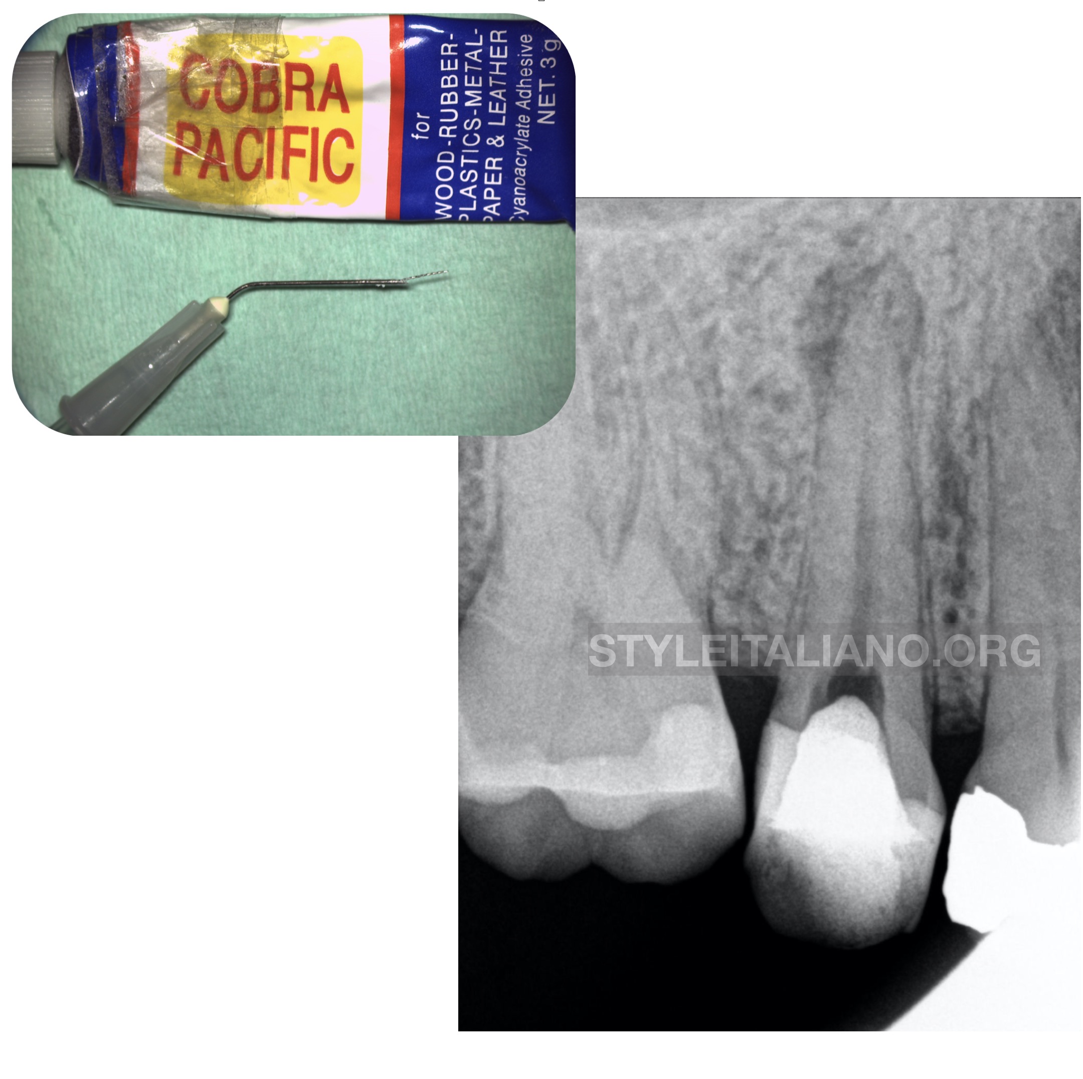
Fig. 2
The bypass of the instrument was attempted with no success, as well as its removal using several systems: IRS (Instrument Removal System, Dentsply Endodontics, Tulsa, OK, USA), ProUltra Tips (Dentsply Tulsa Dental, Tulsa, Oklahoma) and K files (Dentsply Maillefer, Ballaigues, Switzerland) coupled to Endo-chuck (SybronEndo, Orange, California).
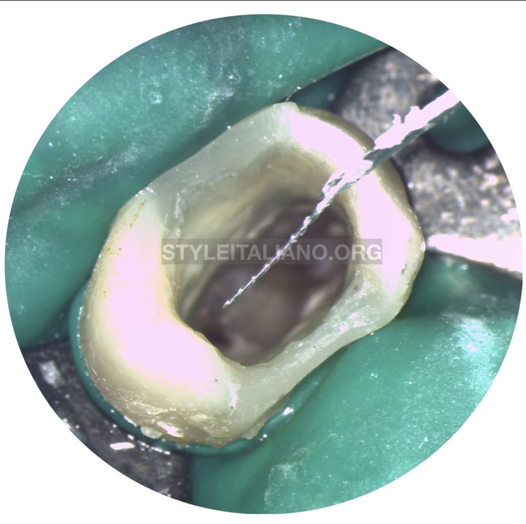
Using a 27-G (BD Microlance ™ 3 Needles 27G X ¾ "- 0.4mm X 19mm) irrigation needle and cyanoacrylate glue (Cobra Pacific Super Glue), it was possible to remove the fragment.
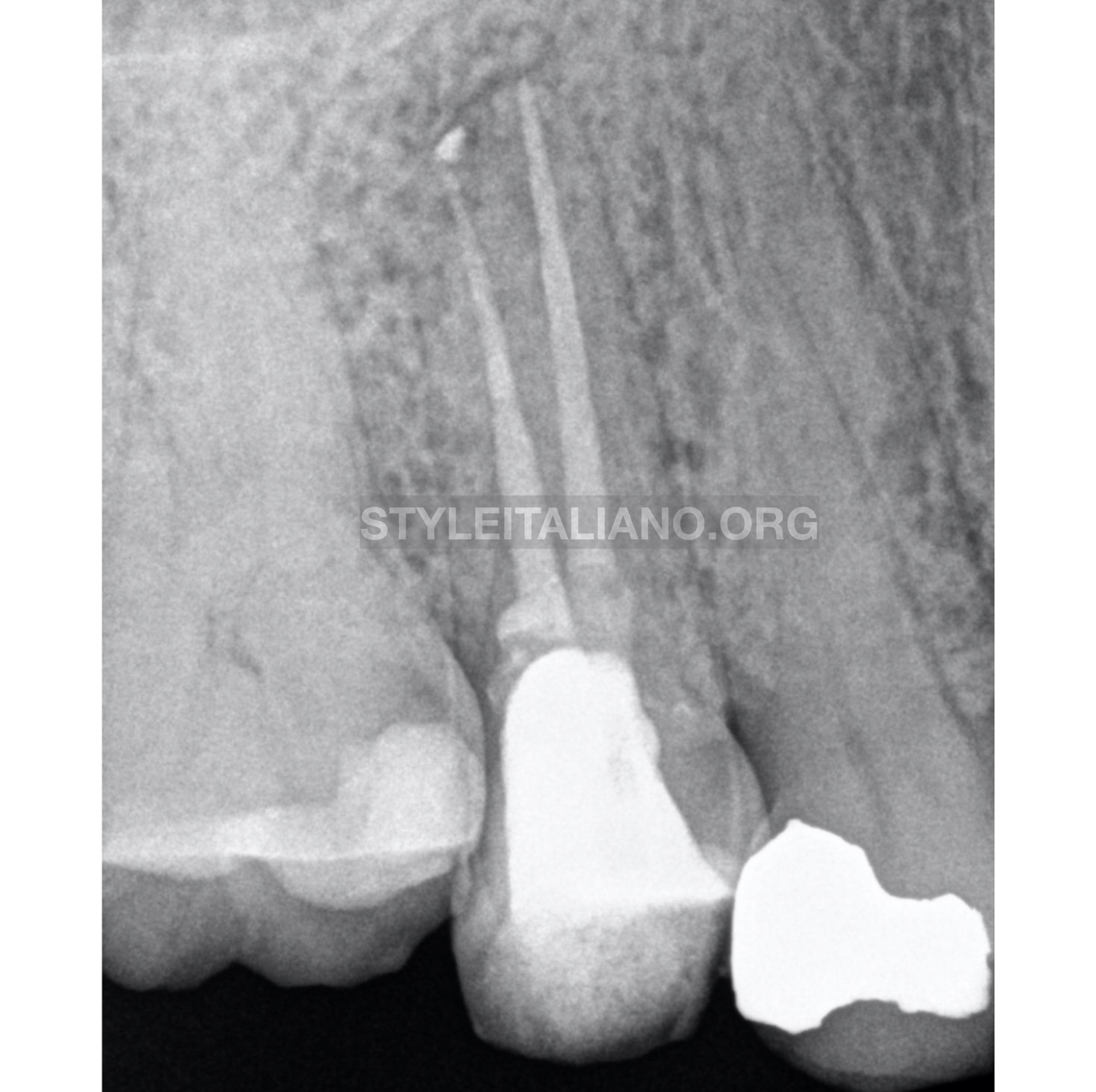
Fig. 3
The tooth was completely instrumented, disinfected, filled with gutta percha and subsequently restored directly and definitively with composite resin.
Conclusions
The method is simple, economical and at the same time can result in predictable success and can be used easily by the general practitioner or specialist in the absence of other instrument removal systems.
Bibliography
1.American Association of Endodontists. Glossary of Endodontic Terms, 8th ed. Chicago: American Association of Endodontists; 2012.
2.Spili P, Parashos P, Messer HH. The Impact of Instrument Fracture on Outcome of Endodontic Treatment. Journal of Endodontics. 2005;31.
3.Parashos P, Messer HH. Rotary NiTi Instrument Fracture and its Consequences. Journal of Endodontics. 2006;32:1031–1043.
4.Madarati AA, Qualtrough AJE, Watts DC. A Microcomputed Tomography Scanning Study of Root Canal Space: Changes after the Ultrasonic Removal of Fractured Files. Journal of Endodontics.. 2009;35: 125–128.
5.Panitvisai P, Parunnit P, Sathorn C, Messer HH. Impact of a Retained Instrument on Treatment Outcome: A Systematic Review and Meta-analysis. Journal of Endodontics.