Extraction or try retention?
15/11/2023
Fellow
Warning: Undefined variable $post in /var/www/vhosts/styleitaliano-endodontics.org/endodontics.styleitaliano.org/wp-content/plugins/oxygen/component-framework/components/classes/code-block.class.php(133) : eval()'d code on line 2
Warning: Attempt to read property "ID" on null in /var/www/vhosts/styleitaliano-endodontics.org/endodontics.styleitaliano.org/wp-content/plugins/oxygen/component-framework/components/classes/code-block.class.php(133) : eval()'d code on line 2
Patient: Female, aged 33 years
Chief complaint: Swelling and pain in the left lower molar for 5 days.
Presenting symptoms: generalized chronic periodontitis in a stable condition. 5 days ago, the left lower tooth felt painful with gingiva swollen, no pain at night, but unable to chew.
Past history: Respiratory system: No history of respiratory disease.
Infectious history: No history of severe infectious disease.
Allergic history: Not allergic to penicillin.
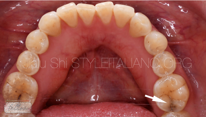
Fig. 1
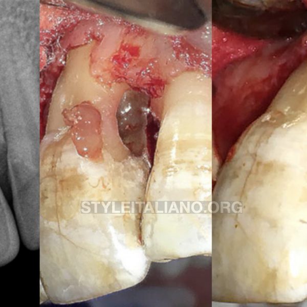
Examination: On examination, the tooth was found to be responsive to cold tests and tender to pressure. The tooth 3.6 had hidden cracks, no deep periodontal pockets, no excessive mobility, and no fluctuating buccal swelling.
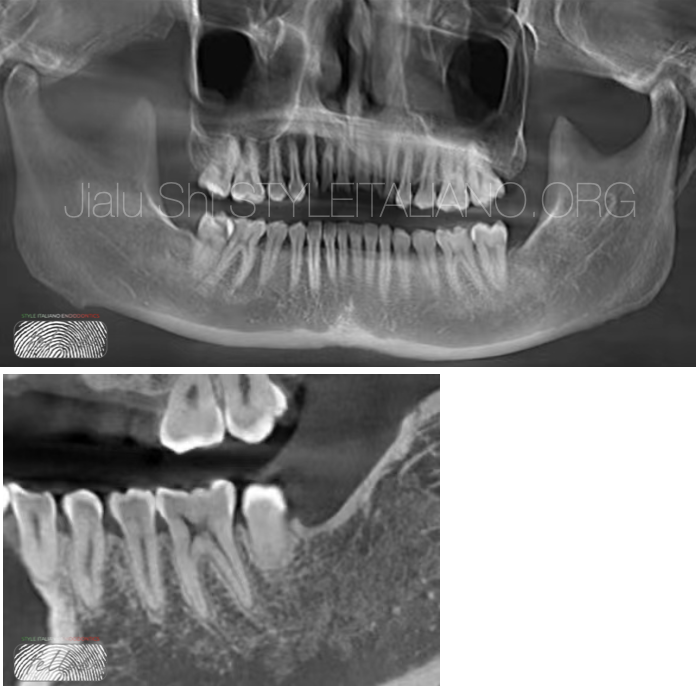
Fig. 2
Pre-operative X-ray
The periapical radiograph shows no radiolucency around the mesial root, distal root as well as into the furcation .
The diagnosis of 36 chronic pulpitis,and cracked tooth syndrome (CTS).
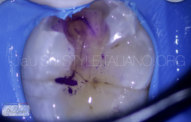
Fig. 3
The patient was informed regarding the guarded overall prognosis of this tooth. Attempts were made to remove the hidden cracks. RCT would be performed if the cryptocrack did not reach the pulp chamber. The tooth would be extracted if the crack reached the bottom of the pulp chamber.
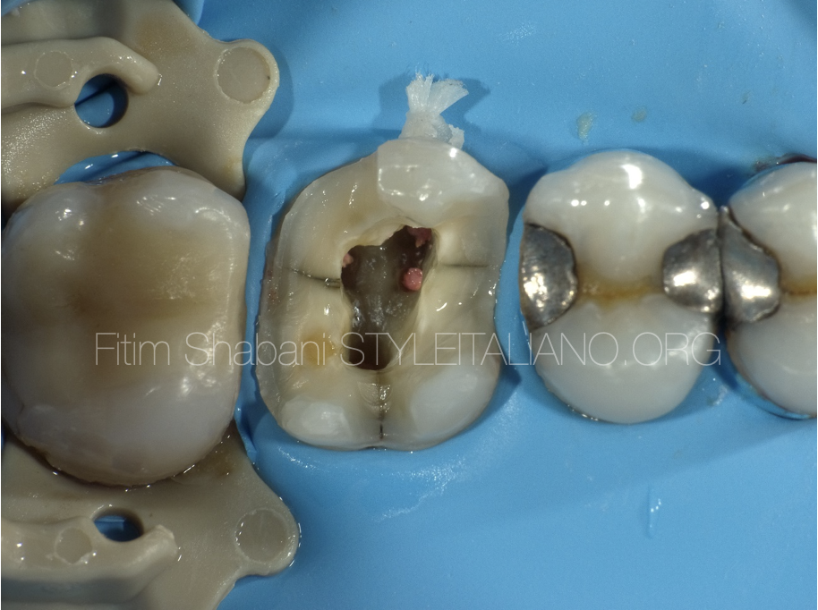
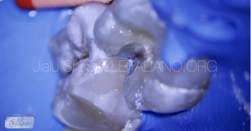
The hidden cracks and caries was removed through adequate local anesthesia and the placement of a rubber dam. Significant staining could be observed in the cracks. It was found that the staining had reached the pulp chamber but not the pulp floor.
The treatment strategy was to remove all tissues from the pulp chamber by 3% NACLO and an activation devices in order to dissolve them all.
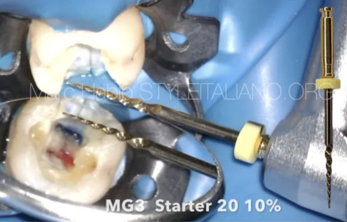
The root canal was prepared by employing the “one stroke, one wipe” method.
In this case, it is imperative to try to retain enough PCD(Peri-cervical Dentin) to ensure tooth resistance
Scout and prepare the “MM”.
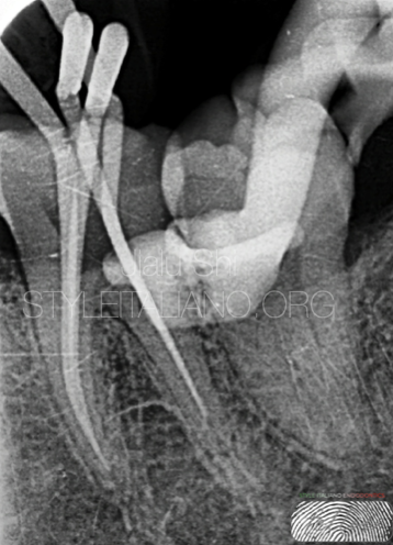
Fig. 4
Cone fit x-ray
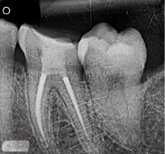
Fig. 5
Post operative X-ray
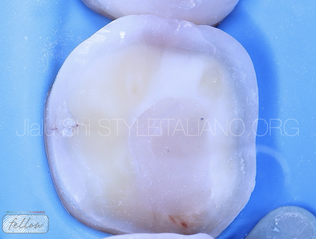
Fig. 6
Overlay preparation
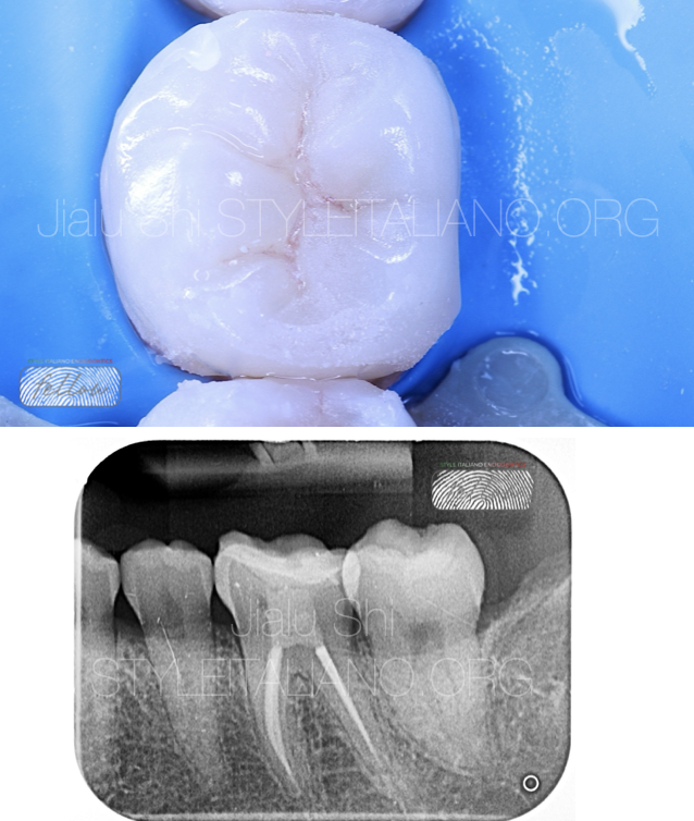
Fig. 7
The final X-ray shows that the overlay was very tightly bonded to the tooth, with no excess binder remaining.

Fig. 8
About the author: Jialu Shi - 施嘉禄
Graduated from Changsha medical University in 2018
Fellow of Style Italiano Endodontics
Conclusions
Teeth diagnosed with cracked tooth syndrome have a guarded prognosis, whereas with proper management,
the success rate after 5 years is relatively high. However, if both marginal ridges are cracked,
the prognosis for teeth diagnosed with cracked tooth syndrome is poor. In the above-mentioned cases, the dentist should inform the patient of the need for a crown/ overlay restoration after endodontic treatment.
Bibliography
1.T Gill , A J Pollard , J Baker , C Tredwin (2021) Cracked Tooth Syndrome: Assessment, Prognosis and Predictable Management Strategies
2, Fei Li(2021). Review of Cracked Tooth Syndrome: Etiology, Diagnosis, Management, and Prevention.
3, Pacquet W, Delebarre C, Gerdolle D.Int J Esthet Dent(2022). Therapeutic strategy for cracked teeth.
4, Angeliki Kakka , Dimitrios Gavriil , John Whitworth (2022)Treatment of cracked teeth: A comprehensive narrative review


