Cracked tooth syndrome
25/01/2021
Marc Habib
Warning: Undefined variable $post in /home/styleendo/htdocs/styleitaliano-endodontics.org/wp-content/plugins/oxygen/component-framework/components/classes/code-block.class.php(133) : eval()'d code on line 2
Warning: Attempt to read property "ID" on null in /home/styleendo/htdocs/styleitaliano-endodontics.org/wp-content/plugins/oxygen/component-framework/components/classes/code-block.class.php(133) : eval()'d code on line 2
Cracked tooth syndrome (CTS) is where a tooth has incompletely cracked meaning without a part of the tooth breaking off. Most of the time it is an incomplete fracture of a vital posterior tooth that occasionally extends
into the pulp.
CTS could be considered a type of dental trauma and also one of the possible causes of dental pain. Cameron in 1964 defines cracked tooth syndrome as being"a fracture plane of unknown depth and direction passing through tooth structure that may progress to communicate with the pulp and/or periodontal ligament”. The symptoms are variable, making it a very difficult condition to diagnose.
There are a variety of habits which predispose patients to CTS including chewing ice, pens, hard sweets and so on. Parafunctional habits such as bruxism are also associated with the development of this condition.
A female patient presented for a continuous pain on her right lower jaw.
She was complaining of a history of discomfort in this area for quite some time witch was increasing while chewing and cold/hot meals.
Tooth #46 was suspected to be the origin of this pain. This tooth was treated with an old occlusal- distal white filling that presented a visible decay and fracture during intra oral examination.
Removing restorations may help to visualise fracture lines but should only be carried out after informing the patient that the tooth might need root canal treatment or extraction.
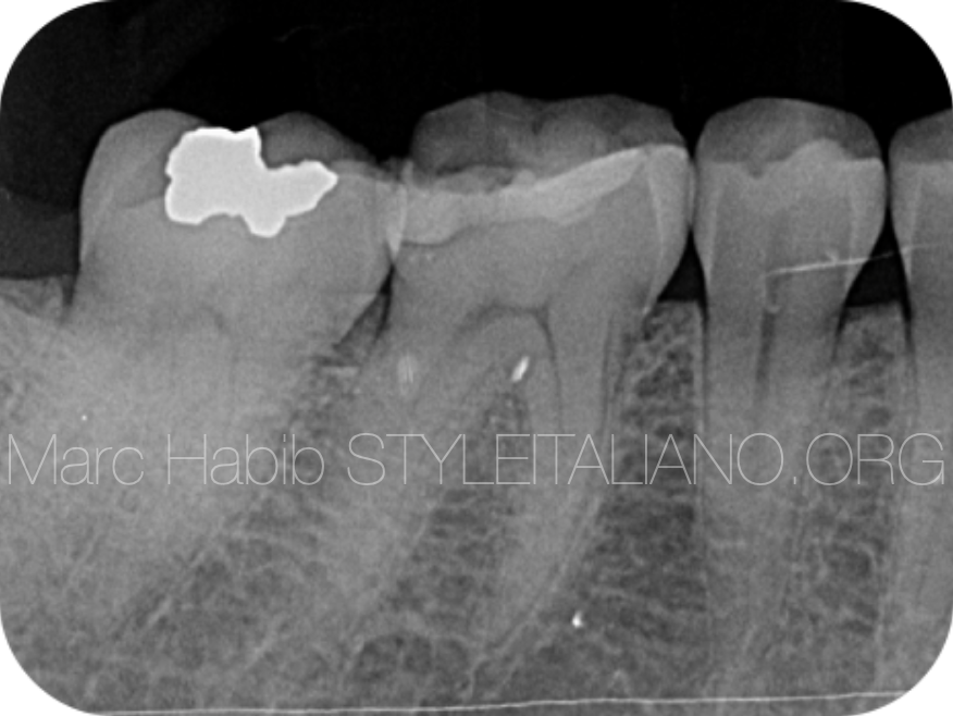
Fig. 1
Pre operative xray showing old white filling with decayed are surrounding its distal aspect.
Radiographs offer little benefit in visualising cracks. This is due to the fact that cracks propagate in a direction which is parallel to the plane of the film however radiographs can be useful when examining the periodontal status.
Once the filling was removed and the crack line was visualized.
Staining the crack with a microbrush then water rinsing to examen the extent of the crack line.
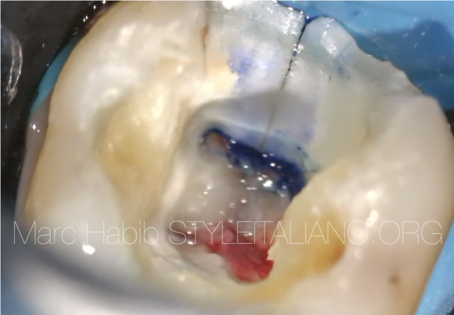
Fig. 2
Dyes may be used to aid visualisation of fractures. In this clinical view we can clearly see crack line stained spreading on the distal wall of the access cavity.
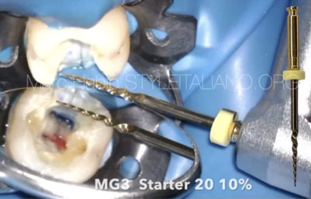
Preflaring using Starter file 20 10% D Perfect®.
All 4 canals orifices were preflared using MG3 Starter continuous rotation file.
Shaping using G1 file 20 4% D Perfect®
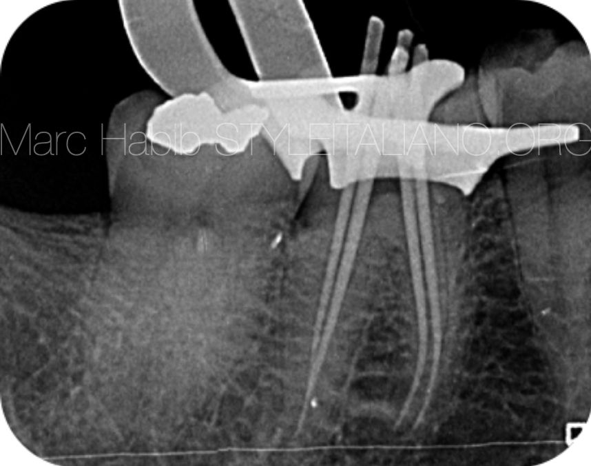
Fig. 3
Shaping was kept as conservative as possible using MG3 G1 20 4% taper from D Perrfect® with some brushing motion while doing the shaping of all 4 canals
Cone fit verification xray showing 4 master cones
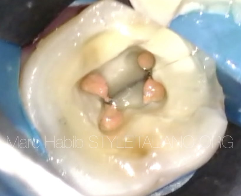
Fig. 4
Final obturation access cavity view showing 4 root canals orifices
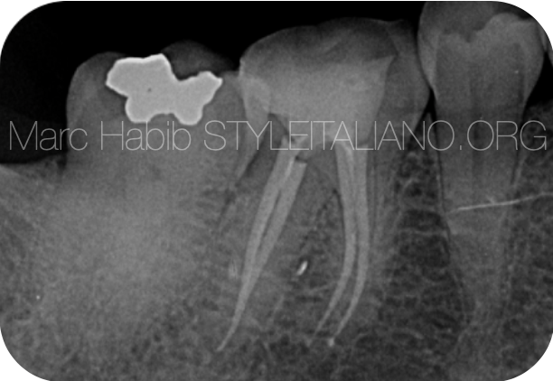
Fig. 5
Final obturation xray showing 4 canals with a resin dual cure build up which will be followed by a crown prosthesis.
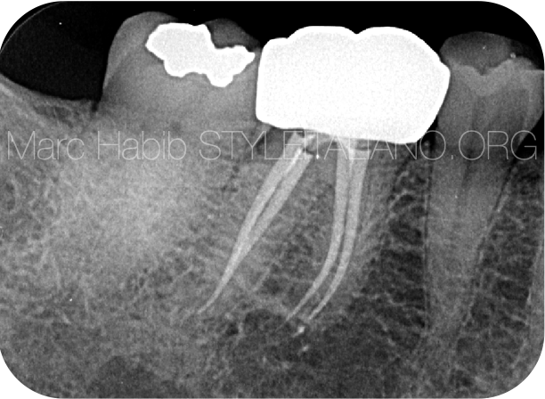
Fig. 6
X ray of Crown prosthesis to protect the tooth from propagation of the crack line.
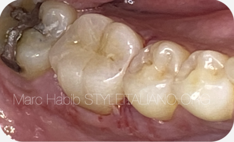
Fig. 7
Intra oral photo of Crown prosthesis to protect the tooth from propagation of the crack line.
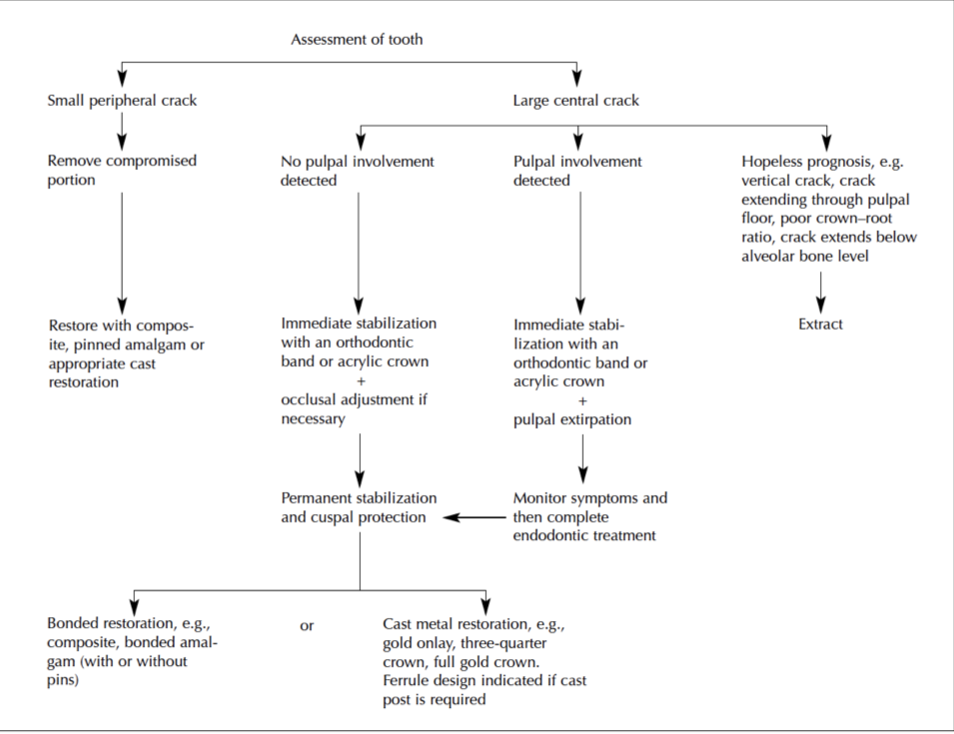
Fig. 8
Lynch C D. , McConnell R J. The Cracked Tooth Syndrome. J Can Dent Assoc 2002; 68(8):470-5
Full case video using D Perfect® MG3 system
Conclusions
Different tools can be used during diagnoses, like carrying out a bite test which produce symptoms associated with cracked tooth syndrome. Bite test is done by asking the patients to bite down on a cotton roll followed by sudden release of pressure. CTS diagnosis is confirmed by pain on release of pressure. Management options vary according to clinical need, from replacement of the fractured cusp with a simple restoration to placement of a crown for cuspal protection.
Teeth originally presenting with CTS may subsequently require root canal therapy followed by a crown prosthesis to protect the tooth from propagation of the crack line.
Bibliography
Cameron CE. Cracked-tooth syndrome. J Am Dent Assoc 1964; 68(March):405-411.
Millar, B. J.; Mehta, S. B.; Banerji, S. (May 2010). Cracked tooth syndrome part 1 : aetiology and diagnosis. British Dental Journal. 208 (10): 459–463.
Bailey O, Whitworth J (2020). Cracked tooth syndrome diagnosis part 1 : integrating the old with the new. Dental Update. 47 (6): 494–499.
Lynch C D. , McConnell R J. The Cracked Tooth Syndrome. J Can Dent Assoc 2002; 68(8):470-5