Expanding the limits of Tooth Restorability
10/02/2024
Fellow
Warning: Undefined variable $post in /home/styleendo/htdocs/styleitaliano-endodontics.org/wp-content/plugins/oxygen/component-framework/components/classes/code-block.class.php(133) : eval()'d code on line 2
Warning: Attempt to read property "ID" on null in /home/styleendo/htdocs/styleitaliano-endodontics.org/wp-content/plugins/oxygen/component-framework/components/classes/code-block.class.php(133) : eval()'d code on line 2
Nowadays, it is a common thing to have patients with extensive dental tissue loss because of caries. However, high magnification and the use of new technology and techniques help us to save teeth that not long ago would be considered for extraction.
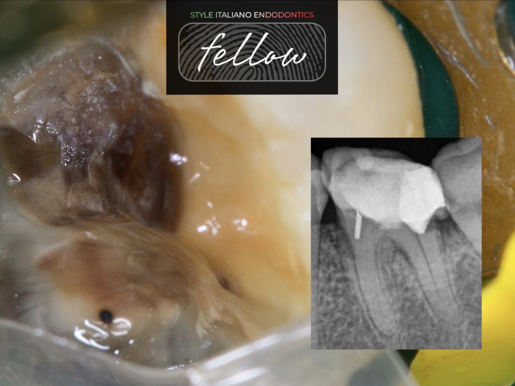
A patient, with clear medical history, was referred for evaluation of restorability and the endodontic treatment of #36. The diagnosis was irreversible pulpitis. The preoperative xray reveals that distal caries have infiltrated the root. After informing the patient about tooth prognosis and treatment plans, we decided to try saving the tooth.
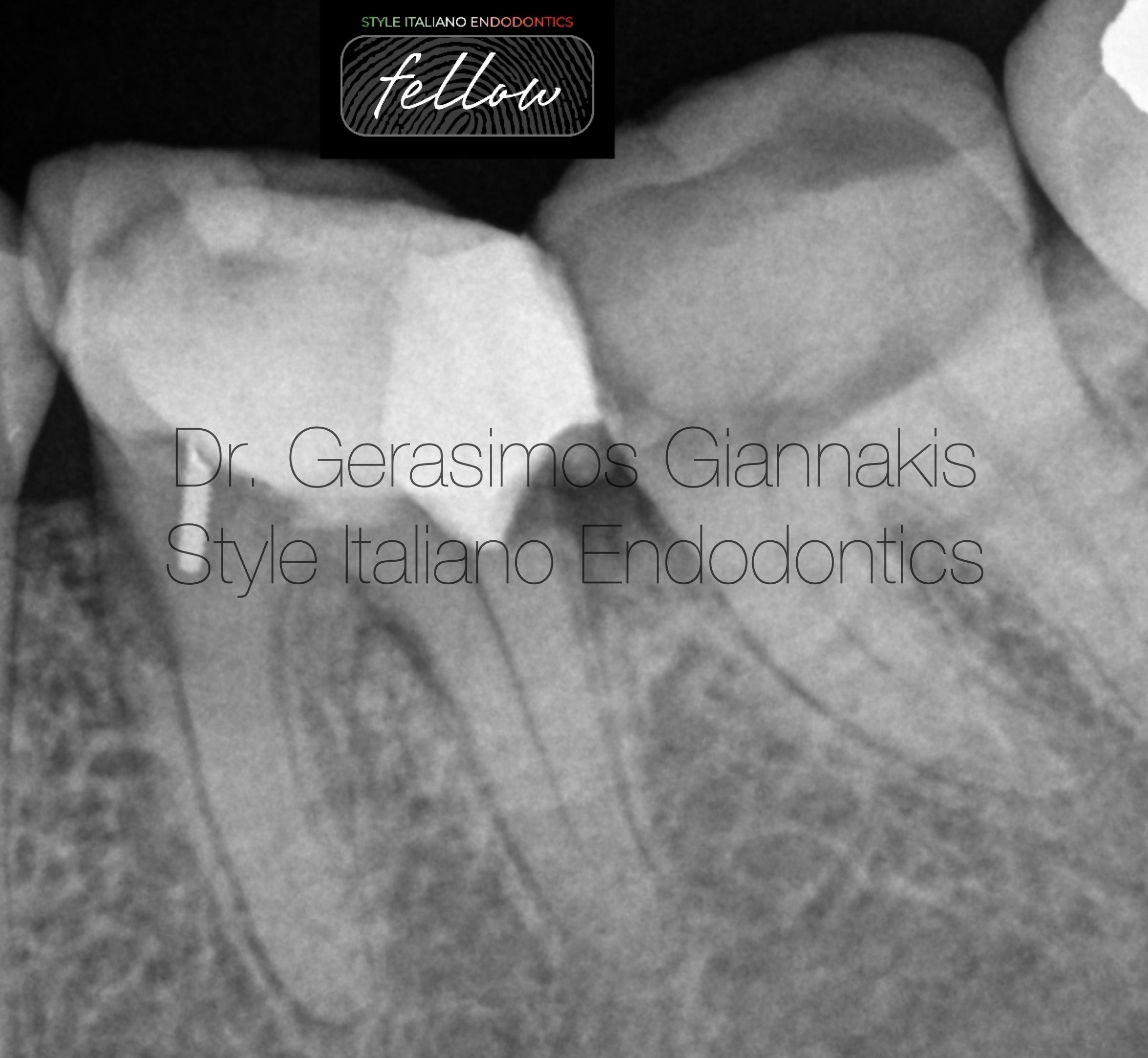
Fig. 1
The preoperative xray reveals:
-Total calcification of mesial canals
-Distal caries infiltrating the root
-A retention pin in the mesial area with false orientation
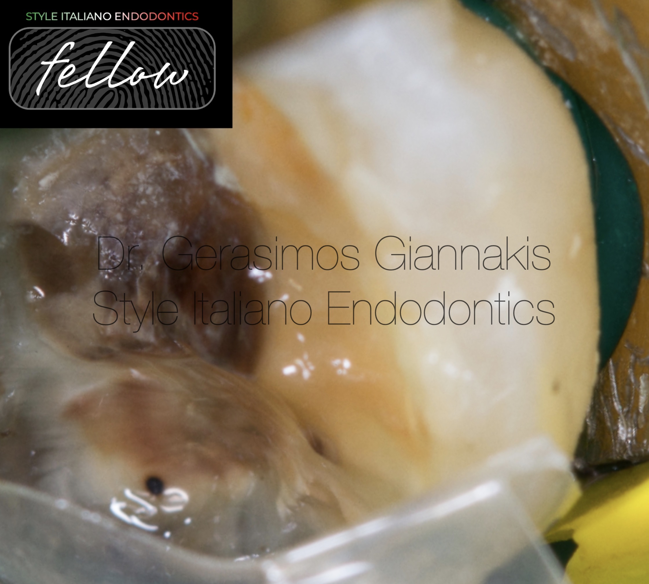
Fig. 2
After caries removal and inflamed soft tissue removal with laser, we isolated the area with rubber dam. Then, a Bioclear matrix and wedge were applied.
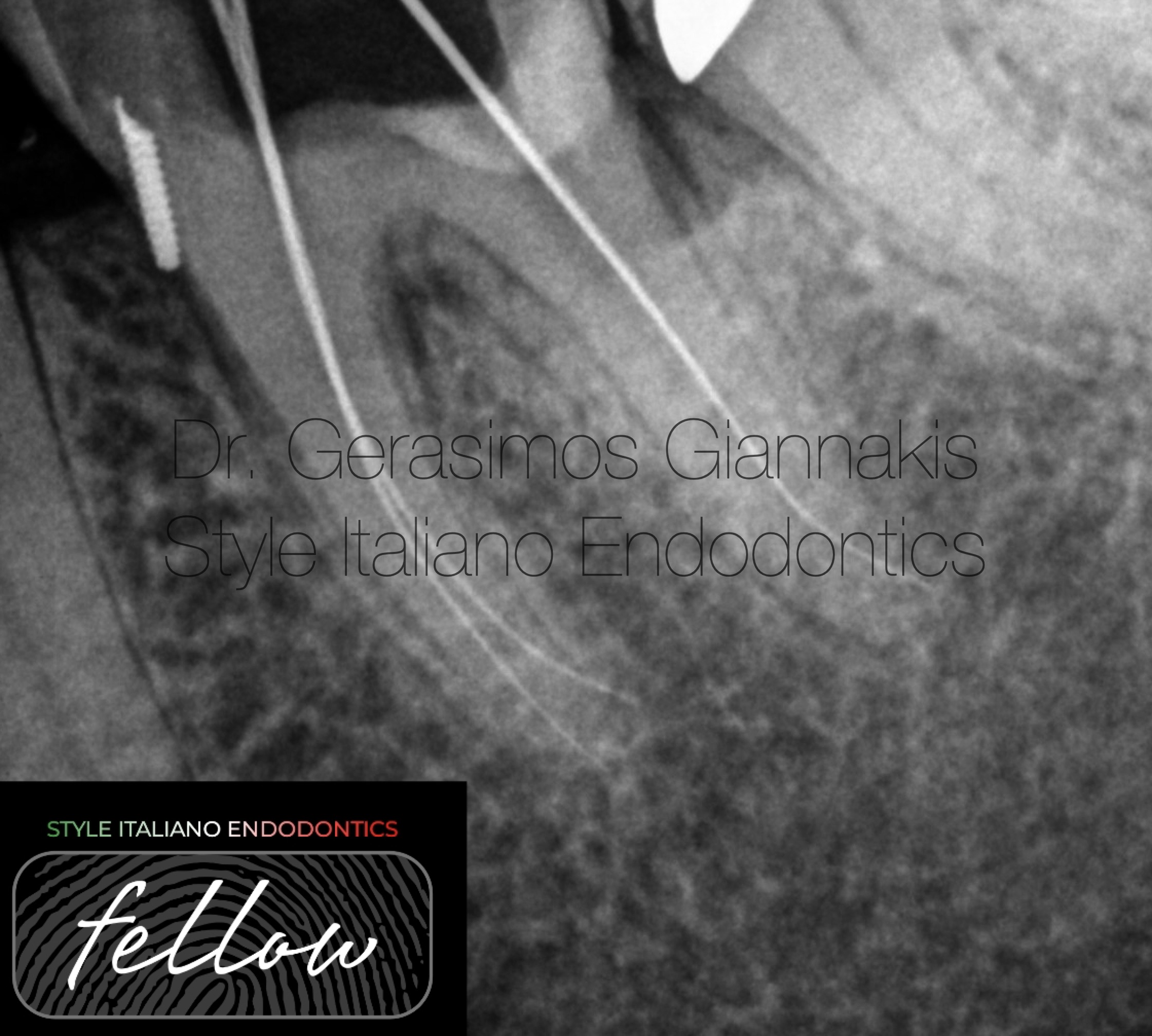
Fig. 3
After selective etching and bonding, the distal wall was restored using increments of Shofu Flow plus F00, which has high compressive and flexural strength.
Working length determination in the same appointment.
We can appreciate the tight adaptation of the restorative material
and the false insertion of the pin. We ended the first appointment at this point.
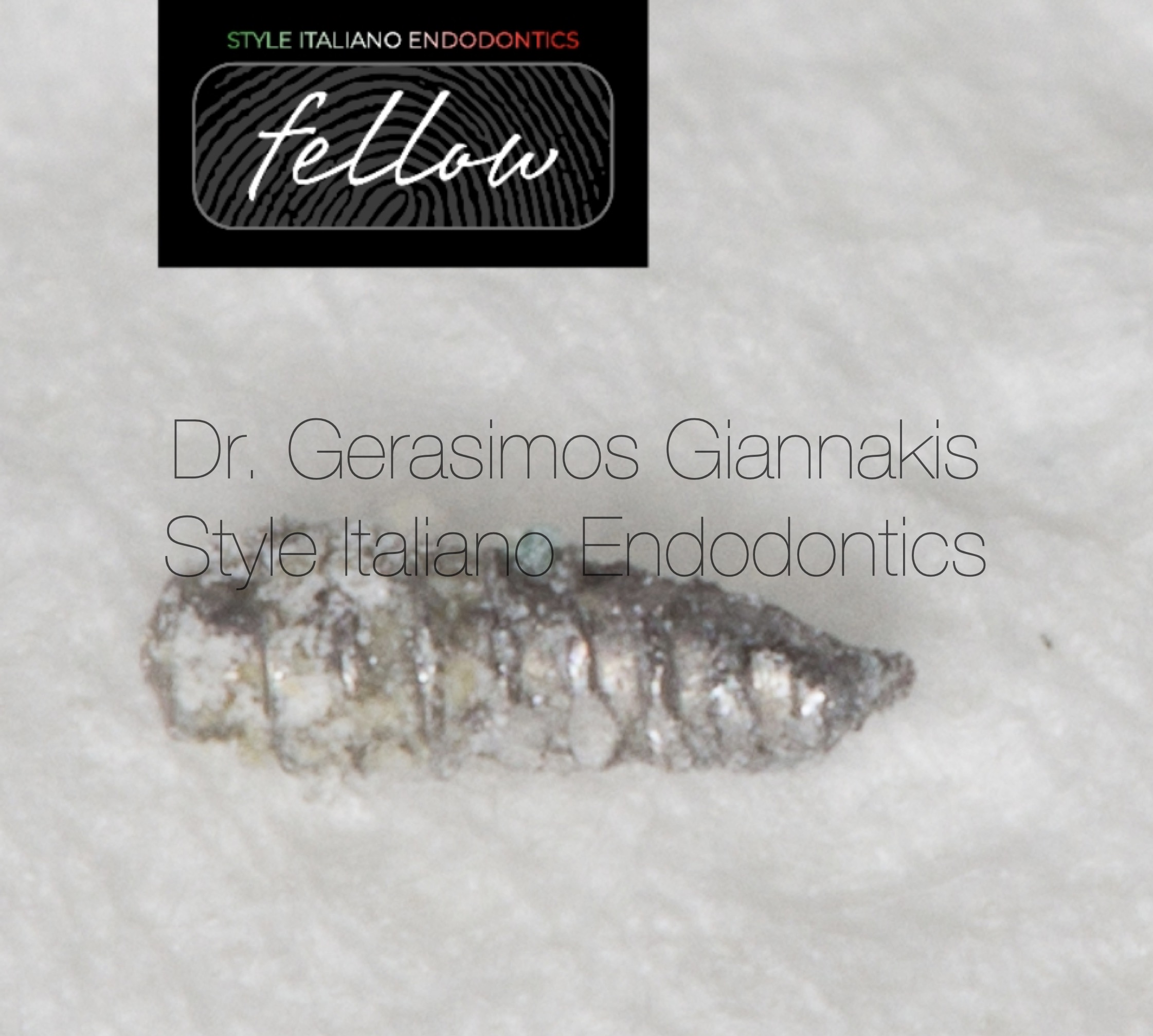
Fig. 4
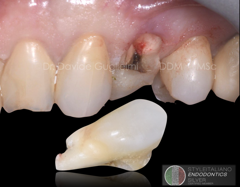
During the second appointment, the pin was removed. Inspection under high magnification. It is shown in literature that pins induce microleakage, thus considering also the malposition removing it was necessary.
Fig. 5
Master cone radiograph. The shaping protocol as follows:
-Coronal preflaring
-Crown-Down until we can reach w.l. with a hand file #10 with just small apical pressure. This ensures a first manual glidepath.
-Then Hyflex EDM 15.03, 10.05 and 20.05. This mechanical preparation is adequate in this case because debris was found in apical part of the finishing file.
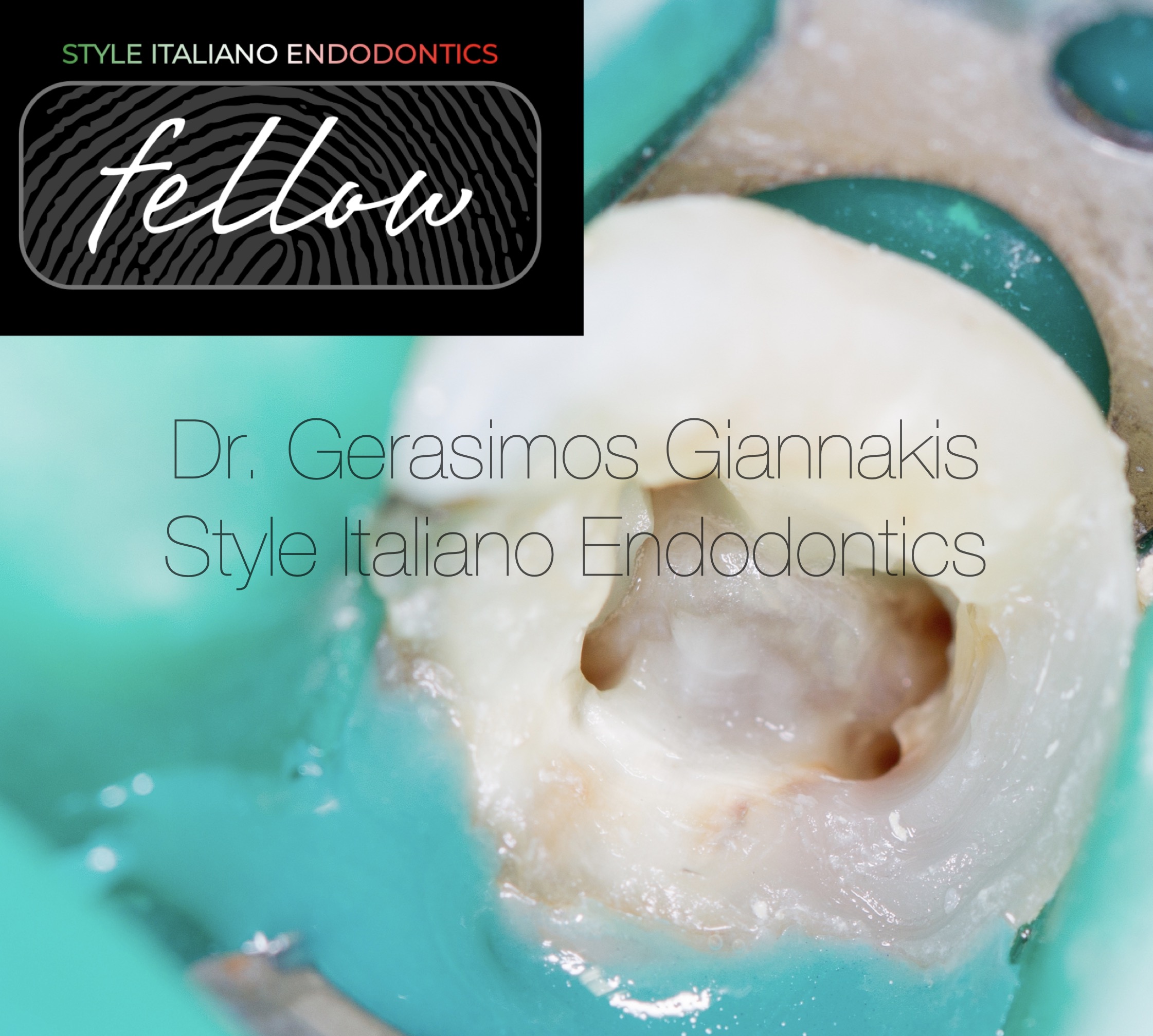
Fig. 6
After BioMechanical preparation and drying of the canals.
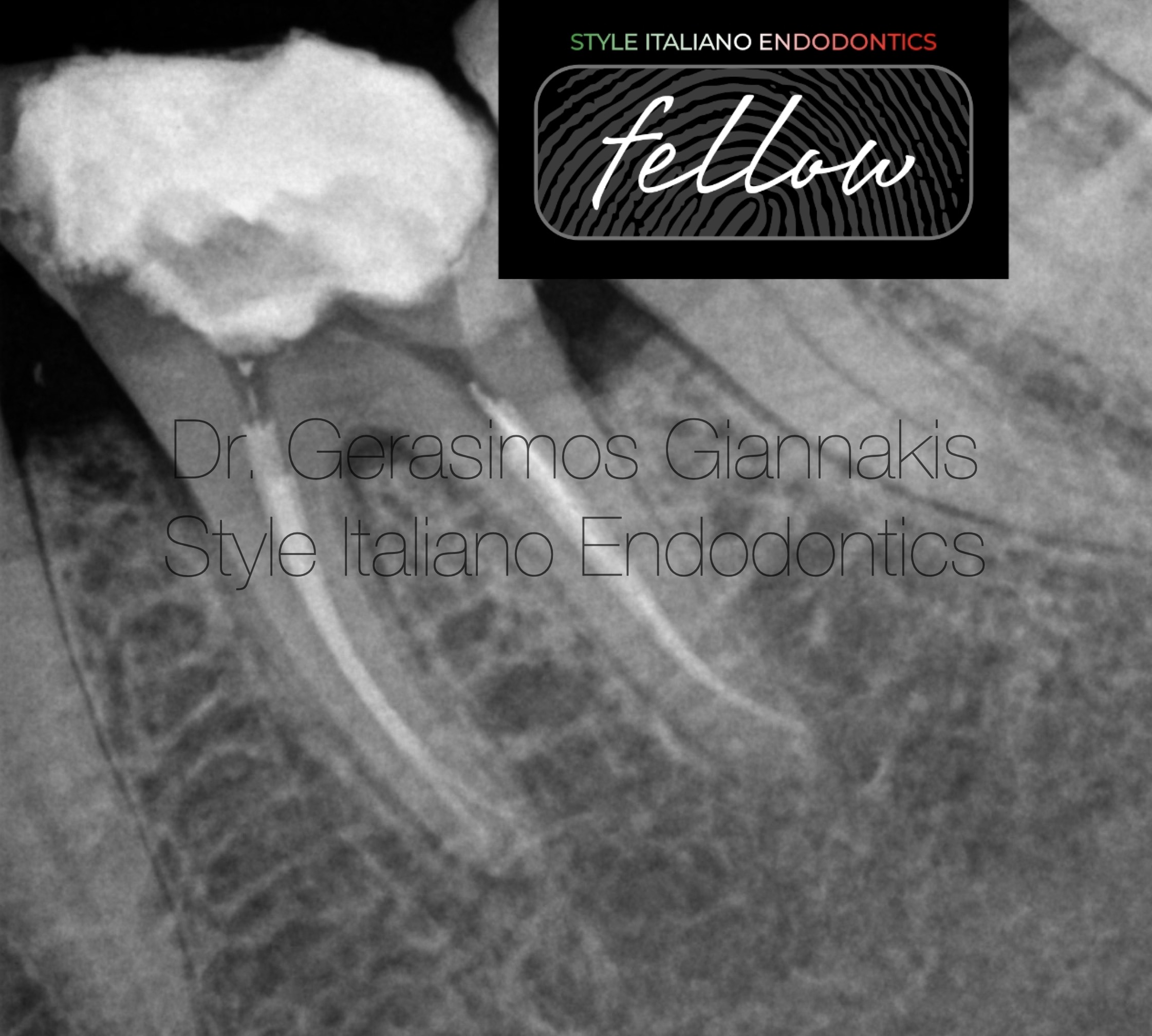
Fig. 7
Post operative radiograph. We can appreciate the conservative shaping, respecting the root canal anatomy.
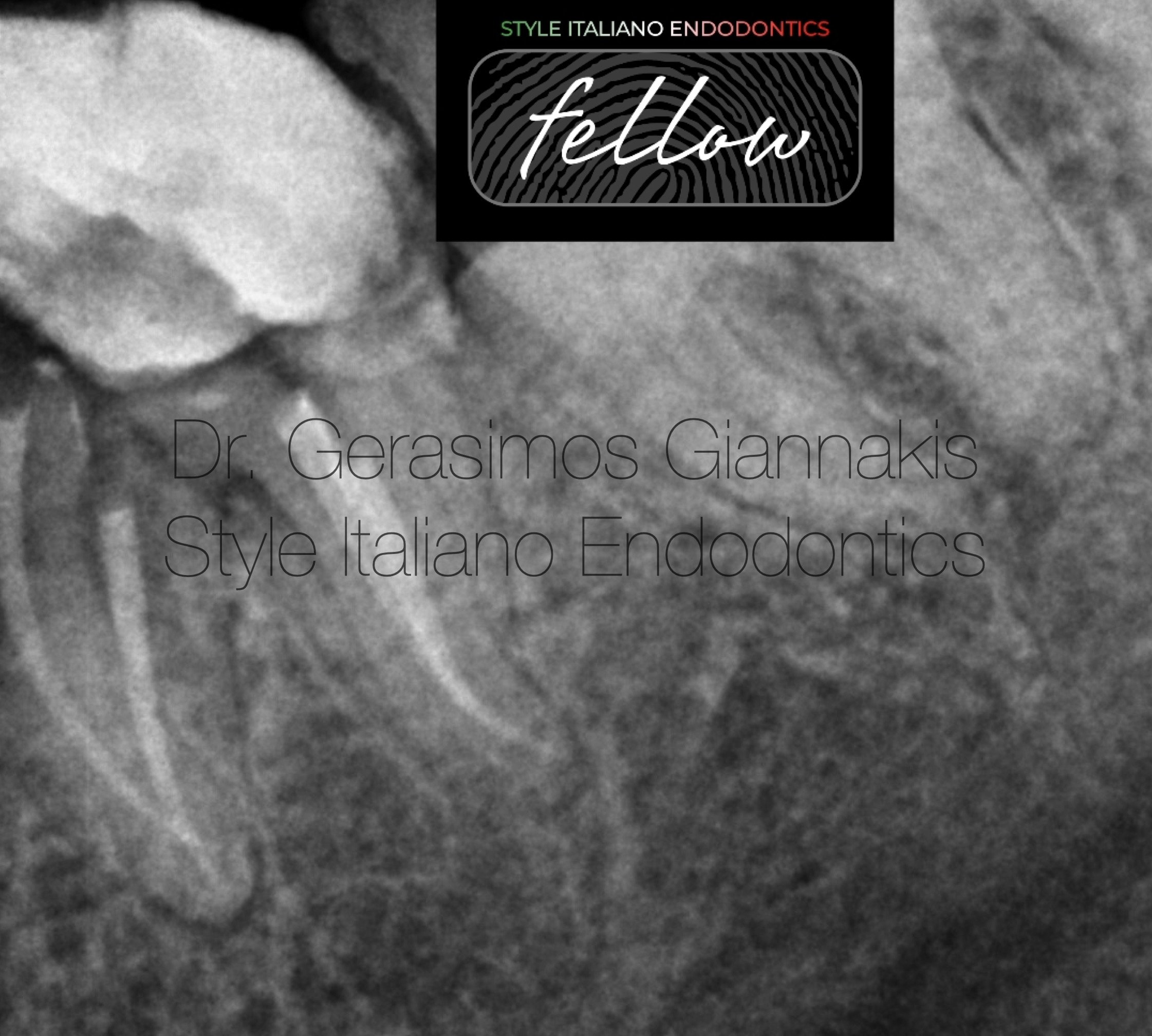
Fig. 8
Post operative radiograph with distal shift. We can appreciate the curvature of mb canal and the apical curvature of the distal system.

Fig. 9
Gerasimos Giannakis received the Doctor of Dental Surgery degree from the University of Athens in 2018.
He has special interest in the field of endodontics, restorative dentistry and microsurgical techniques.
Since then has completed
STYLE ITALIANO Daily menu 40th edition , Milan 2020
TOOTH WEAR MASTER COURSE, by Didier Dietschi 2021
EXPERT COURSE IN MASTERING SOFT TISSUE SURGERY,
In cooperation with the University of Cologne 2022
MICROSCOPE SIMULATION COURSE ON CANAL BLOCKAGE MANAGEMENT by Chaniotis, Sousa Dias, 2020
He is working as an endodontics associate in a clinic, responsible also for the restoration of endodontically treated teeth.
Working exclusively under the microscope and rubber dam, has the opportunity to document the cases.
In the Style Italiano Congress in Greece 2023, was part of the bootcamp and became fellow of the Style Italiano Endodontics community.
Conclusions
The use of dental microscope and new technologies and techniques have made possible to save teeth with extensive tissue loss. Let’s not forget that implants have problems of their own, so keeping a natural tooth as much as possible is advisable.
The deep margin elevation technique has shown high success rates over the period of years. So before considering extraction, we should be certain that the tooth has bad prognosis.
Bibliography
The pin-amalgam restoration. Part I. A review Joseph R. Evans, D.D.S.,* and Jon H. Wetz, D.D.S.** Tufts University School of Dental Medicine, Boston, Mass.
Deep Margin Elevation: A Literature Review
Theodora Kalliopi Samartzi,1,* Dimokritos Papalexopoulos,2 Panagiotis Ntovas,3 Christos Rahiotis,3 and Markus B. Blatz4
M-i-M for DME: matrix-in-a-matrix technique for deep margin elevation
Pascal Magne 1