Endo-restorative treatment of discolored upper incisor
13/11/2023
Fellow
Warning: Undefined variable $post in /home/styleendo/htdocs/styleitaliano-endodontics.org/wp-content/plugins/oxygen/component-framework/components/classes/code-block.class.php(133) : eval()'d code on line 2
Warning: Attempt to read property "ID" on null in /home/styleendo/htdocs/styleitaliano-endodontics.org/wp-content/plugins/oxygen/component-framework/components/classes/code-block.class.php(133) : eval()'d code on line 2
Traumatic dental injuries are common cases reported in dentistry and may result in damage of hard dental tissues, pulpal complex, and periradicular structures.
The anterior tooth, involving enamel and dentin, is the most frequent tooth affected, in which the coronal fractures represents 65-75% of all dental traumas.
The tooth can be discolored by deposition of pigments in its internal structure and on its surface
Importantly, the accurate diagnosis of discoloration is a condition with multifactorial etiology; it is classified as extrinsic and intrinsic
For anterior teeth, the origin of a discoloration is frequently associated with a loss of tooth vitality or with an endodontic treatment. The many chemical products used during root canal disinfection and obturation can be identified as potential causes of discoloration (irrigating solutions, root canal cements, intracanal medicaments)
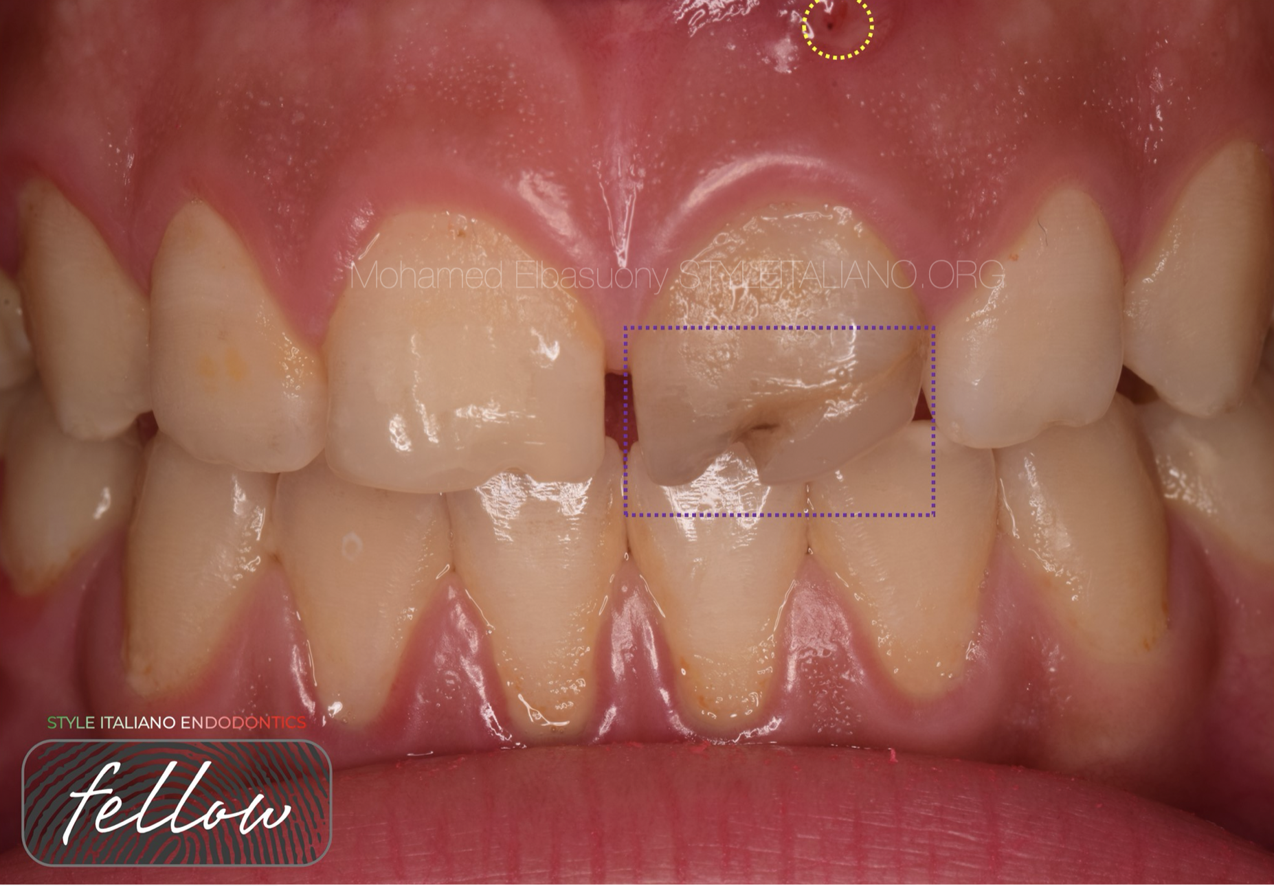
Fig. 1
Fig 1
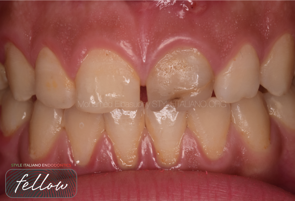
A 16-year-old female patient came to my clinic with a discolored tooth, a maxillary left central incisor, which was compromising the aesthetic of her smile.
On elaborating the dental history of the patient, it was noted that the same tooth had been traumatized previously.
The tooth has an old improper root canal therapy, observed through pre-treatment X-ray.
By clinical examination sinus tract was detected.
Failure of the direct resin restoration by tooth discoloration was detected.
A poor marginal adaptation and mismatching color of a class IV resin composite restoration was observed, which was placed due to a traumatic dental injury.
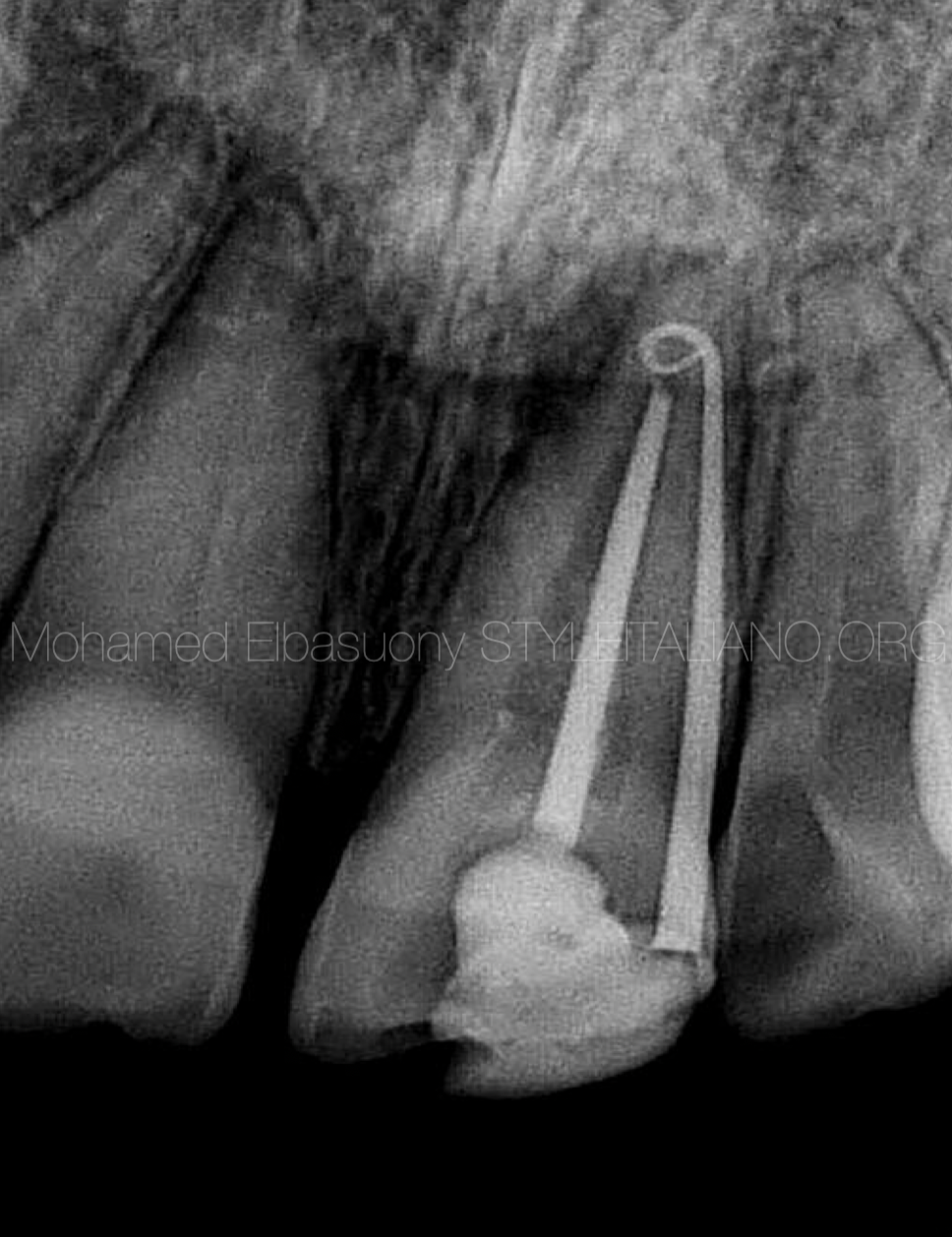
Fig. 2
From the above mentioned findings, in agreement with the patient, a conservative aesthetic rehabilitation was decided.
The treatment was divided into two stages:
1-Endodontic retreatment stage
2-Restorative stage
The diagnosis for retreatment was made according to the signs and symptoms reported by the patient and the preoperative radiographs showing apical radiolucency.
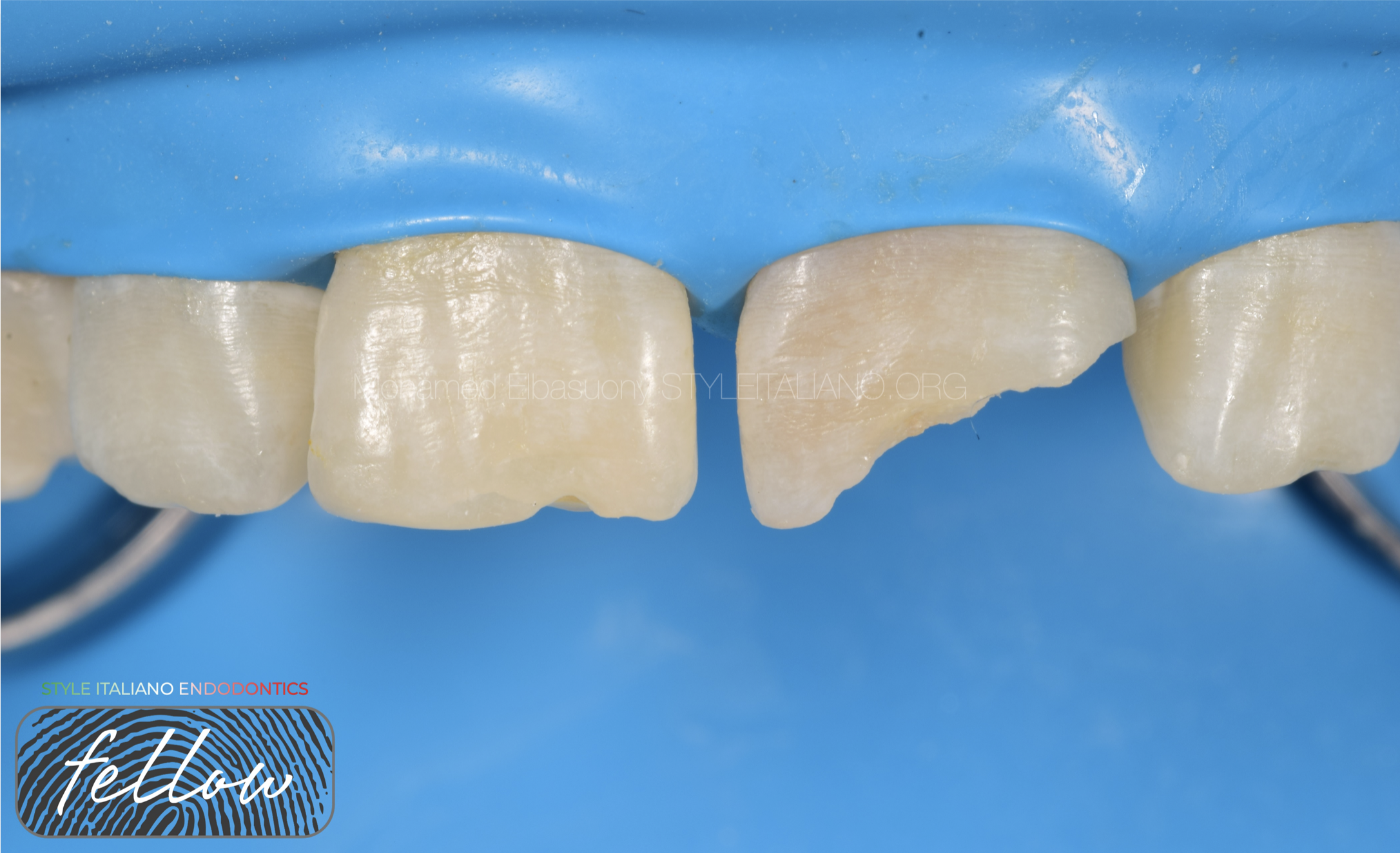
Fig. 3
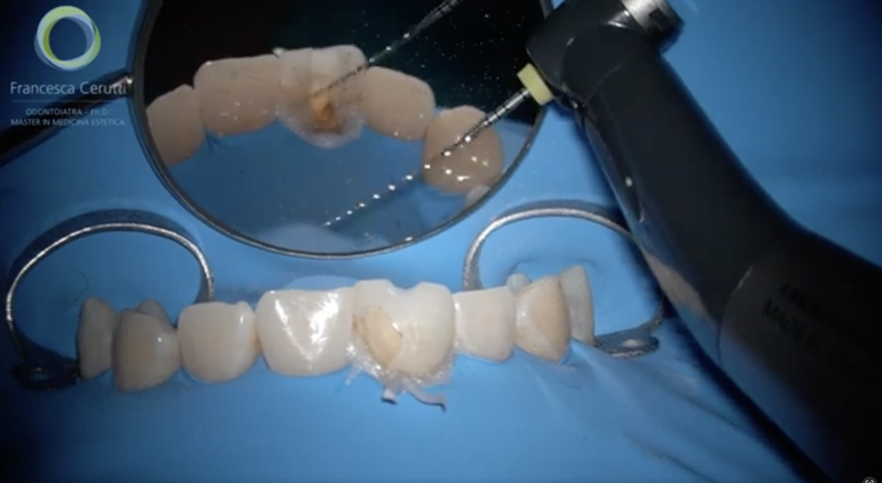
After local anesthesia and rubber dam application, the old restoration is removed first by using a tapered-cylinder, round-end diamond bur.
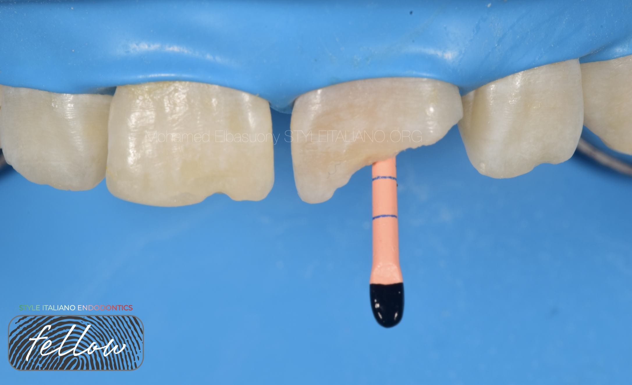
Fig. 4
The crucial factor for achieving successful retreatment is thorough reshaping and cleaning of the canals to eliminate bacteria.
Hand files and ultrasonic device were used to remove old gutta-percha and sealer.
Subsequently the tooth was prepared with rotary Ni-Ti instruments to working length and irrigated with 5.25% NaOCl.
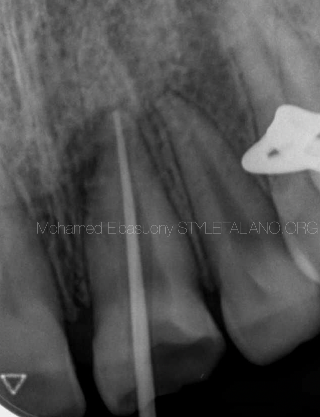
Fig. 5
Master cone radiograph
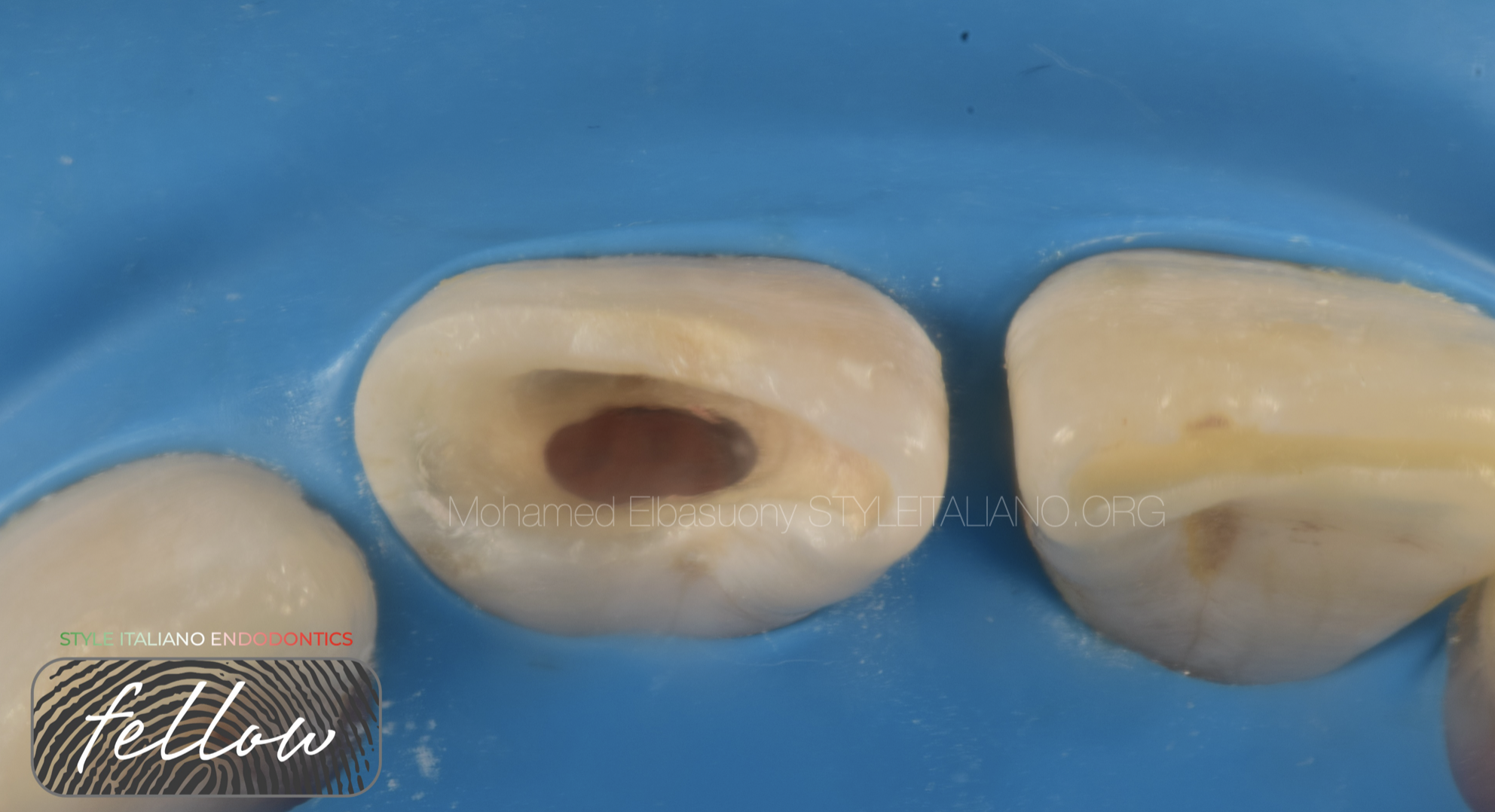
Fig. 6
The obturation was done to level below cementoenamel junction
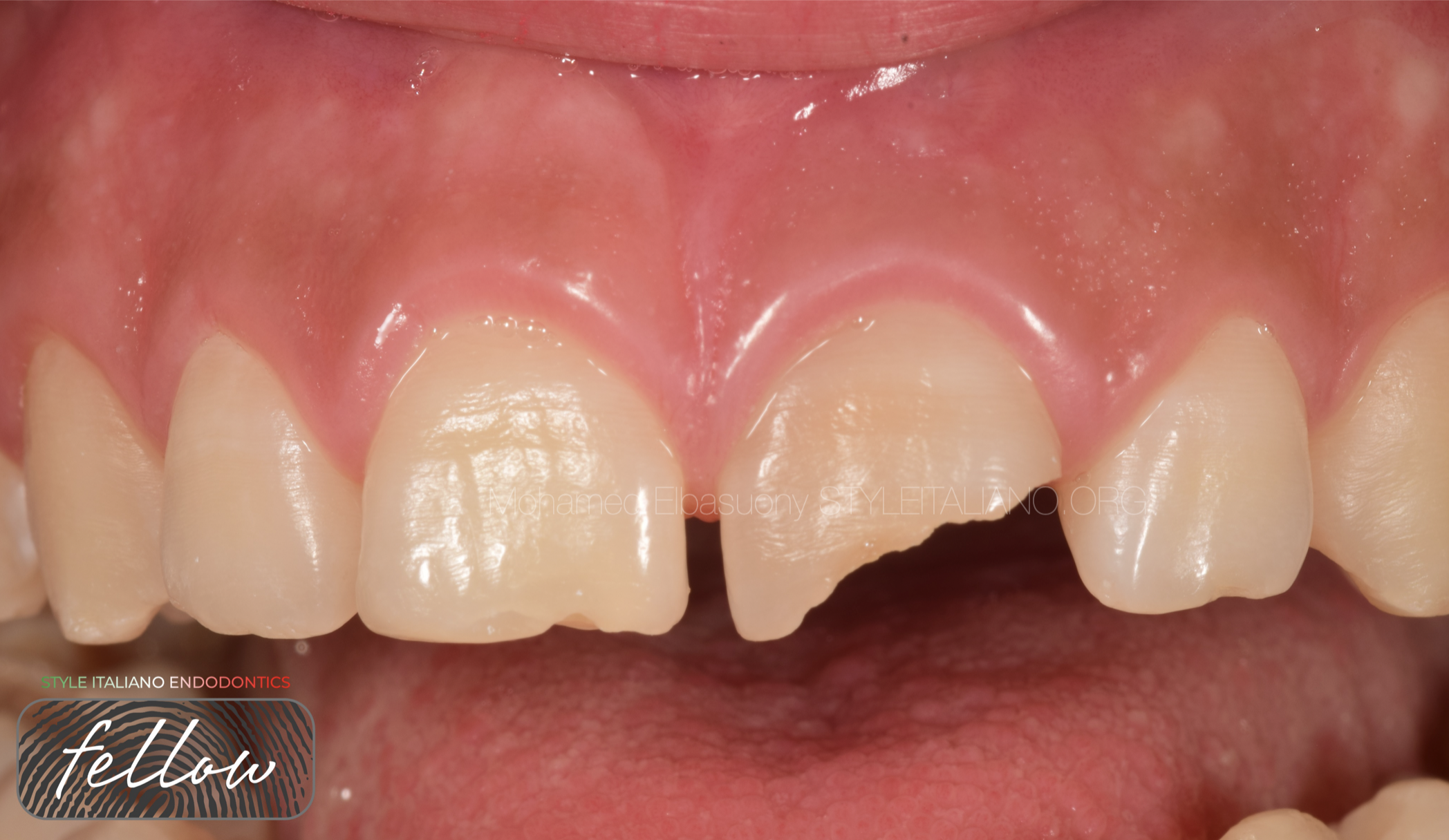
Fig. 7
The walking bleach technique was carried out.
The bleach agent is collocated inside the pulp chamber, after putting a seal with glass-ionomeric cement at the coronal portion of the canal.
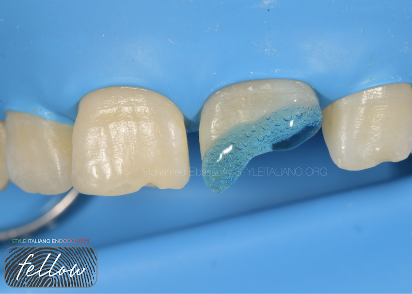
Fig. 8
Restorative stage
1-Shade and size selection
2-Tooth margins and prepared surfaces were then etched for 30 seconds with a 37% phosphoric acid etchant gel.
The tooth were rinsed and dried, and thereafter separated by interproximal matrices. A dental bonding agent was applied to all prepared surfaces, followed by gentle air-drying from the cervical to the incisal aspect and light-curing for 20 seconds.
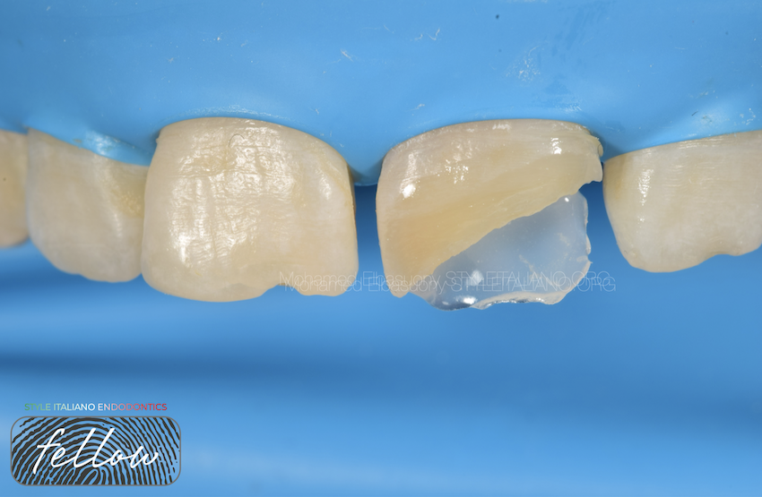
Fig. 9
Palatal shell
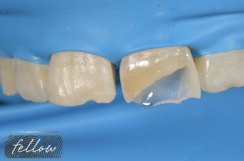
Fig. 10
Distal wall build up
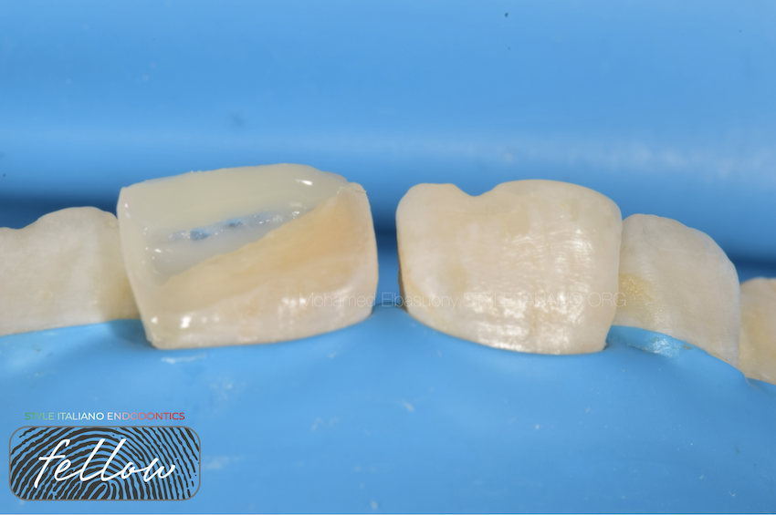
Fig. 11
Halo effect
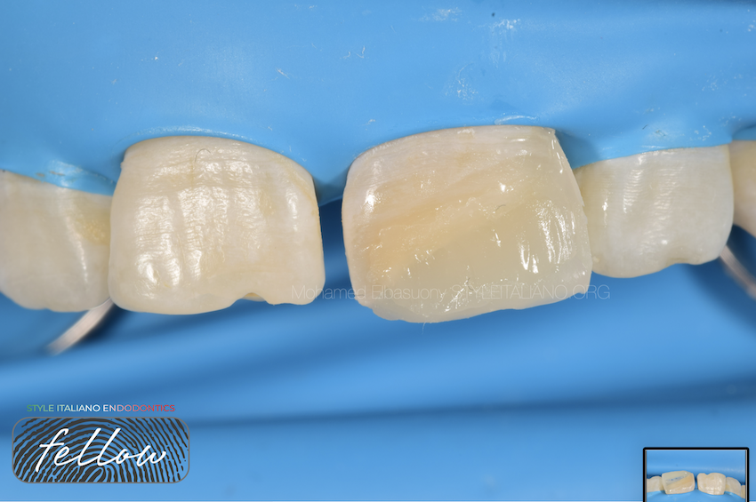
Fig. 12
1-Dentin shaded composite was free-handed and then used to restore with the corresponding preparations of a conventional direct composite restoration and light-curing for 40 seconds.
2-Enamel Layer
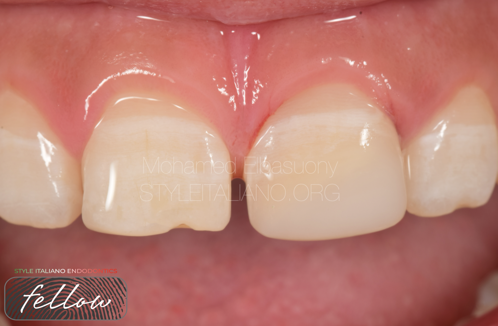
Fig. 13
Contours and occlusal interferences were adjusted with a high speed bur and margins were refined and polished by strips for interproximal areas, flexible aluminum oxide discs, which are ideal to adjust the incisal angles, and silicone rubber polishers were used for the polishing steps.
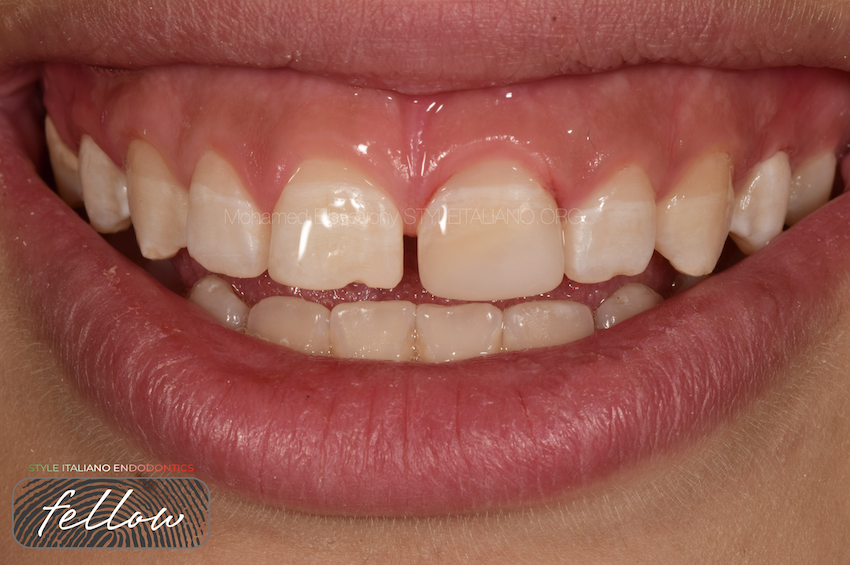
Fig. 14
The final smile
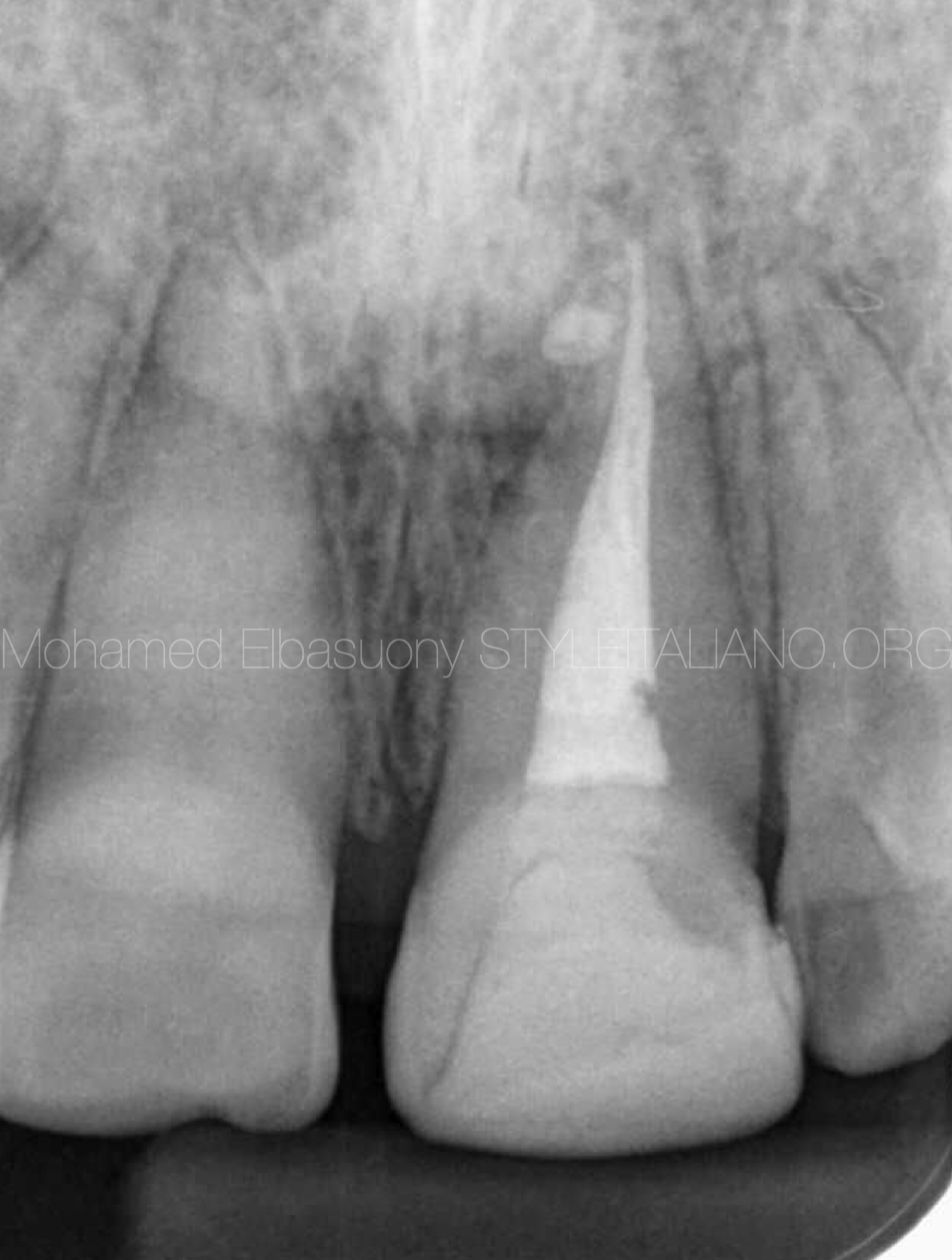
Fig. 15
The final radiograph

Fig. 16
About the author:
Mohamed Elbasuony
BDS from faculty of dentistry, Mansoura university in 2013
Diploma in Endodontics at faculty of dentistry, Mansoura university in 2018.
Enrolled in the MSc in Endodontics at faculty of dentistry, Mansoura university in 2020
StyleItaliano Endodontics Fellow
Endodontic Specialist
Conclusions
In anterior aesthetic problems it’s important to understand the etiology of the discoloration
A multidisciplinary approach is to consider it necessary to resolve the problem: at first endodontic retreatment, successively bleaching and coronal restoration.
To improve the aesthetic result a proper adhesive composite restoration was applied.
Bibliography
- Sjogren U, Figdor D, Persson S, Sundqvist G. Influence of infection at the time of root filling on the outcome of endodontic treatment of teeth with apical periodontitis. Int Endod J. 1997;30:297–306.
- Fasanaro TS. Bleaching teeth: history, chemicals and methods used for common tooth discolorations. J Esthet Dent. 1992;4:71–78.
- Amato M, Scaravilli MS, Farella M, Riccitiello F. Bleaching teeth treated endodontically: longterm evaluation of a case series. J Endod. 2006;32:376–378.
- Krastl G, Allgayer N, Lenherr P, Filippi A. Tooth discoloration induced by endodontic materials: a literature review. Dent Traumatol. 2013;29:2–7