Endo-resto in a lower first molar
04/05/2026
Warning: Undefined variable $post in /home/styleendo/htdocs/styleitaliano-endodontics.org/wp-content/plugins/oxygen/component-framework/components/classes/code-block.class.php(133) : eval()'d code on line 2
Warning: Attempt to read property "ID" on null in /home/styleendo/htdocs/styleitaliano-endodontics.org/wp-content/plugins/oxygen/component-framework/components/classes/code-block.class.php(133) : eval()'d code on line 2
Post endodontic restoration is a critical phase in the long-term success of root canal–treated teeth. While endodontic therapy effectively eliminates infection and preserves the tooth, it often leaves the remaining structure weakened due to caries, previous restorations, and access cavity preparation. Without proper restoration, these teeth are significantly more prone to fracture and failure. A well-designed post-endodontic restoration not only reinforces the remaining tooth structure but also restores function, aesthetics, and coronal seal—preventing reinfection. Therefore, the restorative phase should be considered an integral extension of endodontic treatment, directly influencing the prognosis and longevity of the tooth.
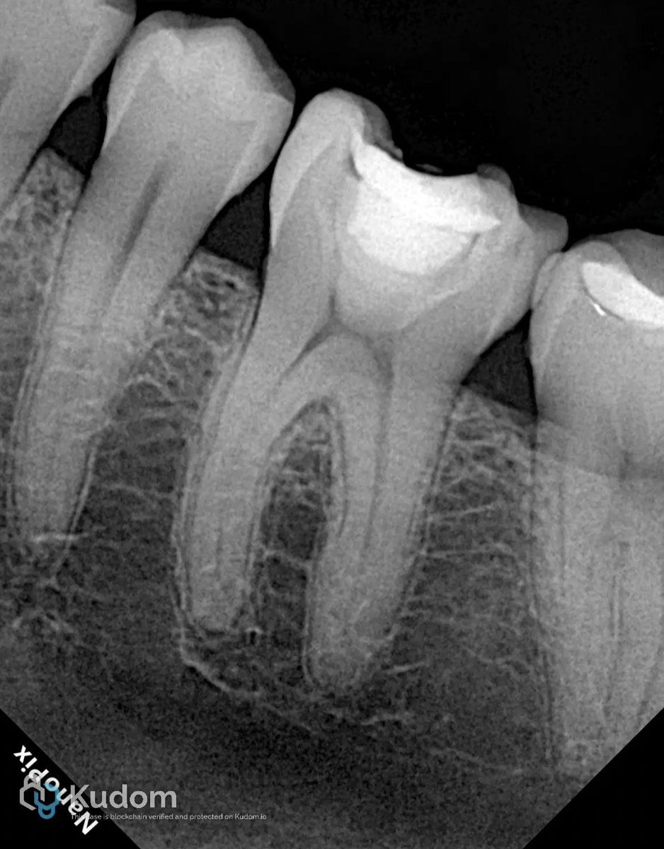
Fig. 1
A 42-year-old woman is referred for endodontic treatment of a mandibular first molar.
The initial radiograph reveals very narrow canals.
Pulp diagnosis: symptomatic irreversible pulpitis.
Periapical diagnosis: normal periapical tissues.
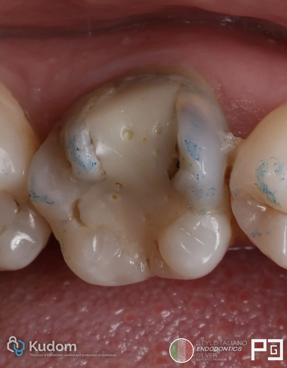
Fig. 2
Initial situation.
As can be seen in the image, microleakage and the presence of caries are observed.
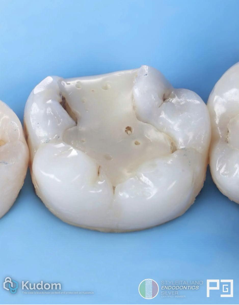
Fig. 3
Initial situation after anesthesia and rubber dam placement.
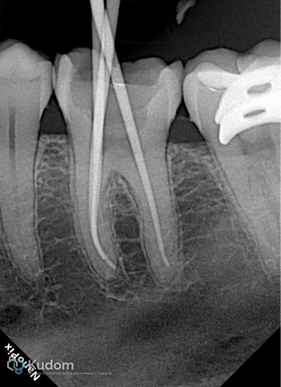
Fig. 4
Shaping and cleaning with Eflex blue and ultrasonic activation with Ultra X.
Cone Fit X-ray
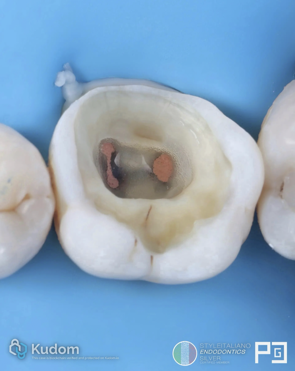
Fig. 5
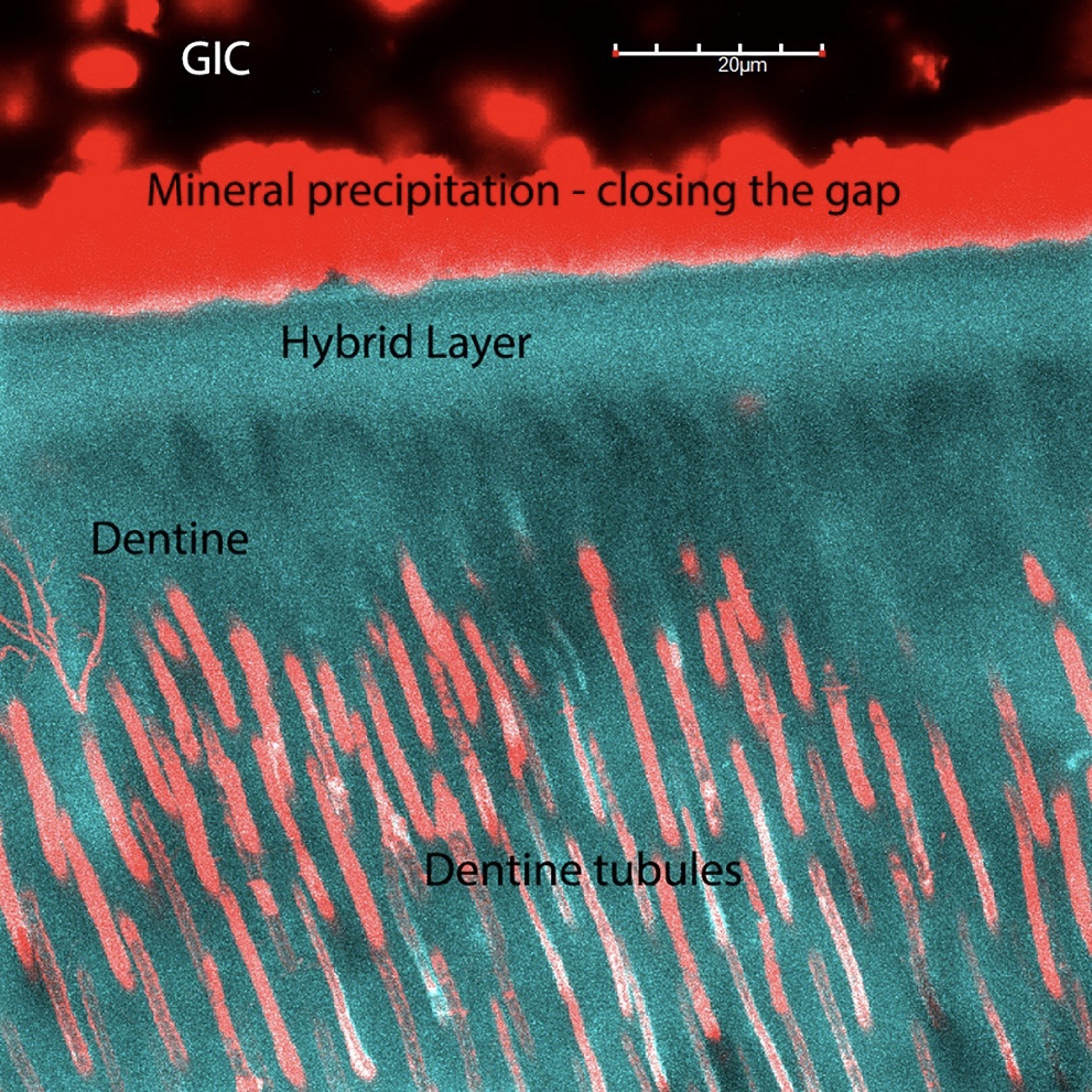
Endo done, cleaning of pulp chamber is a mandatory step, in order to post endo restoration.
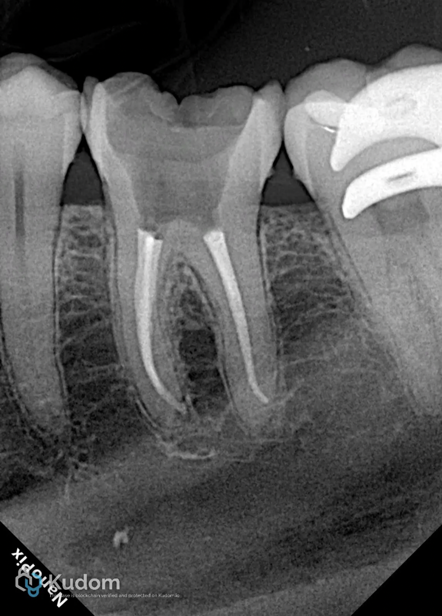
Fig. 6
Obturation: Single cone with bioceramic sealer and final X-ray.
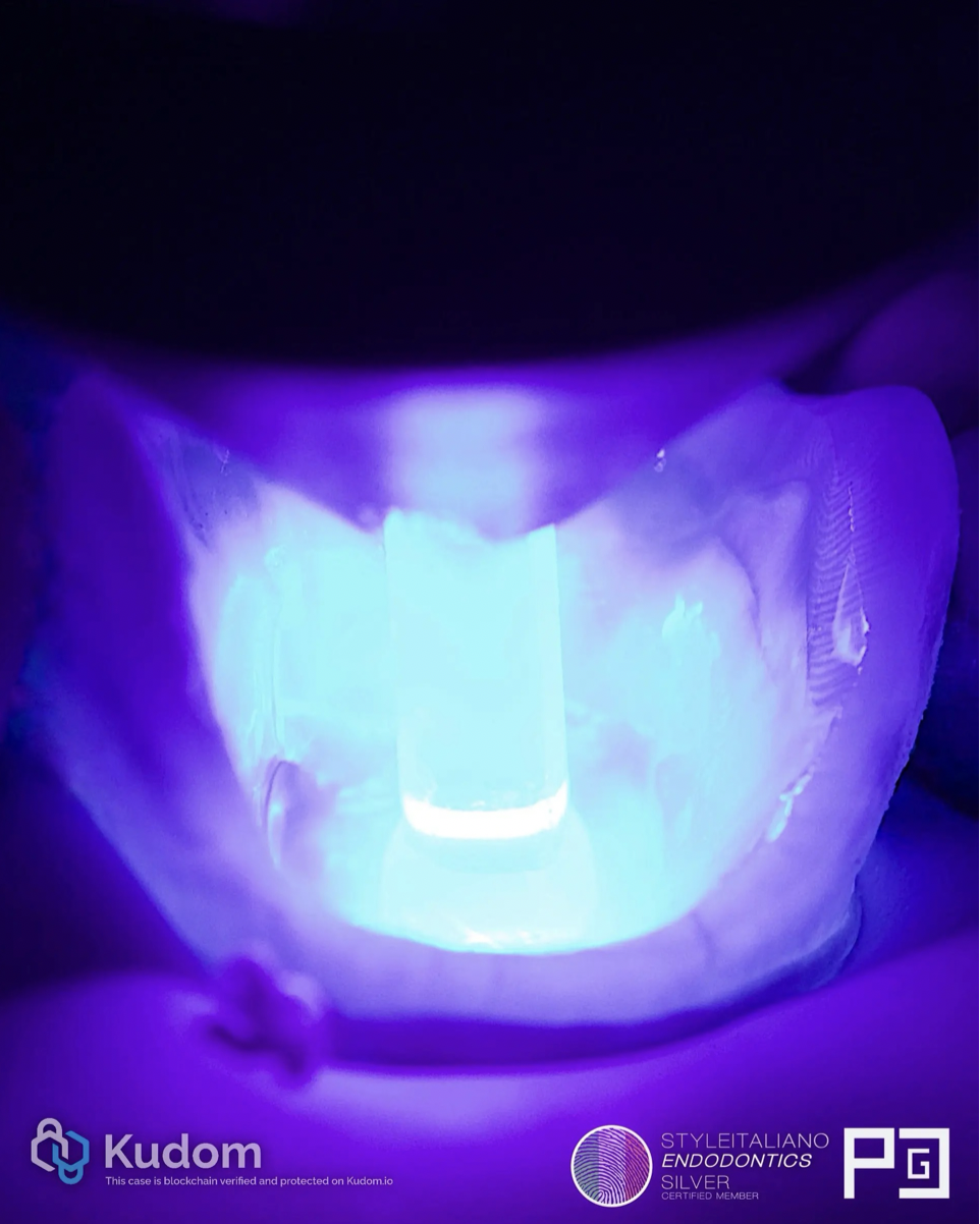
Fig. 7
Mandibular molar conditioned with 3-step adhesive, total etch, Optibond FL and cured with Valo Grand and special tip to deep areas.
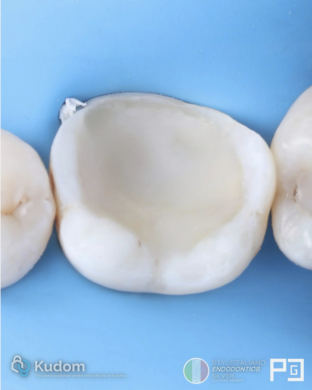
Fig. 8
Build up completed and preparation refined.
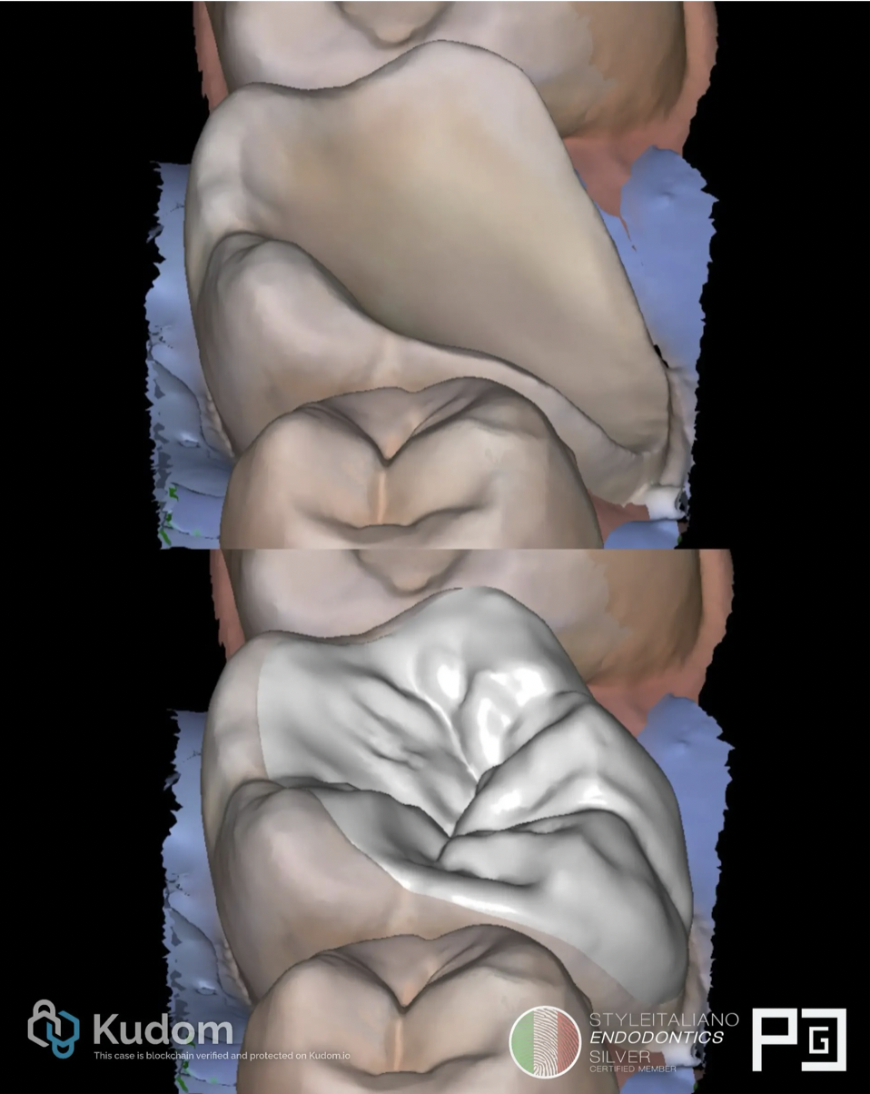
Fig. 9
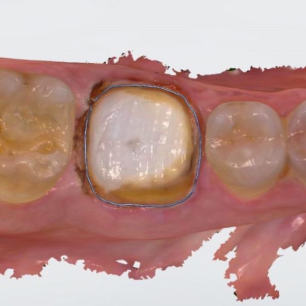
Conservative prep, scanned and designed with Exocad.
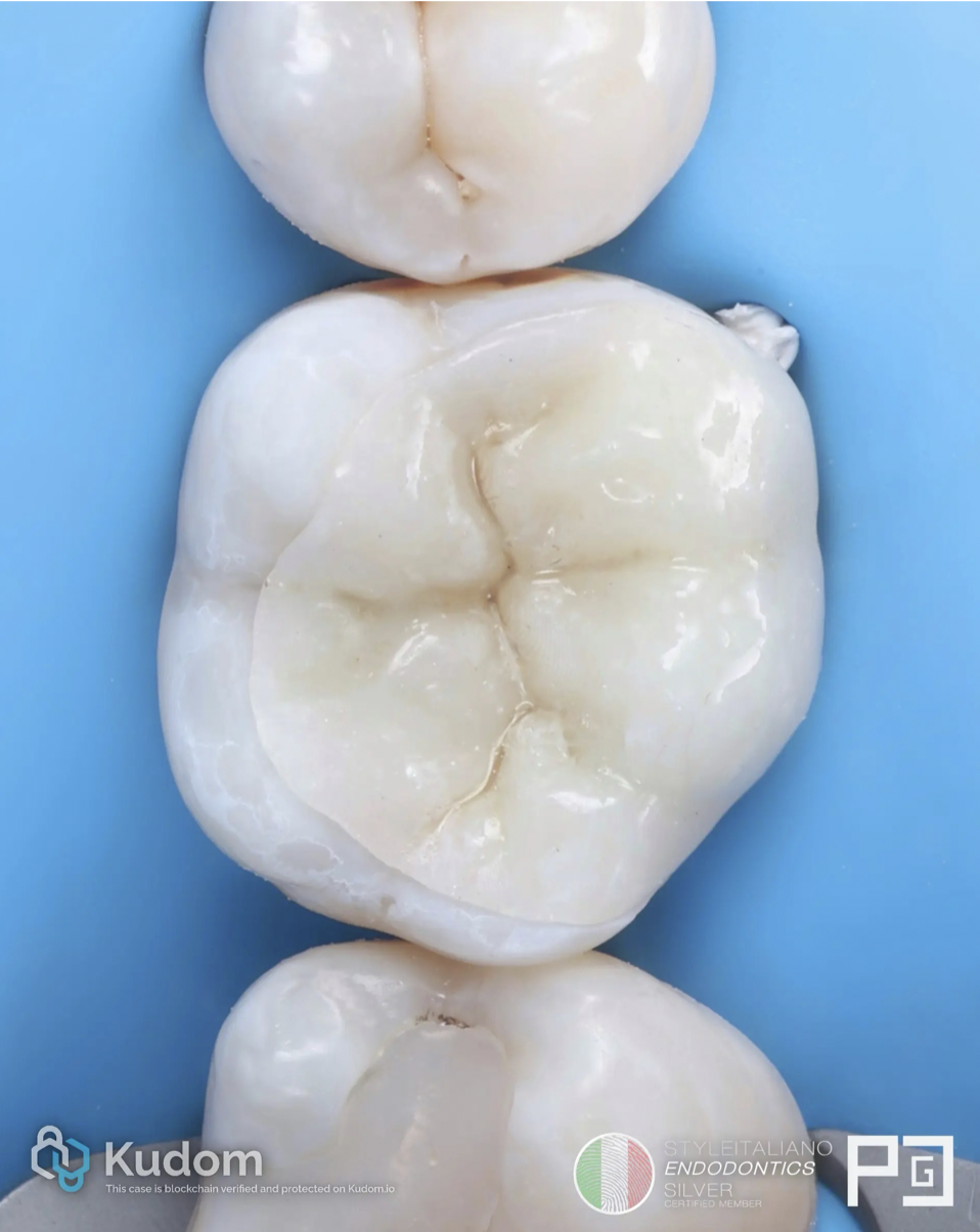
Fig. 10
At the second appointment, rubber dam isolation and try-in of a ceramic restoration.
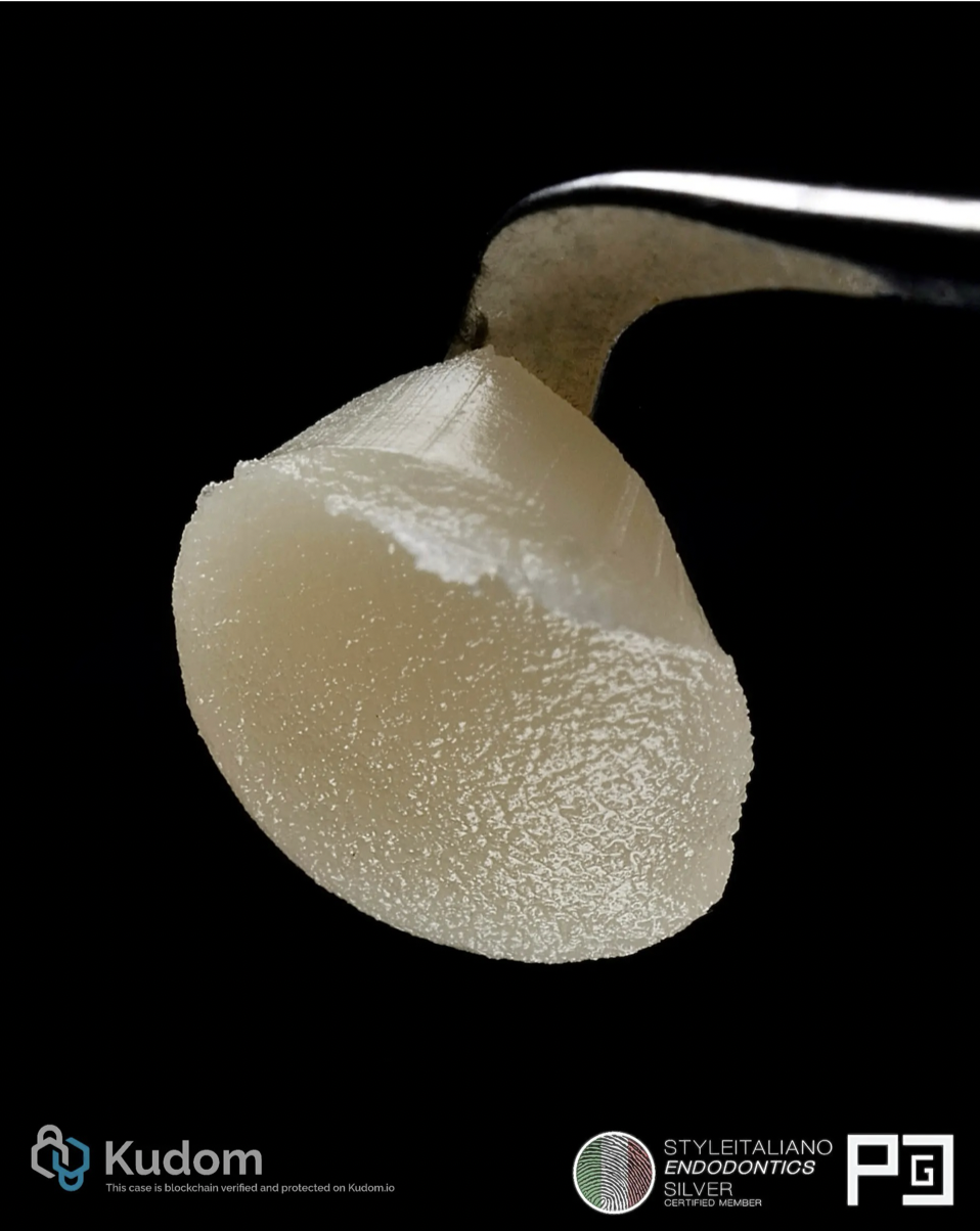
Fig. 11
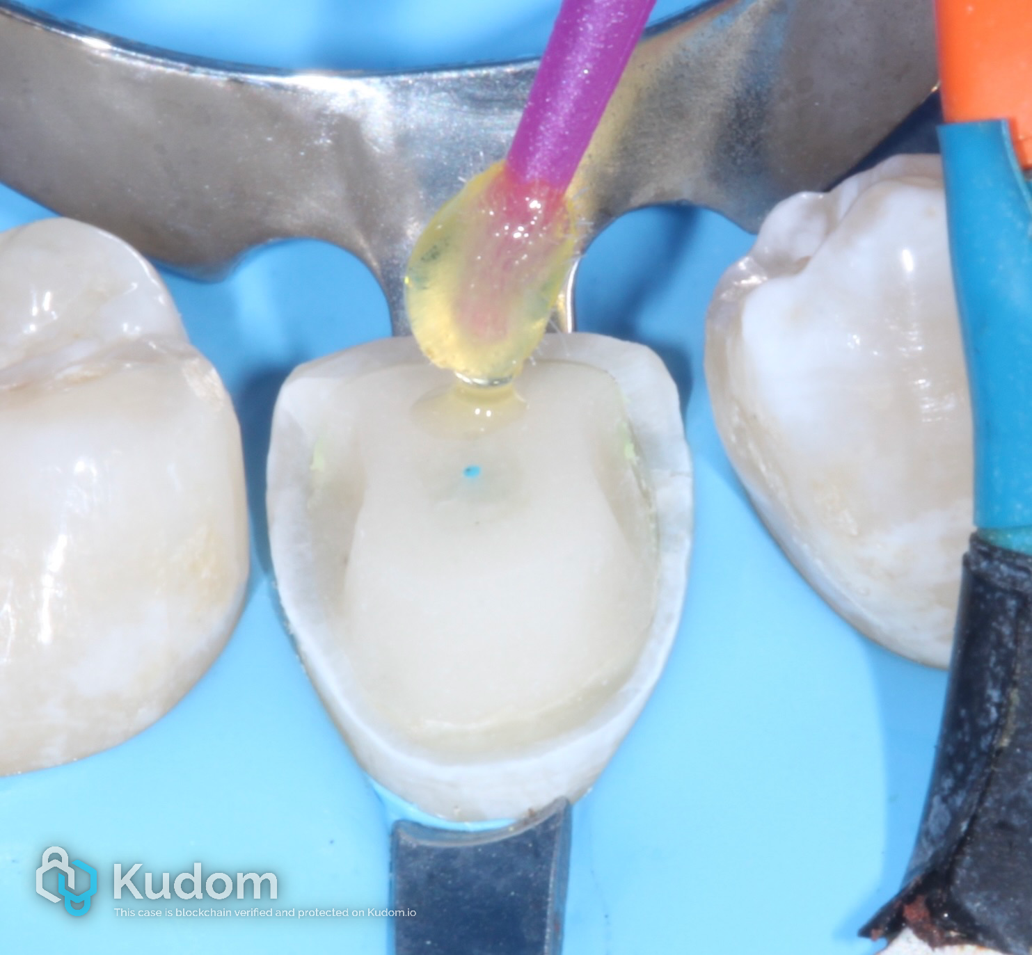
Preheated composite as a luting cement.
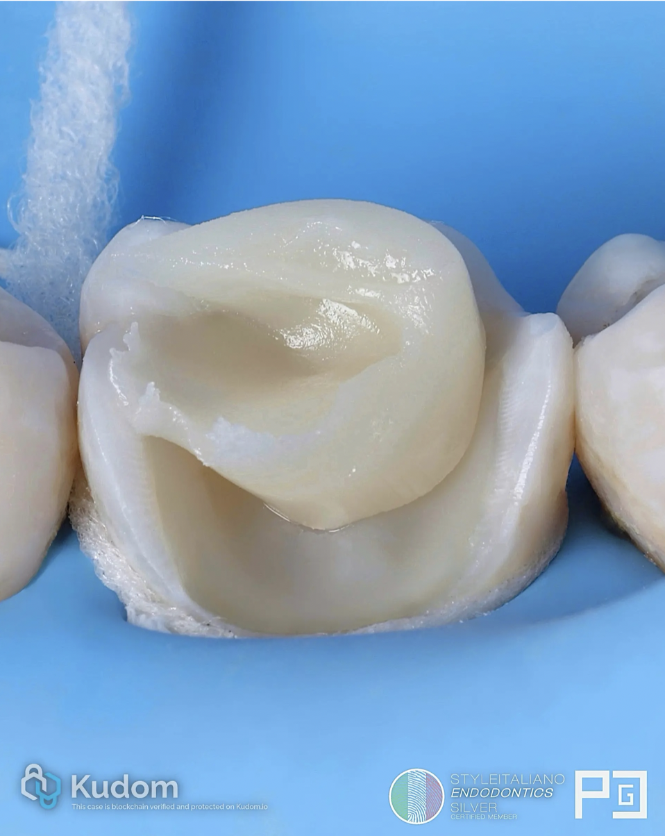
Fig. 12
Preheated composite.
The viscosity is excellent and allows for easy removal of excess material.
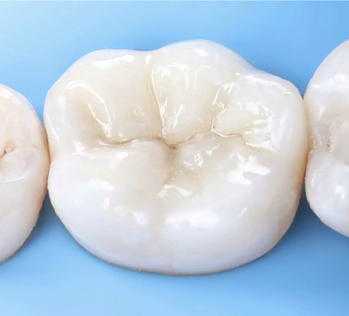
Fig. 13
Thanks to this technique, an imperceptible union of the restoration margin with the tooth is possible.
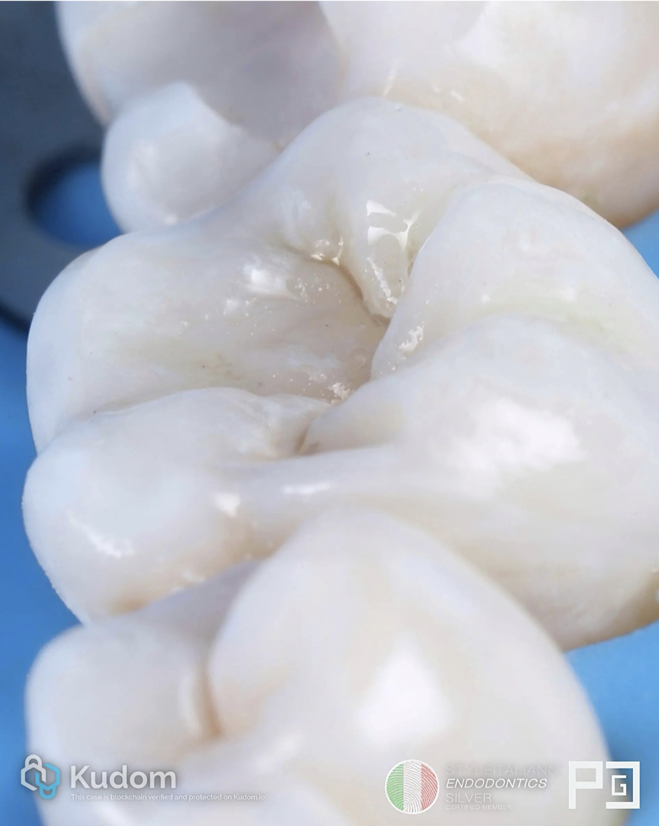
Fig. 14
Other view.
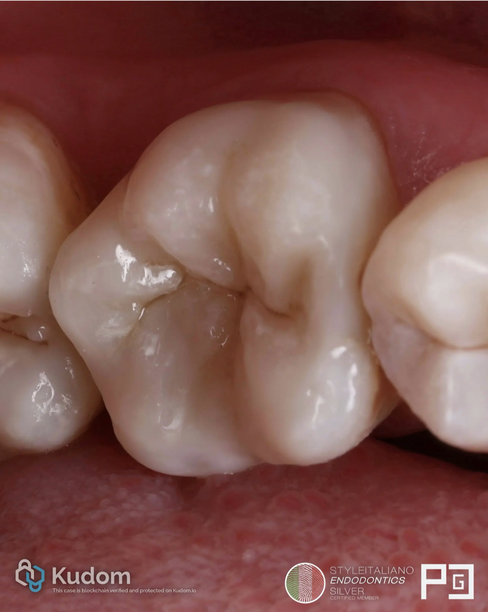
Fig. 15
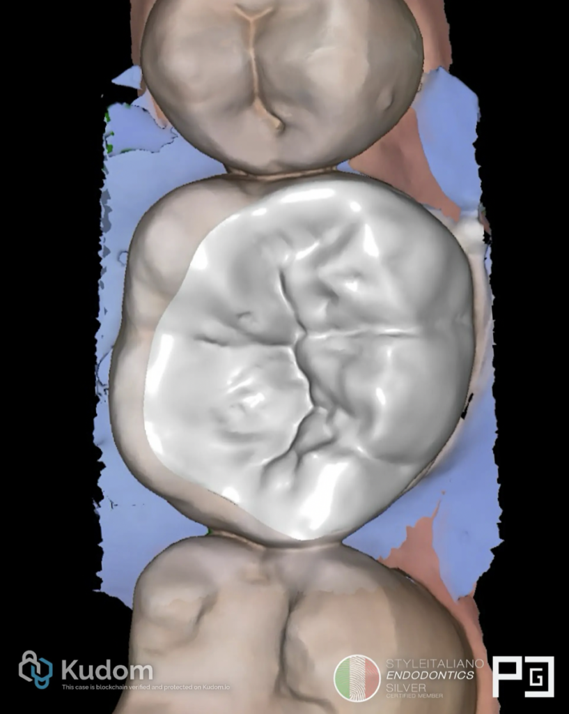
Final situation after cementation of the ceramic restoration.
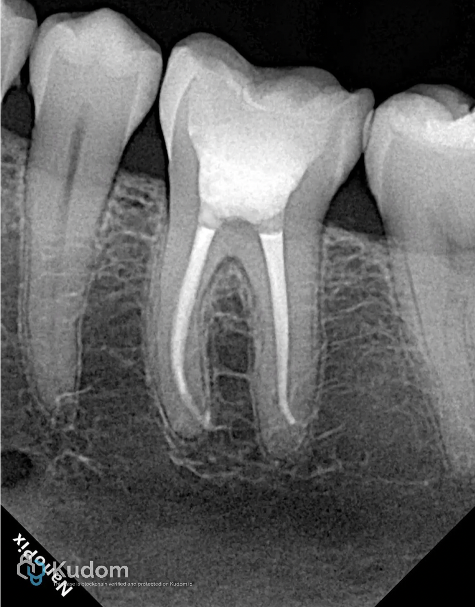
Fig. 16
Post endodontic restoration X-ray
Conclusions
In conclusion, the success of endodontic treatment does not rely solely on proper canal disinfection and obturation, but also on an effective post-endodontic restoration. Restoring the tooth appropriately enhances its structural integrity, prevents reinfection, and ensures long-term function. Therefore, a well-planned restorative approach is essential to maximize the prognosis and longevity of endodontically treated teeth.
Bibliography
- Sedgley, C. M., & Messer, H. H. (1992). Are endodontically treated teeth more brittle? Journal of Endodontics, 18(7), 332–335. https://doi.org/10.1016/S0099-2399(06)80483-8
- Linn, J., & Messer, H. H. (1994). Effect of restorative procedures on the strength of endodontically treated molars. Journal of Endodontics, 20(10), 479–485. https://doi.org/10.1016/S0099-2399(06)80043-9
- Dietschi, D., Duc, O., Krejci, I., & Sadan, A. (2007). Biomechanical considerations for the restoration of endodontically treated teeth: A systematic review of the literature—Part I. Composition and micro- and macrostructure alterations. Quintessence International, 38(9), 733–743.
- Dietschi, D., Duc, O., Krejci, I., & Sadan, A. (2008). Biomechanical considerations for the restoration of endodontically treated teeth: A systematic review of the literature—Part II. Evaluation of fatigue behavior, interfaces, and in vivo studies. Quintessence International, 39(2), 117–129.
- De Andrade, G. S., Saavedra, G. de S. F. A., Augusto, M. G., Leon, G. A., Brandão, H. C. B., Tribst, J. P. M., & Dal Piva, A. M. de O. (2023). Post-endodontic restorative treatments and their mechanical behavior: A narrative review. Dentistry Review, 3(1), Article 100067. https://doi.org/10.1016/j.dentre.2023.100067