Coalescence Phenomena of MB2 Canal
10/07/2025
Fellow
Warning: Undefined variable $post in /home/styleendo/htdocs/styleitaliano-endodontics.org/wp-content/plugins/oxygen/component-framework/components/classes/code-block.class.php(133) : eval()'d code on line 2
Warning: Attempt to read property "ID" on null in /home/styleendo/htdocs/styleitaliano-endodontics.org/wp-content/plugins/oxygen/component-framework/components/classes/code-block.class.php(133) : eval()'d code on line 2
The Presence of Second Mesio-buccal canal in Upper Molars is One of the most common scenarios of Anatomical Variations. According to Studies from Different Middle Eastern Countries, The prevalence of the MB2 Ranged from 57% to 74% In Upper First and Second Molars ( Ghobashy et al, 2017) with Vertucci Type II Configuration as the most common Type. The joining of The MB1 and MB2 canals in the coronal, middle, and apical third, respectively can range from 23 to 58 % (Al-Habib M et al, 2021 ) While Coalescence of MB1 and MB2 , as with our case , can occur after the process of cleaning and shaping turning The Vertucci Type II into Type I; Two Canals Merging into one .( Karobari et al, 2021)
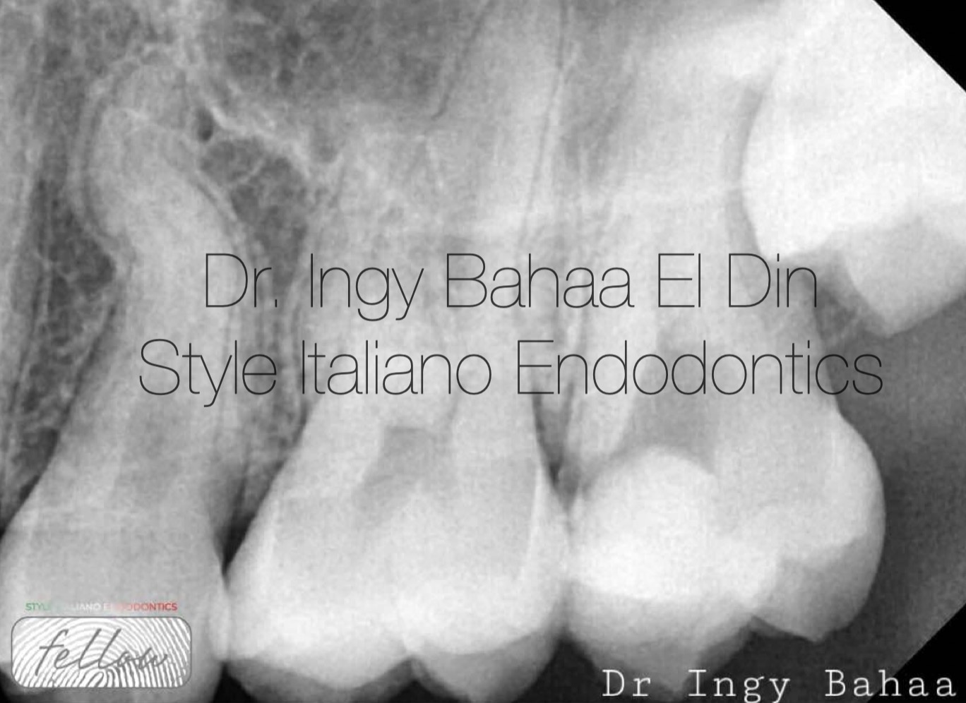
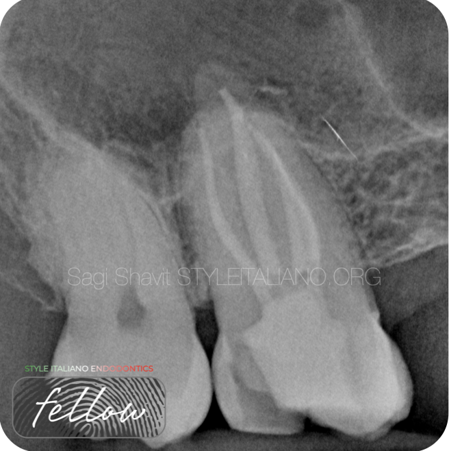
Fig. 1
Case: Referred 32 years old patient came to our clinic with lingering pain in her upper left area where she had an emergency access cavity done previously. we diagnosed her with irreversible pulpitis; cold testing and sensibility test was positive along with pain on percussion was also positive. A Periapical Radiographic Xray ( fig1 ) showed obvious widening in lamina dura around mesial and distal root and normal periapical tissue.
Non Surgical Root canal Treatment was decided and orthograde filling for UL7
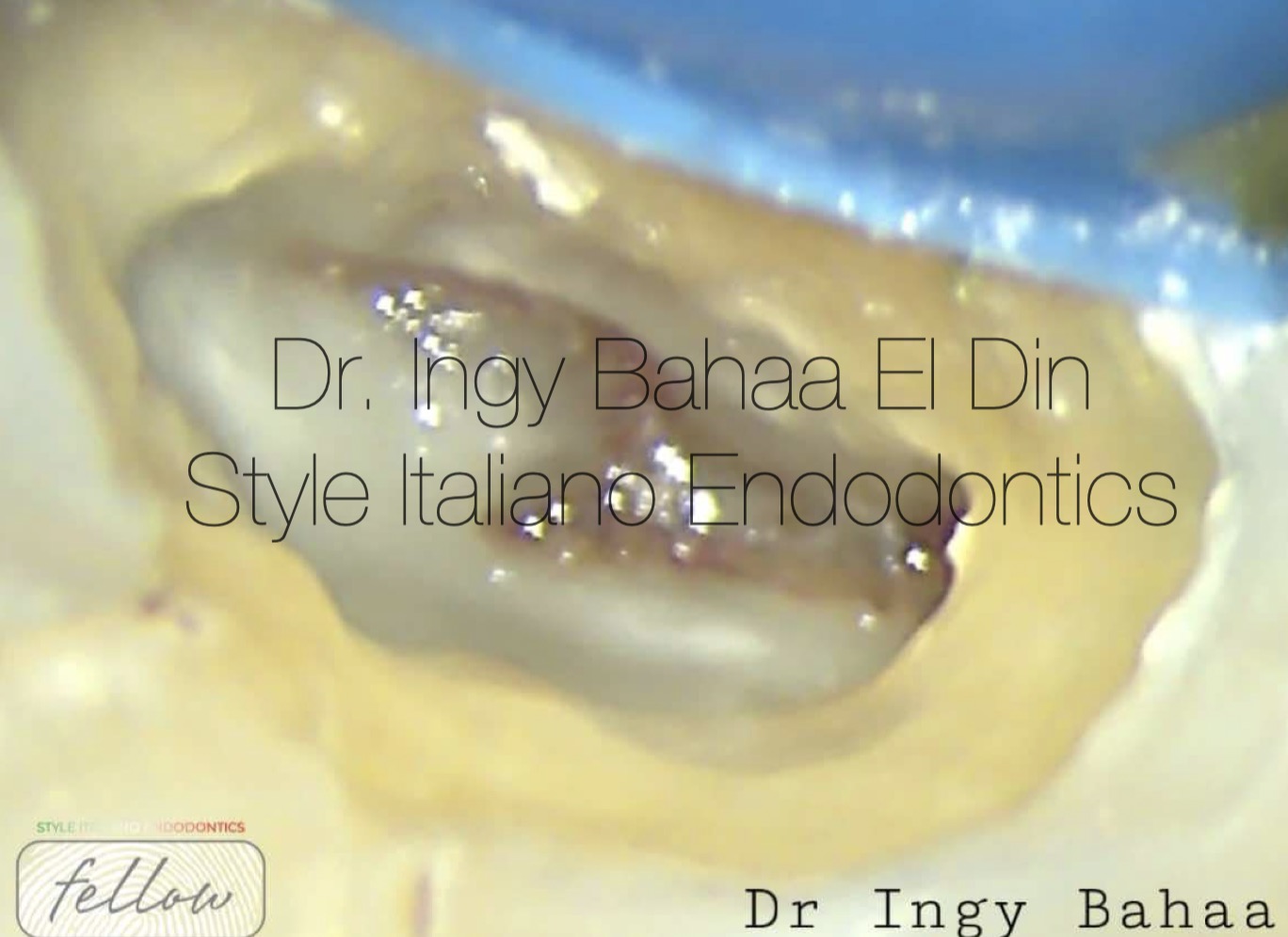
Fig. 2
Upon inspection using Semorr Microscope, Obvious pulp chamber roof needed more troughing to expose the dentin map
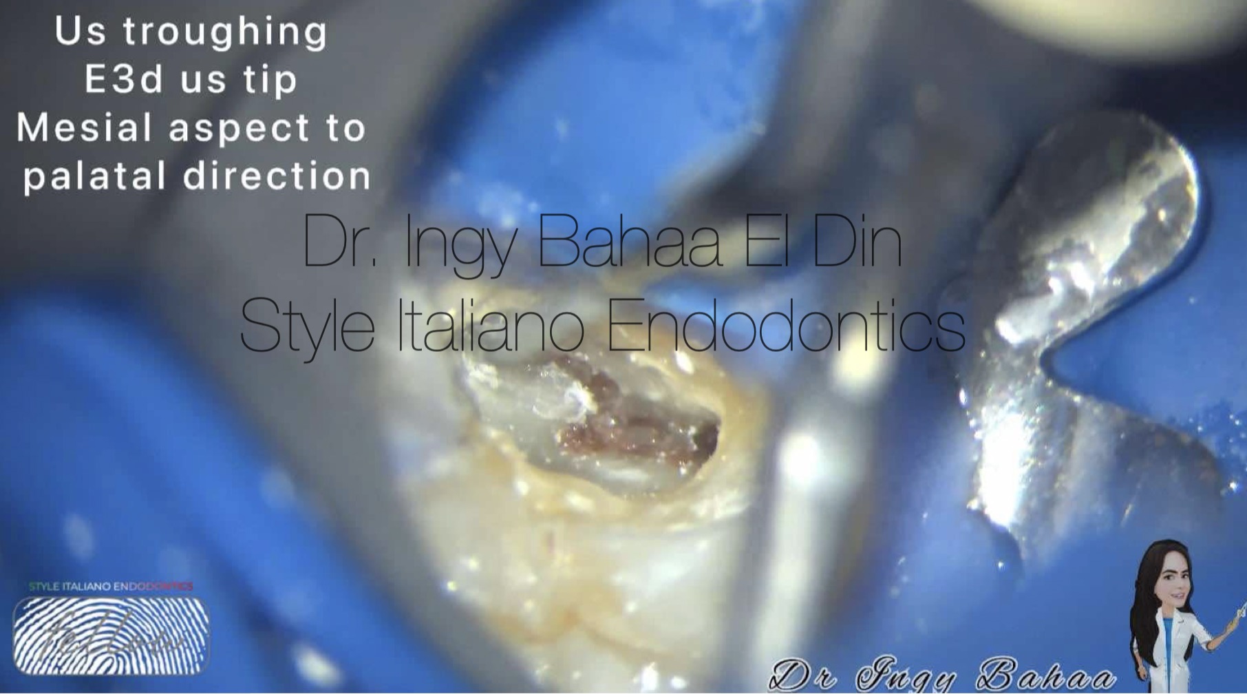
Fig. 3
Diamond Cutting Ultrasonic Tip from Woodpecker (E3 D)with brushing motion in the mesial aspect to Palatal Direction was used to ensure safe deroofing .
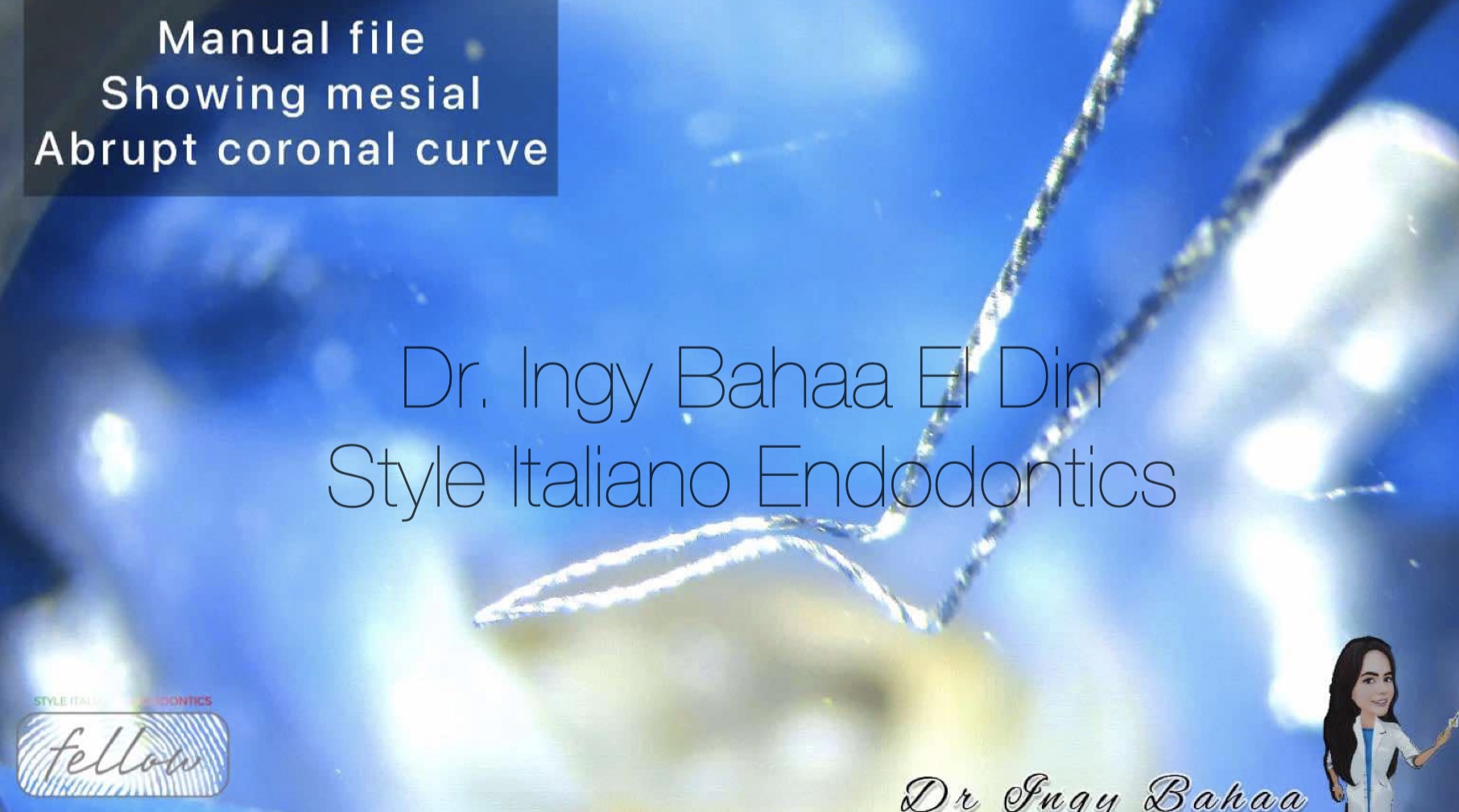
Fig. 4
Upon insertion of 10 k File manually inside Mb2 Orifice, File took an impression of the abrupt curve present coronally which is a common state in most Mb2 canals.(Tauby et al , 2012)
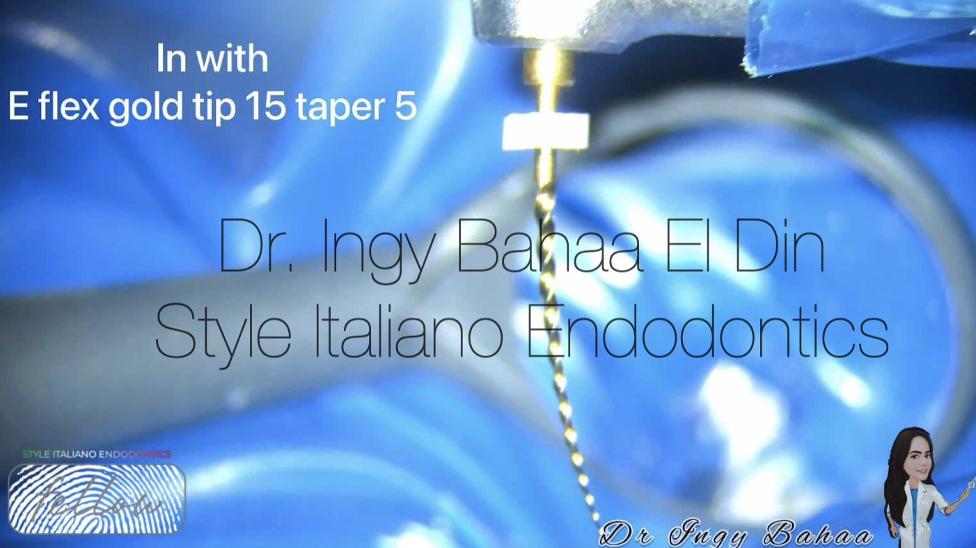
Fig. 5
To Overcome This dilemma, Flexible Niti Orifice Opener Taper 5 Size 15 ( Eflex Gold from Eighteeth ) was used in rotation motion in short strokes to allow for deeper gauging beyond the coronal curve
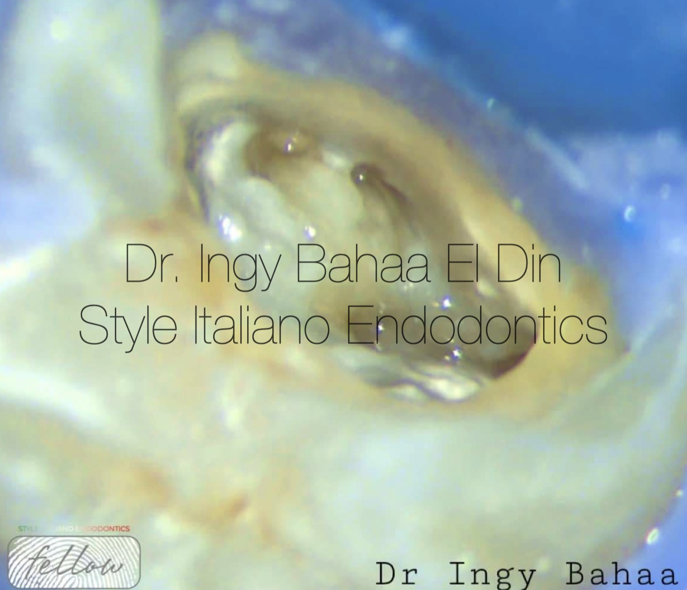
Fig. 6
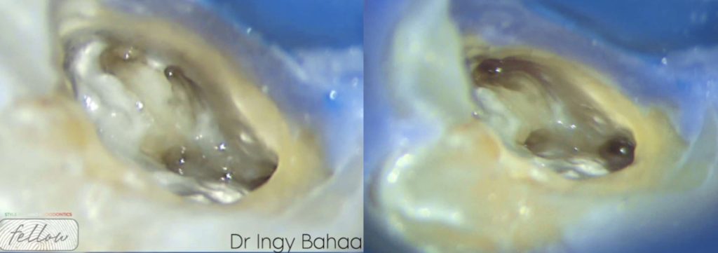
Orifices more clear, Four orifices present of all canals
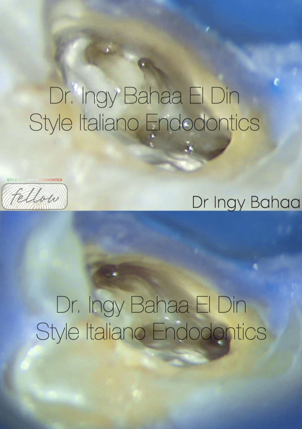
Fig. 7
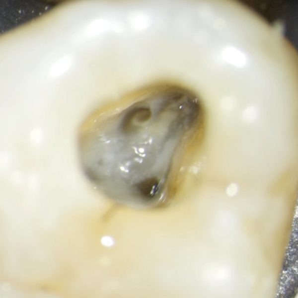
After Complete Cleaning and Shaping, Merging of the Two Canals ( Mb1 and Mb2 ) Occurred in the form of coalescence .
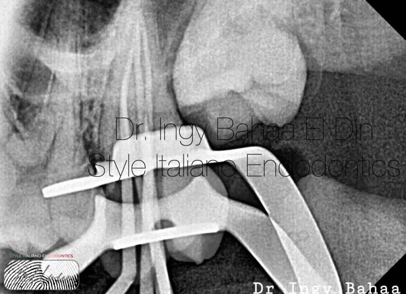
Fig. 8
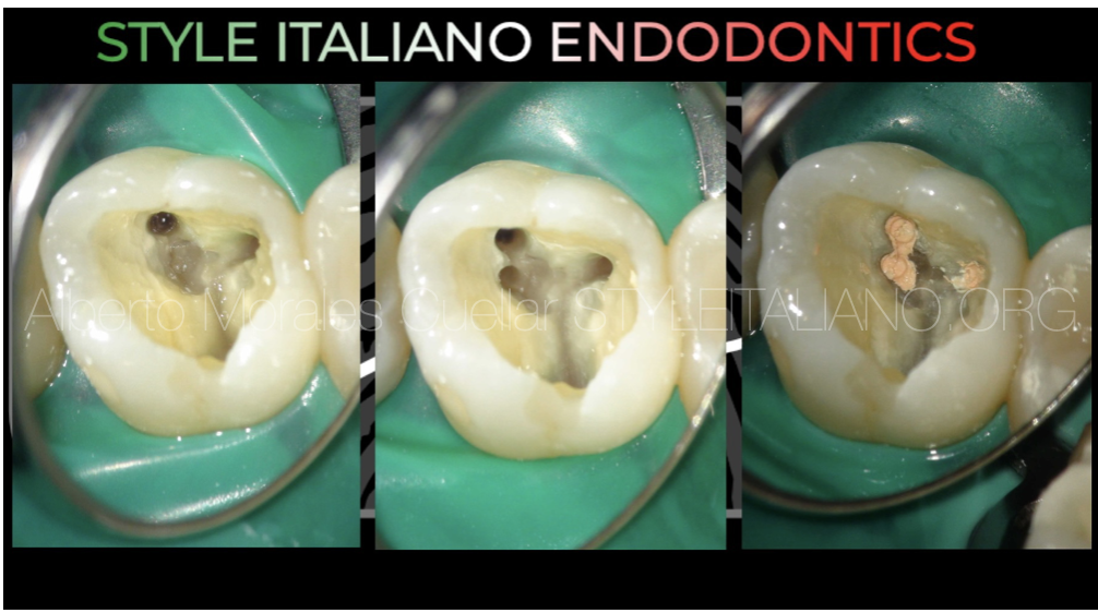
Master Cones adjustment
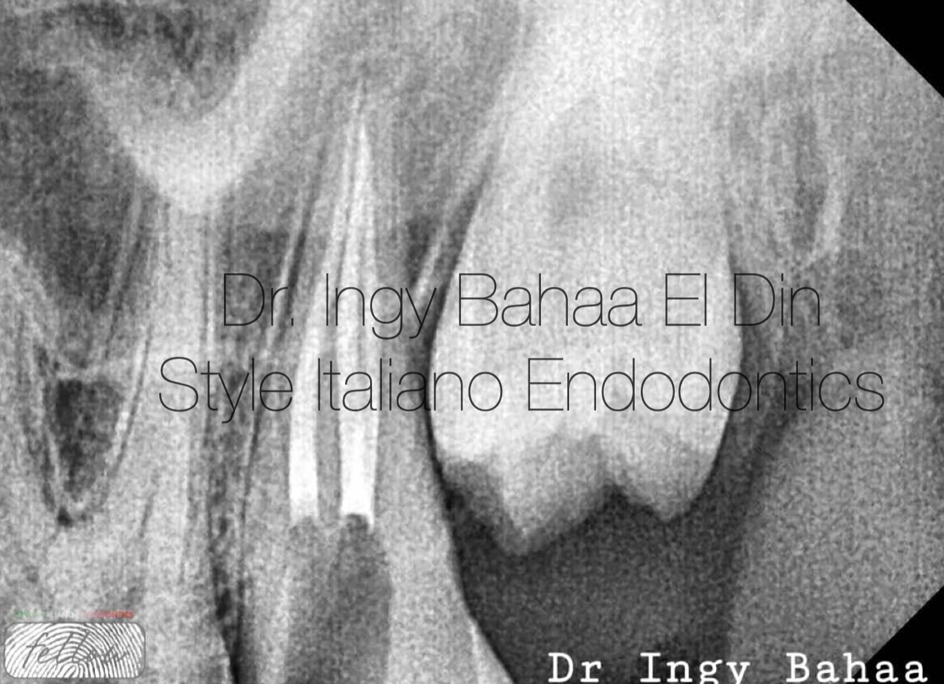
Fig. 9
Obturation was done using Warm vertical Compaction Technique with Bioceramic Sealer showing Palatal and Disto-Buccal Canal as Vertucci Type 2 ( One Apex, Two Canals )
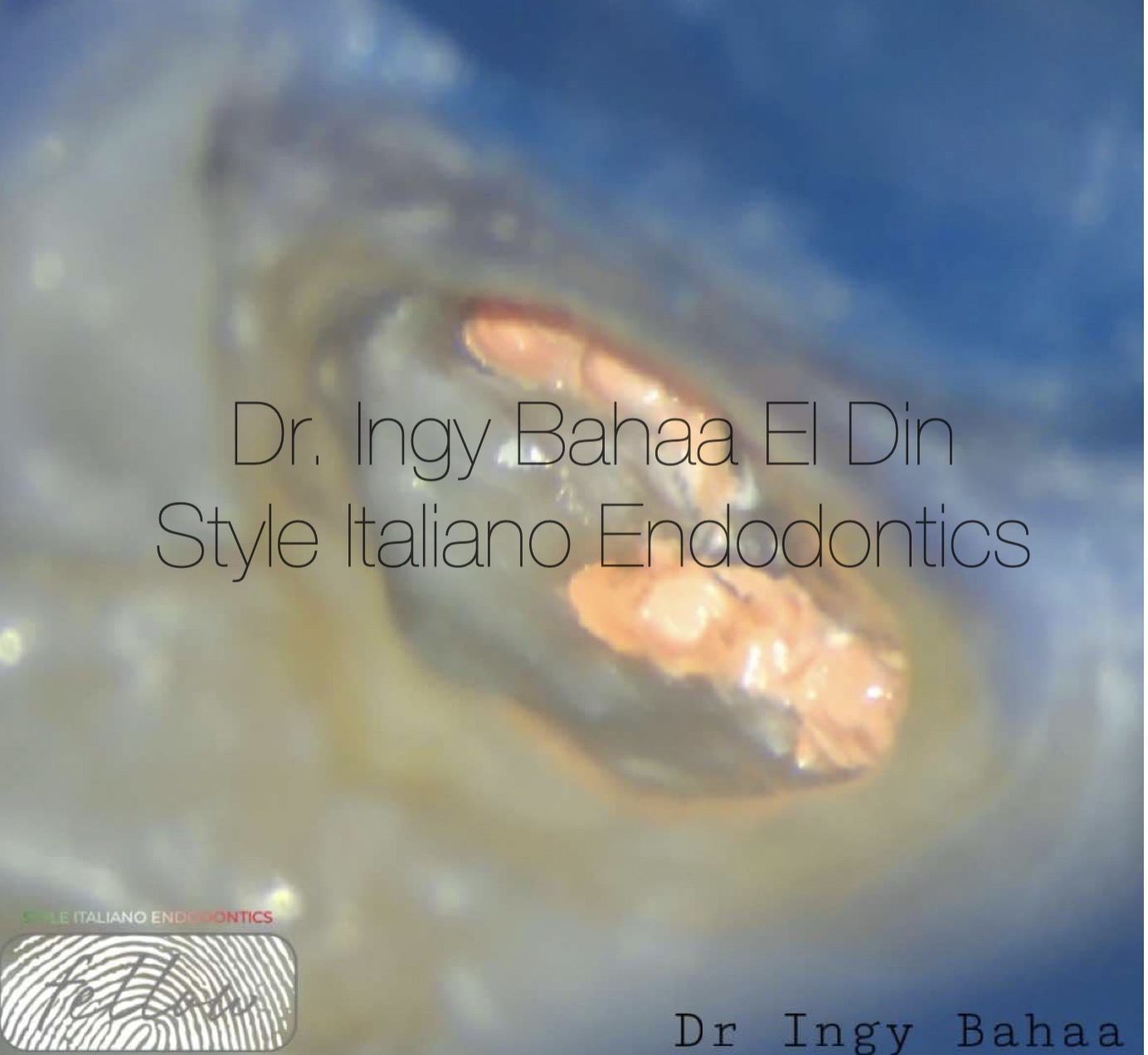
Fig. 10
Immediate Post Operative
Video of the procedure

Fig. 11
Dr. Ingy Bahaa El Din
Passion for Endodontics and Aesthetic Dentistry . Graduated with Bds from Future University in Egypt, 2011. Masters Degree Holder of Endodontics from Ain-shams University in Cairo, 2022. Co,owner and Head Operator of Beyond Dental Clinic in Cairo since 2015. Endodontic Specialist at EgyptAir Hospital. Conducting many One to One Endodontic Courses and Speaker in Multiple National and International Conferences.
Conclusions
Magnification along with Counter-thinking techniques can be of great assistance to the clinician in locating Second Mesio-Buccal Canals (Pattanshetti et al , 2008). Achieving Patency can be more of a challenge than locating the canal but with the right understanding of anatomy and how to negotiate curvatures this can be achievable. Complete cleaning and shaping of anatomical variations can lead the outcome of endodontic treatment into success as most failures occur due to missed canals (Wolcott J et al , 2005)
Bibliography
Wolcott J, Ishley D, Kennedy W, Johnson S, Minnich S, Meyers J. A 5 yr clinical investigation of second mesiobuccal canals in endodontically treated and retreated maxillary molars. J Endod. 2005 Apr;31(4):262-4
Pattanshetti N, Gaidhane M, Al Kandari AM. Root and canal morphology of the mesiobuccal and distal roots of permanent first molars in a Kuwait population--a clinical study. Int Endod J. 2008 Sep;41(9):755-62.
Tauby de Souza Coutinho-FilhoI; Eduardo Diogo Gurgel-FilhoII; Francisco José Souza-FilhoIII; Emmanuel João Nogueira Leal da SilvaIV. Preliminary investigation to achieve patency of MB2 canal in maxillary molars. Braz. J. Oral Sci. vol.11 no.3 Piracicaba Jul./Set. 2012
Ghobashy AM, Nagy MM, Bayoumi AA. Evaluation of Root and Canal Morphology of Maxillary Permanent Molars in an Egyptian Population by Cone-beam Computed Tomography. J Endod. 2017 Jul;43(7):1089-1092.
Vyas, Tarun. (2020). DEMYSTIFYING MB2 : A REVIEW. 8. 38-41
Karobari MI, Parveen A, Mirza MB, Makandar SD, Nik Abdul Ghani NR, Noorani TY, Marya A. Root and Root Canal Morphology Classification Systems. Int J Dent. 2021 Feb 19;2021:6682189.
Al-Habib M, Howait M. Assessment of Mesiobuccal Canal Configuration, Prevalence and Inter-Orifice Distance at Different Root Thirds of Maxillary First Molars: A CBCT Study. Clin Cosmet Investig Dent. 2021 Mar 24;13:105-111.