Below an old amalgam restoration. A case report
11/08/2020
Francesca Cerutti
Warning: Undefined variable $post in /home/styleendo/htdocs/styleitaliano-endodontics.org/wp-content/plugins/oxygen/component-framework/components/classes/code-block.class.php(133) : eval()'d code on line 2
Warning: Attempt to read property "ID" on null in /home/styleendo/htdocs/styleitaliano-endodontics.org/wp-content/plugins/oxygen/component-framework/components/classes/code-block.class.php(133) : eval()'d code on line 2
When removing large amalgam restorations we frequently observe that modifications have occurred in the pulp chamber, that often presents a diminished size, with a more difficult finding of the root canal openings.
The use of ultrasonic tips, together with the knowledge of the anatomy, can help in finding the orifices without making iatrogenic damages.
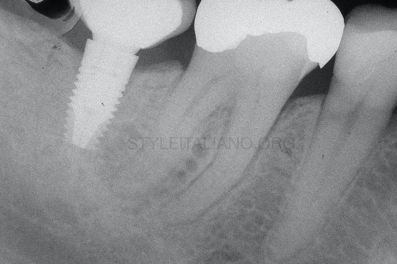
Fig. 1
A 54 years old patient came to me complaining about pain in the lower right area. The clinical examination showed the absence of pathologic probing and tenderness to percussion on the tooth 4.6. A periapical x-ray was taken and showed a slight enlargement of the periodontal space on the mesial root of the tooth 4.6 and the contraction of the pulp chamber.
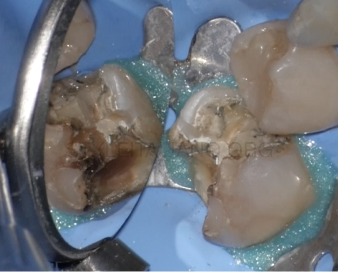
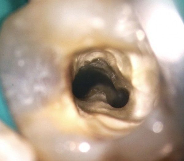
Fig. 2
After removing the amalgam, the cavity presented decayed tissue and several pigmented areas
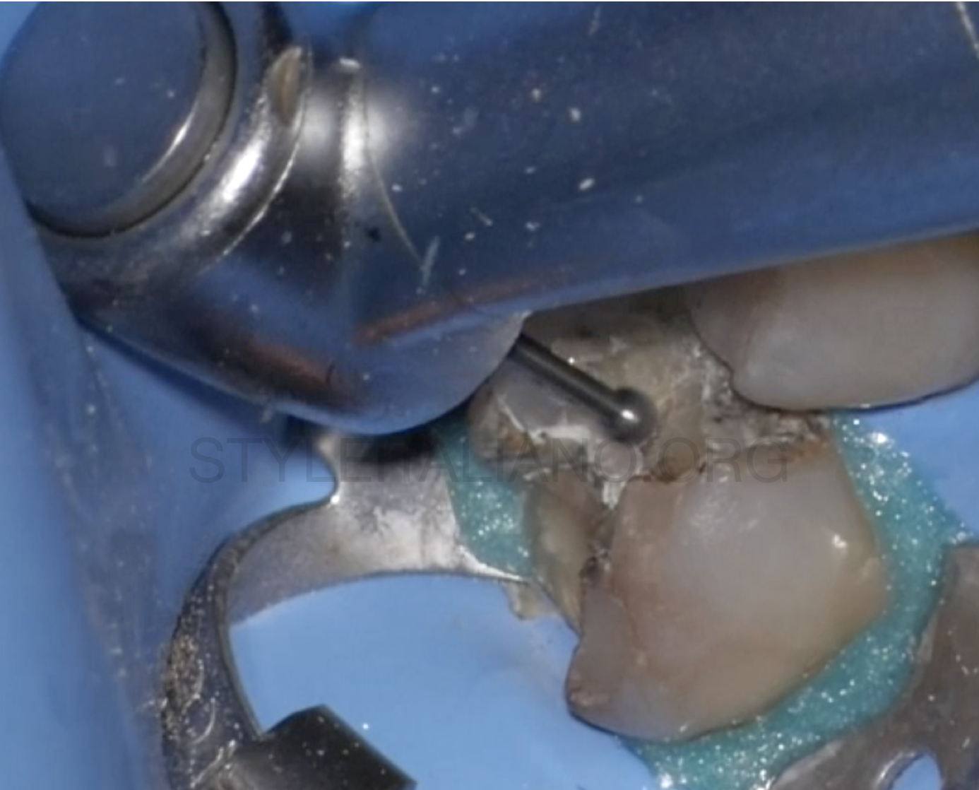
Fig. 3
The first step was to remove the decayed tissue with a low speed handpiece and a multiple blade bur
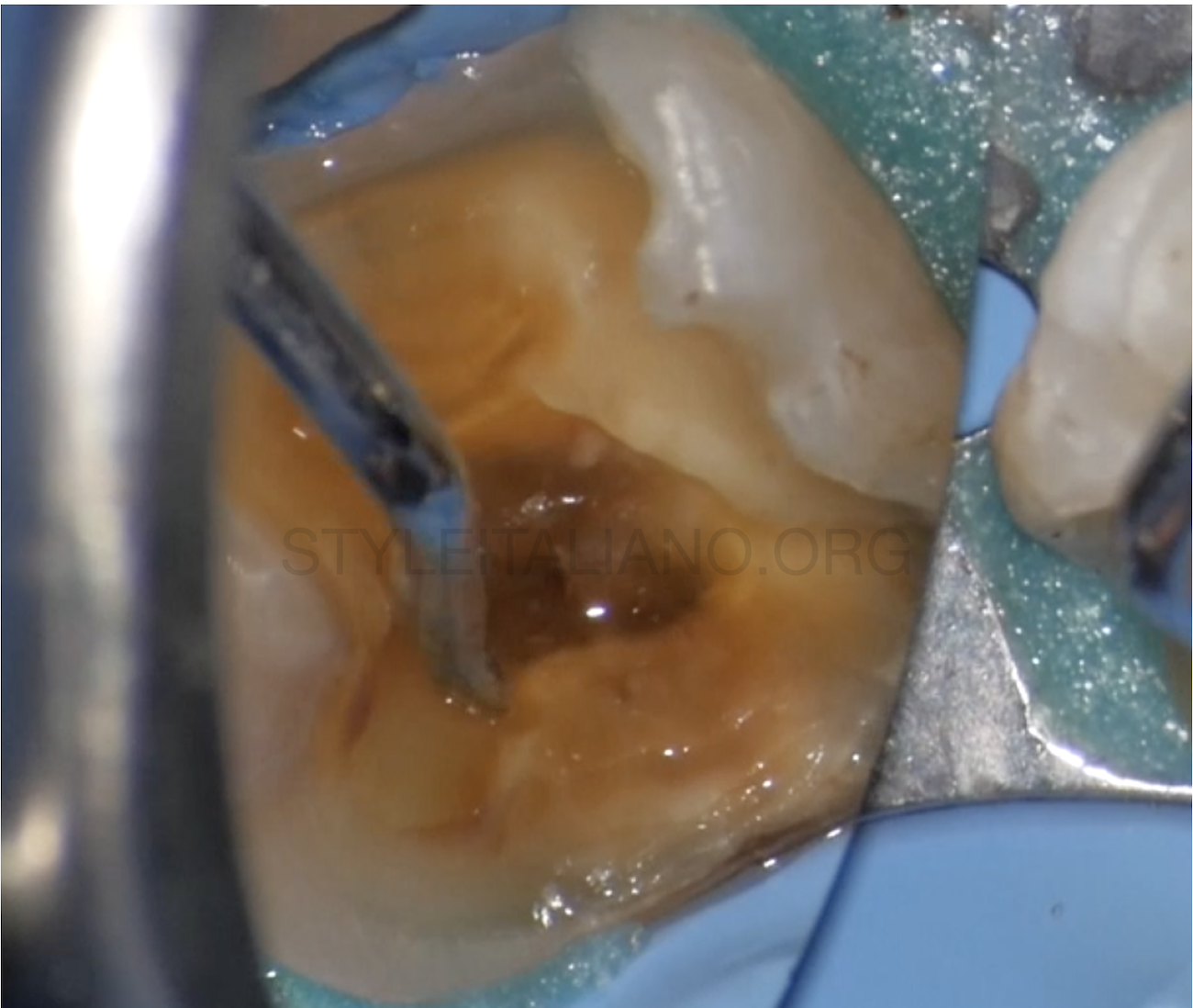
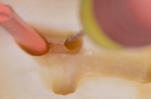
Fig. 4
After removing the majority of the pigmented and decayed tissue, an excavator was used to assess the quality of the dentin
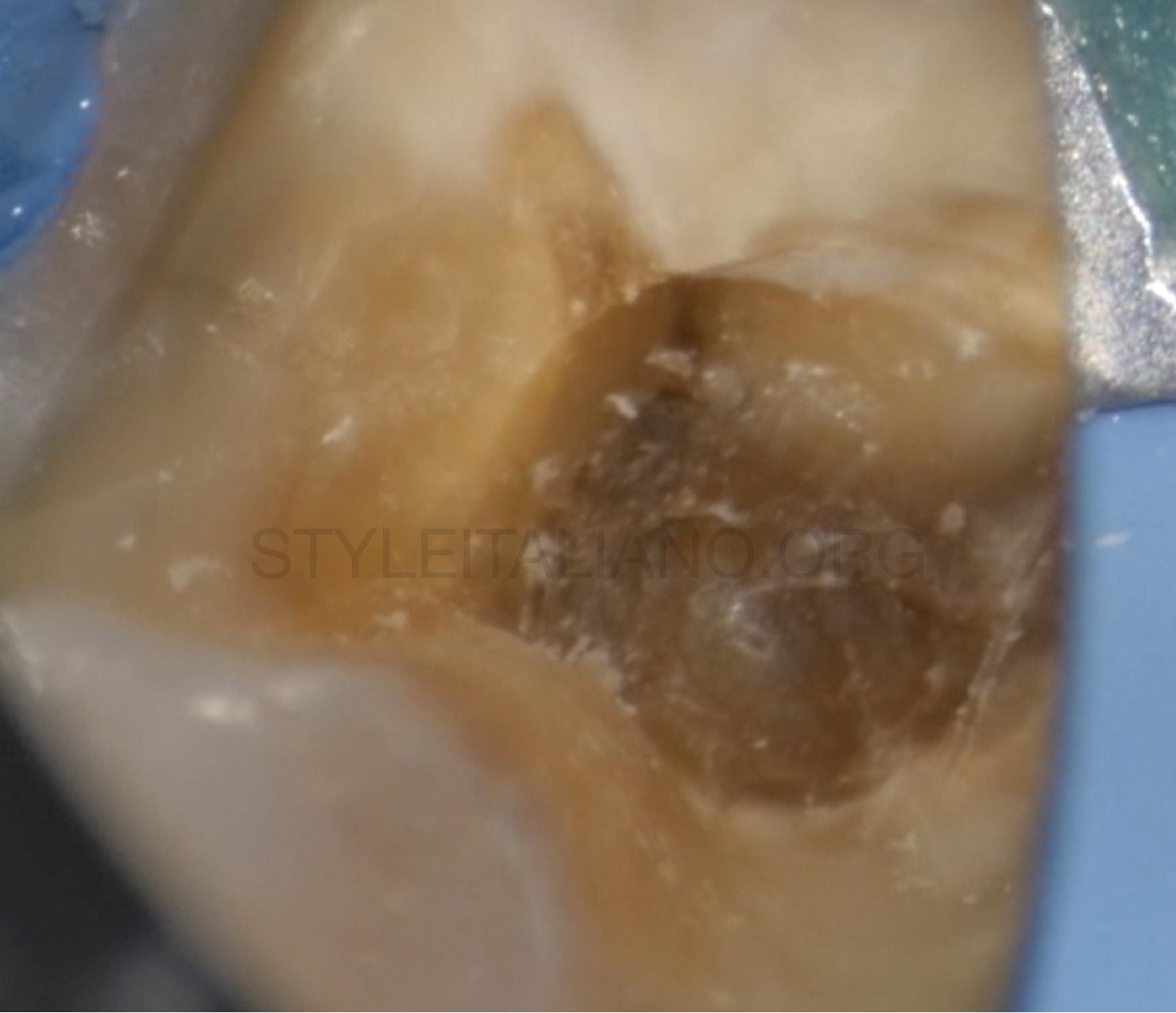
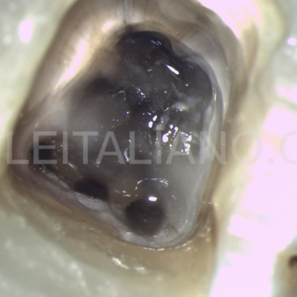
Fig. 5
The pulp chamber presented deposits of mineralized tissue on the root canal openings, even if the mesio-lingual and the distal canal openings can be seen.
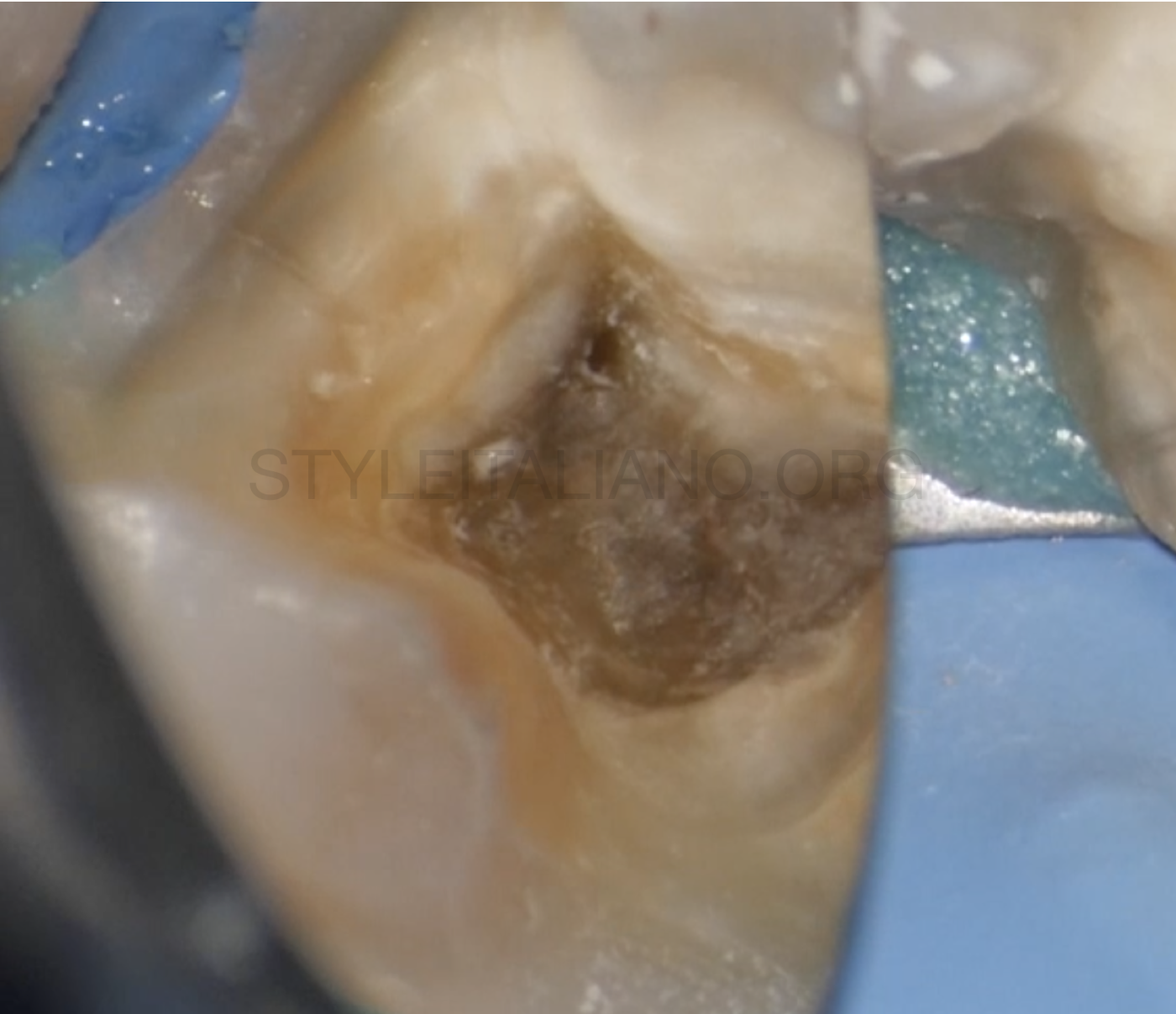
Fig. 6
After refining the access cavity with an ultrasonic diamond-coated tip, the mechanical scouting of the root canals began. The first canal to be approached was the distal, then it was to turn of the mesio-lingual.
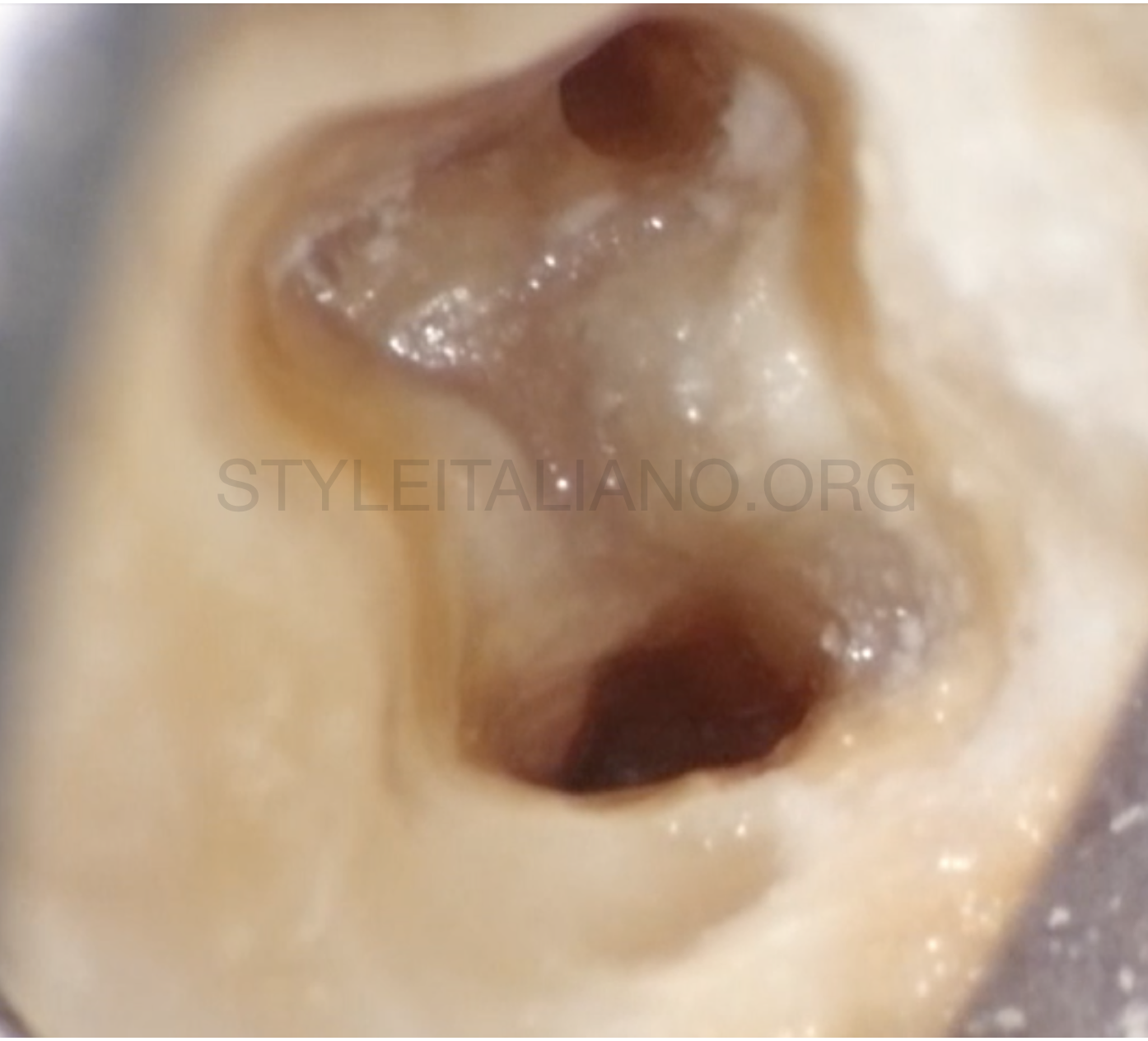
Fig. 7
The distal and the mesio-lingual canals were shaped and were ready to be filled, but the mesio-buccal had not been found yet. The knowledge of the anatomy and the observation of the dark lines present on the pulp chamber floor were the key to find the opening of the missing canal.
The missing canal is located, then it is possible to proceed with shaping and cleaning. Cleaning was done for 15' with 5% sodium hypochlorite activated by ultrasonic K-files.
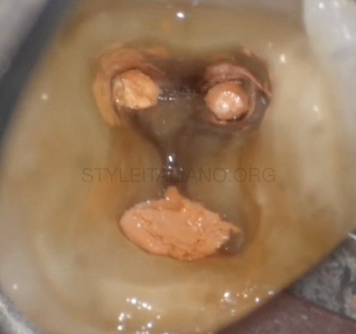
Fig. 8
Then the canals were dried with paper points and filled with warm gutta-percha.
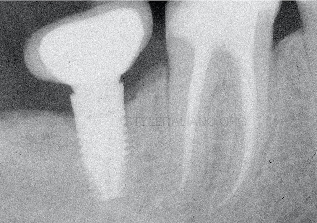
Fig. 9
A composite build-up was done and the final x-ray was taken.
Conclusions
In teeth with large restorations it is frequent to observe alterations of the anatomy of the pulp chamber: these can make it harder to find the root canal openings.
Following the color rules and the symmetry law can help in identifying the anatomy of the root canal system, avoiding iatrogenic damages.
Bibliography
McCabe PS, Dummer PM. Pulp canal obliteration: an endodontic diagnosis and treatment challenge. Int Endod J. 2012;45(2):177-97.
Kiefner P, Connert T, ElAyouti A, Weiger R. Treatment of calcified root canals in elderly people: a clinical study about the accessibility, the time needed and the outcome with a three-year follow-up. Gerodontology. 2017;34(2):164-70.
Mohammadi Z, Asgary S, Shalavi S, P VA. A Clinical Update on the Different Methods to Decrease the Occurrence of Missed Root Canals. Iranian endodontic journal. 2016;11(3):208-13.