A novel method for Fragment Reattachment After Post Placement
26/10/2015
Riccardo Tonini
Warning: Undefined variable $post in /home/styleendo/htdocs/styleitaliano-endodontics.org/wp-content/plugins/oxygen/component-framework/components/classes/code-block.class.php(133) : eval()'d code on line 2
Warning: Attempt to read property "ID" on null in /home/styleendo/htdocs/styleitaliano-endodontics.org/wp-content/plugins/oxygen/component-framework/components/classes/code-block.class.php(133) : eval()'d code on line 2
Fracture by trauma is one of the most common kind of dental injury in the permanent dentition among children and teenagers.If the original tooth fragment is retained following fracture line, the natural tooth structures can be reattached using adhesive protocols.
In this article we report one case of traumatized maxillary permanent central incisors, which were treated with novel method of Fragment reattachment, preserving coronal integrity after post placement.
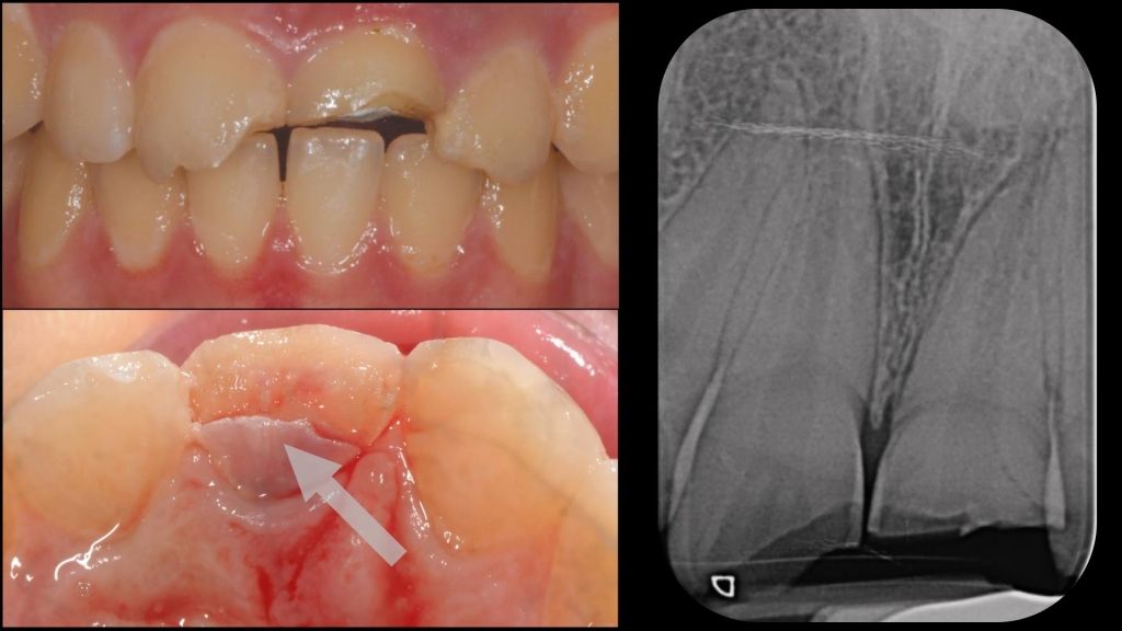
Fig. 1
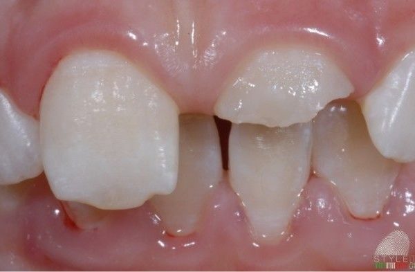
A nineteen year old young woman presented to our attention reporting acute pain at the level of 2.1. On oral examination the patient showed first of all a poor hygiene because pain prevented her from brushing properly.
The trauma had caused the fracture of coronal part of three teeth, 1.1, 2.1 and 2.2. No elements mobility has shown.Especially the 2.1 had a complicated fracture with pulp exposure and a palatal second fragment still present.
On investigating the intraoral periapical radiograph did not appear signs of root fractures and elements 1.1 and 2.2 responded positively to the pulp thermal testing that was performed on the labial surface.
Instead, the left central incisor, which had been treated by another dentist with a partial pulpotomy immediately after trauma, presented acute pain to percussion.
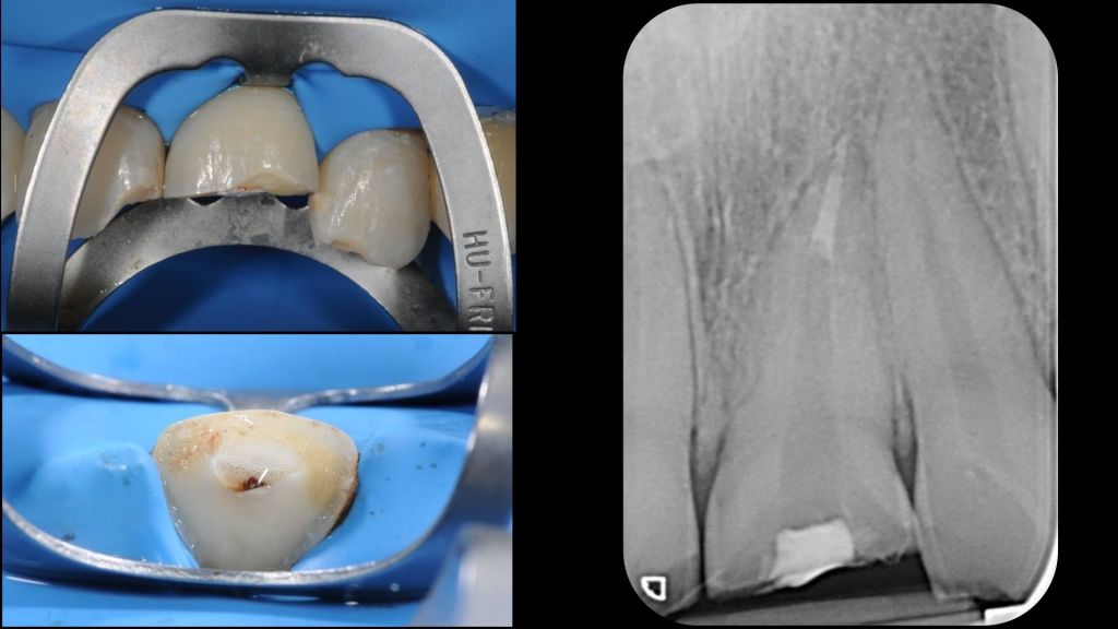
Fig. 2
We decided to treat this case performing the following operative plan:
Tooth 1.1: direct restoration with composite (fragment not available)
Tooth 2.1: Root canal treatment,fragments reattachment and fiber post.
Tooth 2.2: direct restoration with composite (fragment not available)
Especially for 2.1 element we have planned an innovative operative technique which will be described in this article.
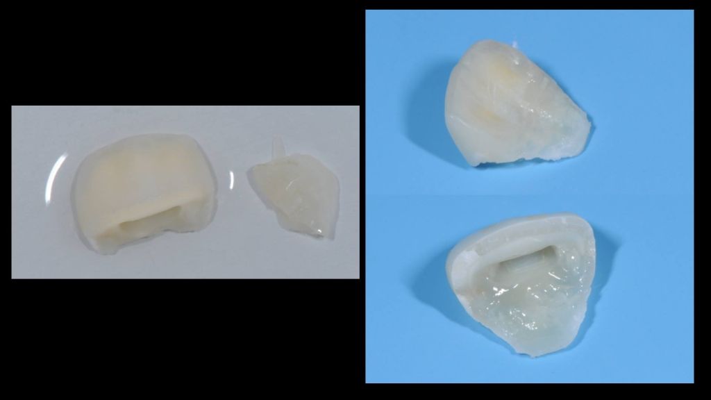
Fig. 3
All fragments were removed,cleaned and stored properly in saline water at 4 degrees.
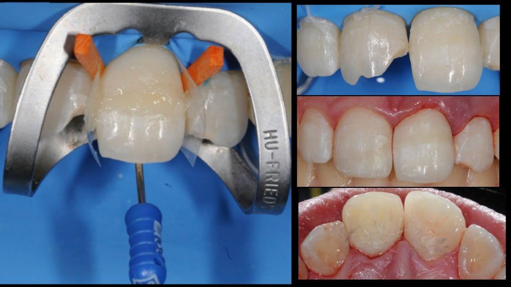
2)All elements were isolated with rubber dam positioning clamp on 2.1 under fracture line exposing all sound enamel.
3) Root canal treatment was performed granted al least 4 mm of apical guttapercha seal.
Shaping: Step back technique,Proglider and Protaper next X1.X2,X3,X4 (Dentsply Maileffer). Irrigation: Chlor Xtra 6% , EDTA 17%. Obturation:AH plus as sealer and Warm Guttapercha condensation.
4) Fragments were treated with conventional 3 steps adhesive technique and joined together polymerizing a thin line of flow composite placed in the between.
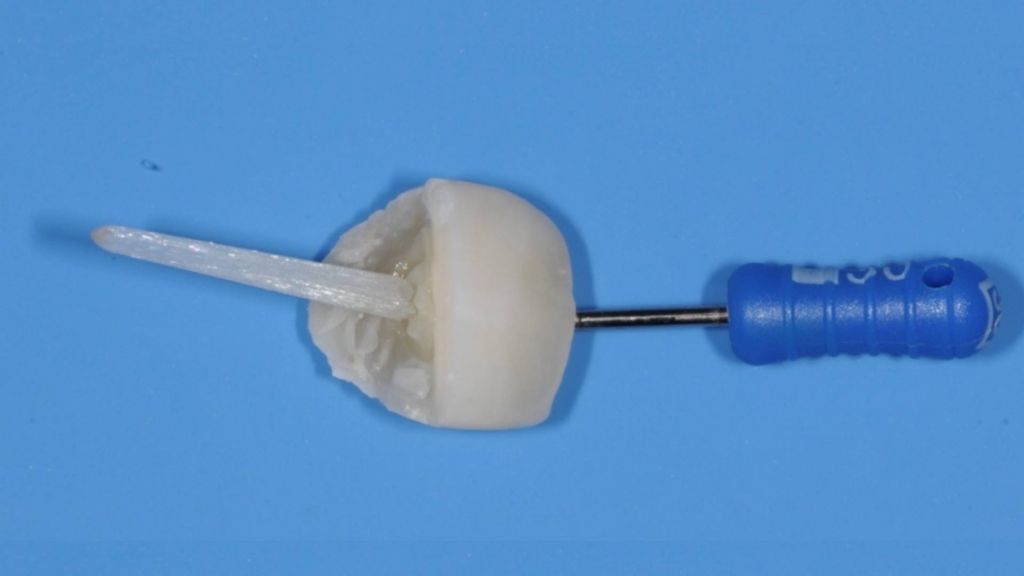
Fig. 4
5) For a better holder of the crown and to avoid any separation during luting technique a small amount of composite was positioned on palatal side, above fracture line, with a K file holder merged inside.
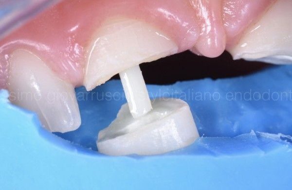
6) Root canal was cleaned and prepared with a largo bur number 3. At this point fiber post length was tested cutting the post head till that crown fragment fitted perfectly with post and tooths margins at same time. After that post was treated with silane,adhesive and bonded by the head inside fragment pulp chamber with composite,without polymerizing and without doing any hole in the crown.

Fig. 5
7)Root canal was prepared with 3 steps adhesive technique and adhesive was mixed with activator for a chemical setting. We selected a dual cure composite and we positioned it with a micro cannula starting from the bottom avoiding in this way any void.
8) Fragments with post were positioned removing all composite excess before polymerization. The k file holder was removed and afterwards,
finishing and polishing steps were performed.
Elements 1.1 and 2.2 were restored with composite during the same session.
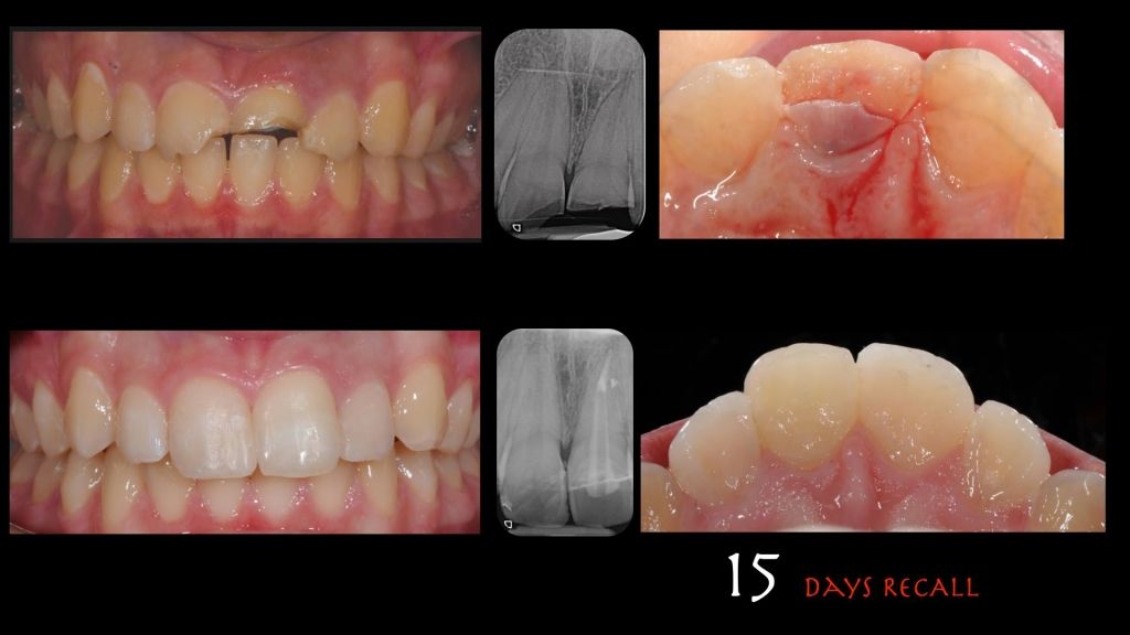
Fig. 6
15 DAYS RECALL
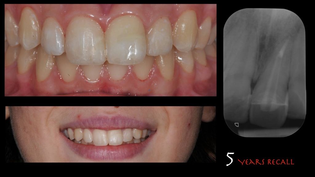
Fig. 7
5 YEARS RECALL


