Treatment of a cracked tooth
06/05/2021
Fitim Shabani
Warning: Undefined variable $post in /var/www/vhosts/styleitaliano-endodontics.org/endodontics.styleitaliano.org/wp-content/plugins/oxygen/component-framework/components/classes/code-block.class.php(133) : eval()'d code on line 2
Warning: Attempt to read property "ID" on null in /var/www/vhosts/styleitaliano-endodontics.org/endodontics.styleitaliano.org/wp-content/plugins/oxygen/component-framework/components/classes/code-block.class.php(133) : eval()'d code on line 2
One of the most undesirable situations for us dentists is the diagnosis of cracked teeth, as well as their treatment.
This happens because diagnosing tooth micro-fractures, which are usually invisible (not even seen on the Xray), when they are below the level of the gums complicates our situation.
If the crack reaches the pulp chamber of the tooth, the pulp tissue becomes exposed to bacteria, and gets inflamed developing a tooth infection.
Do we have to treat a cracked tooth or to extract it? It depends on some conditions which must be clarified before the treatment: the location of the microfracture, its depth, the general condition of the tooth (damaged structure or not, the presence of decay, etc.)
In this article I will present a clinical case where I treated a cracked tooth
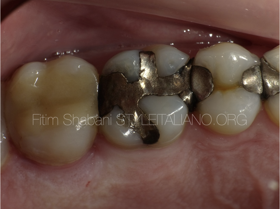
Fig. 1
Clinical data
Tooth: First maxillary left molar
The patient had pain during the bite
Tooth vitality negative
Large amalgam filling
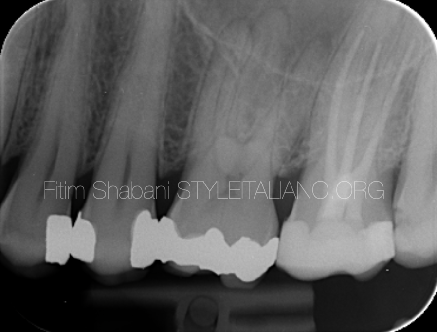
Fig. 2
Xray
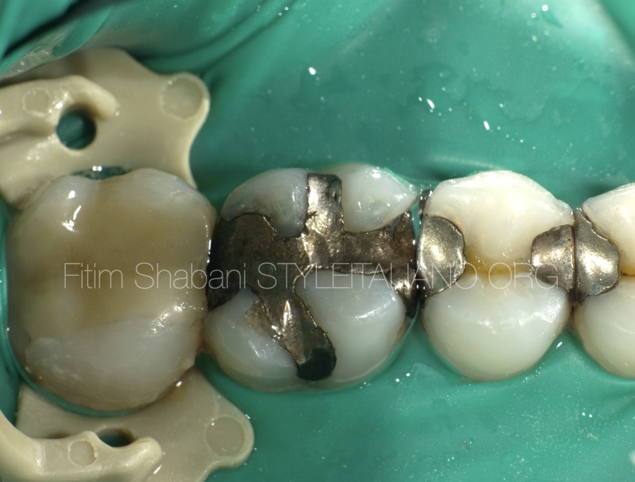
Fig. 3
Isolation
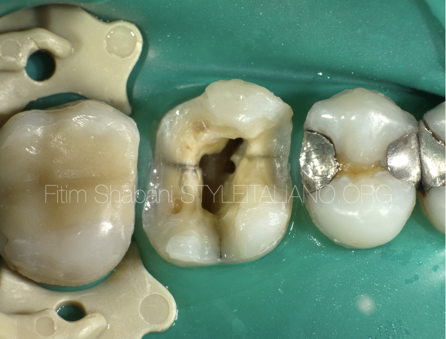
Fig. 4
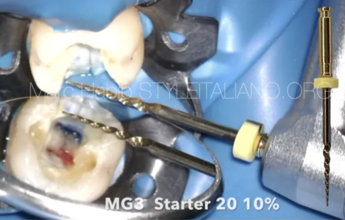
Remove of the amalgam filling and root canal treatment was performed.
A medication was placed in the canals and a provisional filling was executed.
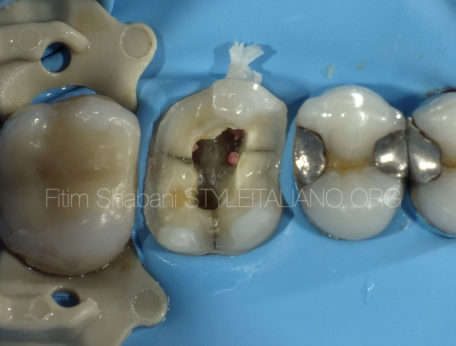
Fig. 5
One month later, when the patient was without complaints, we performed tooth obturation
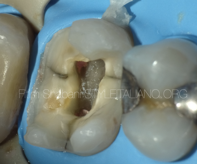
Fig. 6
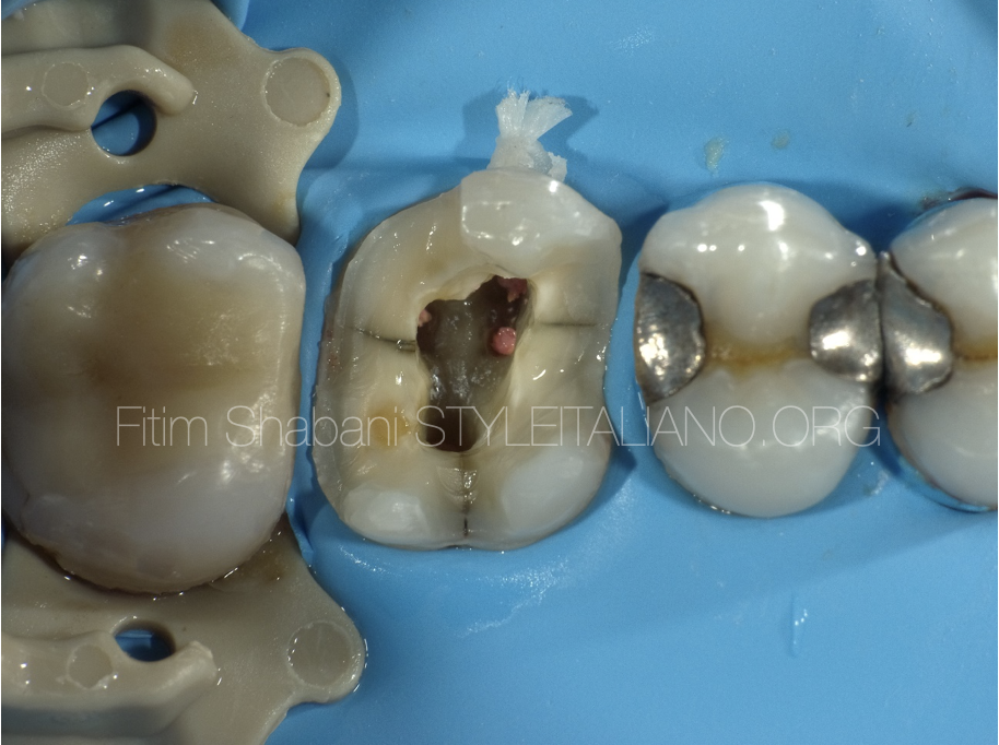
The image shows the distal line of the micro-fracture, which does not reach the pulp chamber of the tooth
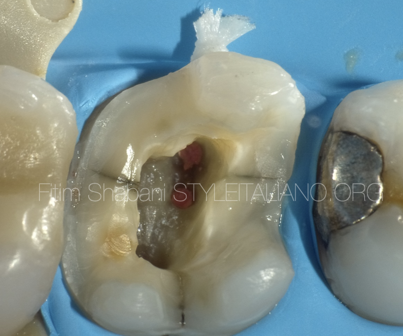
Fig. 7
The mesial line of the micro-fractures does not either reach the pulp chamber.
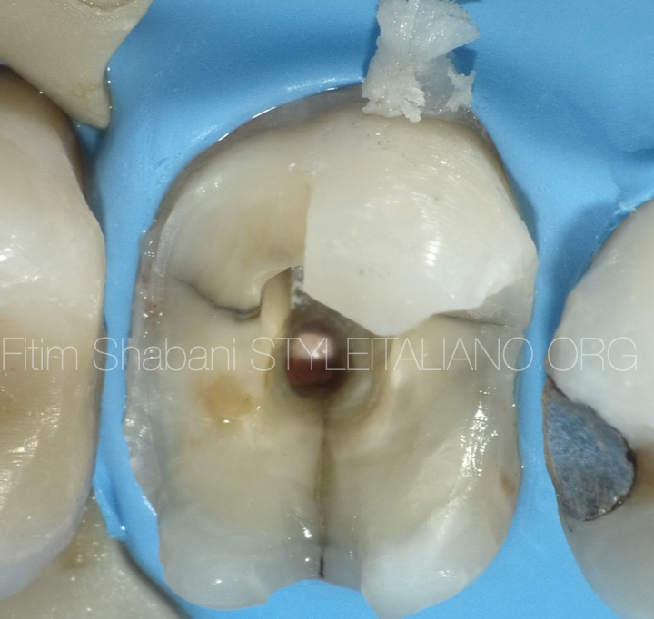
Fig. 8
The palatal line of the microfractures goes a little deeper compared to the mesial and distal line.
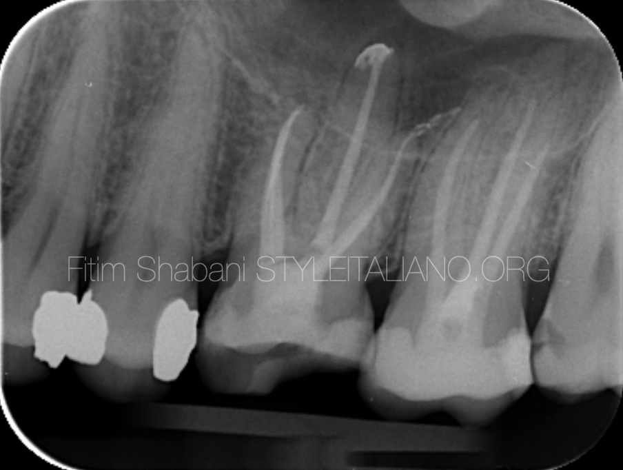
Fig. 9
Xray after obturation
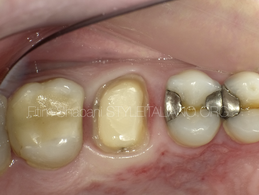
Fig. 10
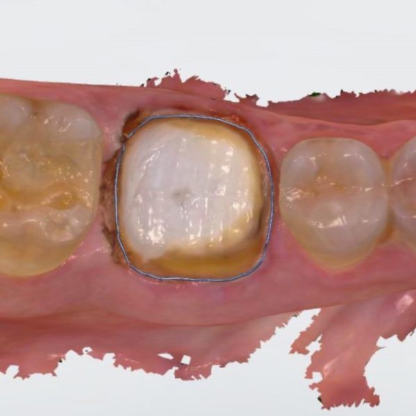
Third visit – Tooth preparation
The microfracture lines are not found in the margin of the prepared tooth
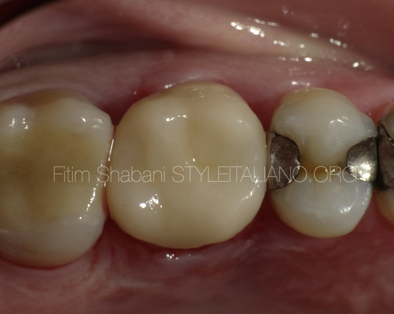
Fig. 11
After cementation of the Crown
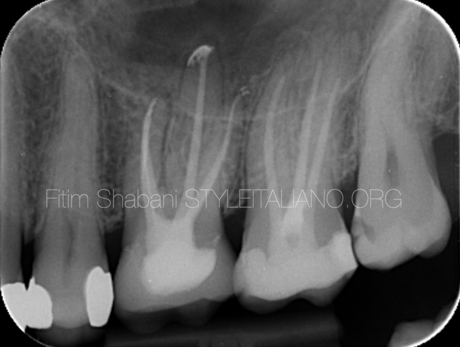
Fig. 12
Xray after cementation
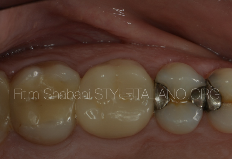
Fig. 13
Follow up at one and half year
The patient is without complaints
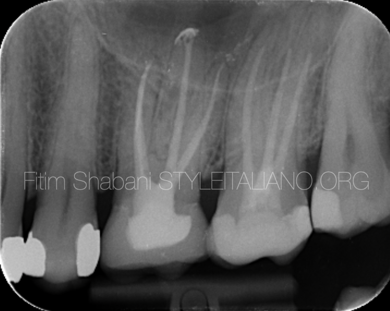
Fig. 14
Xray after one and half year
There are no pathological changes
Conclusions
Cracked tooth syndrome is a challenge for the practitioner.
In order to be successful, the treatment of cracked needs to be preceded by data analysis and proper treatment planning.
The use of the operative microscope is extremely helpful in managing these cases.
Bibliography
Sebeena Mathew, Boopathi Thangavel, Chalakuzhiyil Abraham Mathew, SivaKumar Kailasam, Karthick Kumaravadivel, Arjun Das, Diagnosis of cracked tooth syndrome, Journal of Pharmacy and Bioallied Sciences Vol 4 August 2012
William Khaler, Mscdent, Dclindent, Fracds The cracked tooth conundrum: Terminology, classification, diagnosis, and management, American Journal of Dentistry, Vol. 21, No. 5, October, 2008
John S. Mamoun, Donato Napoletano. Cracked tooth diagnosis and treatment: An alternative paradigm, European Journal of Dentistry, Vol 9/I ssue 2/Apr- Ju n 2015
Christopher D. Lynch, BDS, MFDRCSI, Robert J. McConnell, BDS, PhD, FFDRCSI The cracked tooth Syndrome, Can Dent Assoc 2002; 68(8):470-5


