Challenging Endo-resto of a central incisor
29/01/2021
Federico Ceroni
Warning: Undefined variable $post in /home/styleendo/htdocs/styleitaliano-endodontics.org/wp-content/plugins/oxygen/component-framework/components/classes/code-block.class.php(133) : eval()'d code on line 2
Warning: Attempt to read property "ID" on null in /home/styleendo/htdocs/styleitaliano-endodontics.org/wp-content/plugins/oxygen/component-framework/components/classes/code-block.class.php(133) : eval()'d code on line 2
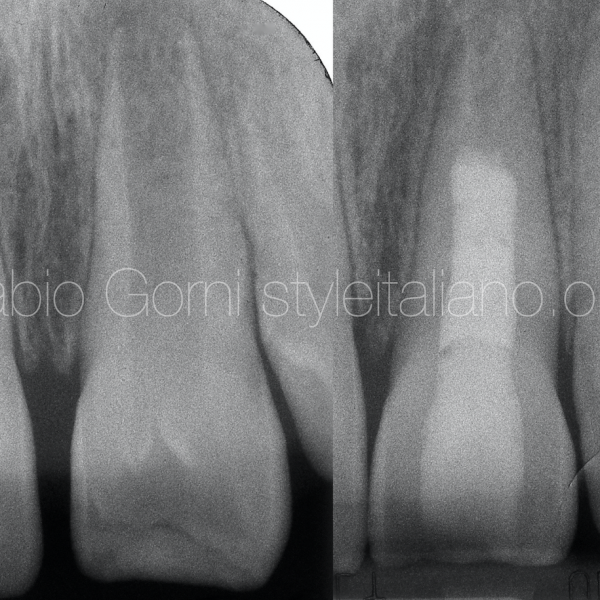
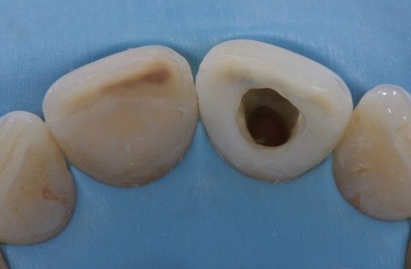
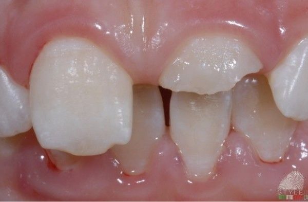
A 10 yrs old female came to our office due to a trauma he had some months before. The involved tooth was a non vital immature permanent upper incisor: the intra-oral examination showed swelling and an unaesthetic restoration, while radiographic examination showed open apex with incomplete root formation.
One of the main objective in case of incomplete root formation is to re-establish the blood flow allowing the continuation of the root development. So a revascularization technique (an alternative to the traditional apexification) was performed, using calcium hydroxide for a period of time in order to induce a formation of calcified apical barrier .
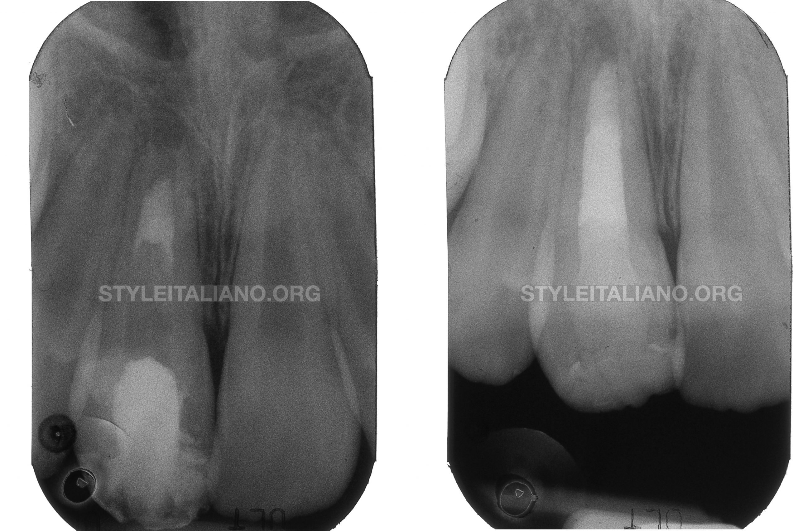
Fig. 1
After one month , in a second appointment , the canal was irrigated with saline solution and the periapical tissue , beyond the apex , instrumented with manual files to encourage the bleeding. Afterwards a fibrin sponge was inserted in the apical one third 3-4 mm to the foramen to allow blood clot formation . After few minutes the protocol was followed by the application of a mta plug over the clot . A flow composite temporary filling closed the access.
Unfortunately at 1 year control the apex wasn’t closed so we decided to remove the mta and finish the treatment closing the apex with an MTA apical plug and two day after the coronal part of the root was filled with guttaperca. The last appointment was dedicated to replace the old restoration.
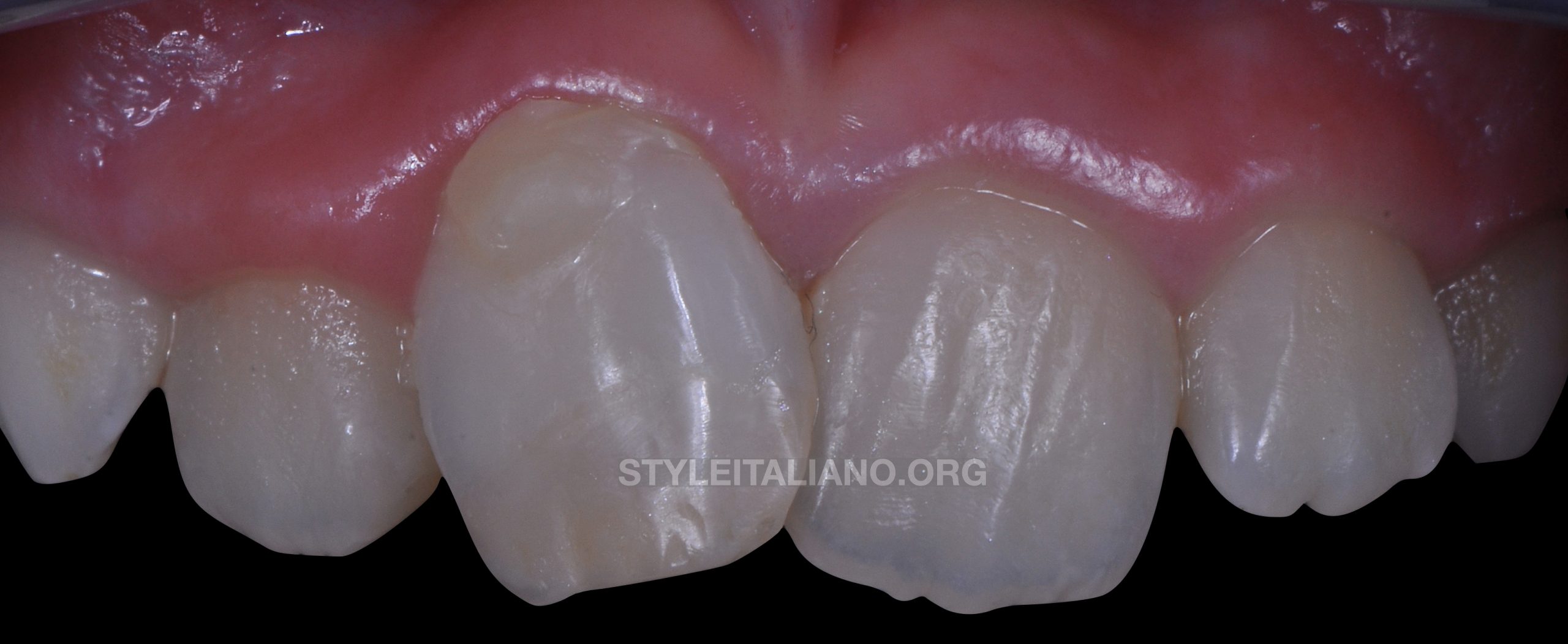
Fig. 2
The restoration of endodontically treated anterior teeth presents in general different mechanical and esthetic challenges. Several aspects have to be considered, such as:
Remaining Tooth structure ( Presence of enamel, number of remaining walls, quality of dentin )
Patient age
Functional loads
Esthetic demands
Direct or indirect restorations with or without post placement should be chosen considering all these aspects.
In this particular case I decide to restore the tooth with a direct restoration without any post, this because the remaining structure was enough to resist at the functional loads and, considering the age of the patient, was the least invasive solution
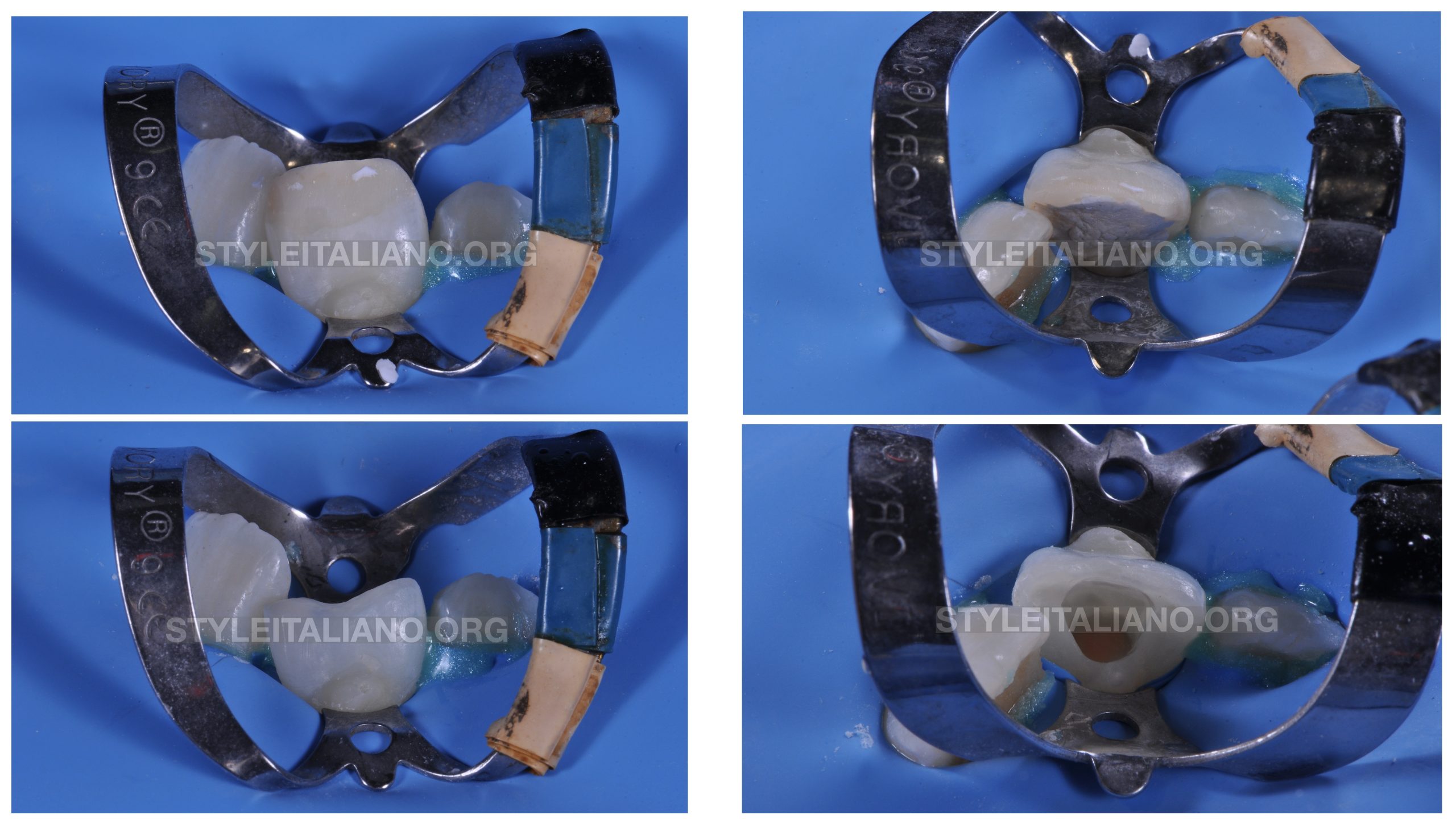
Fig. 3
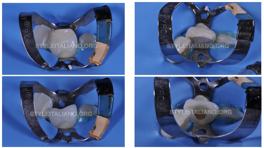
After the rubberdam placement the old composite restoration was carefully removed using a cylindrical medium-grain bur and the margins were clean and smoothened using a silicon bur.

Fig. 4
A total etch adhesive procedures was performed,
Etching for 30 sec the enamel and 15 sec the dentin.
Then a universal adhesive was placed
lightcuring for at least 30 sec.
After that we can start with the stratification technique following the principles of the one described by Lorenzo Vanini.
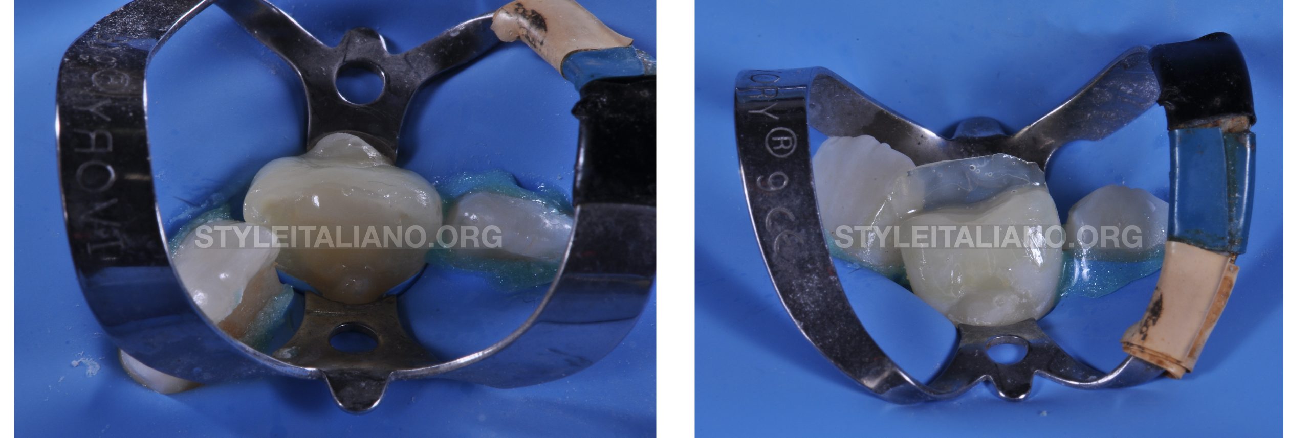
Fig. 5
The pulp chamber was filled with a dentin composite to replace the chromatic effect given in a natural tooth by the dentin
The palatal shape was built using a silicone key packaged on a diagnostic wax-up and an enamel composite was used for this step.

Fig. 6
A dentin composite layer was used to recreate the mamelons, leaving the space for the characterizations.
A trasparent composite and some stains were used to recreate all the effect of the incisal margin of the tooth.
And in the end a translucent composite was used to recreate the enamel layer. That’s because the enamel structure has a highly translucent appearance.
The final light curing is 60 seconds long, and is better performed using an air-blocking system. This procedure will provide the composite resins’s complete polymerization, improving surface resistance and giving better polishing performances.
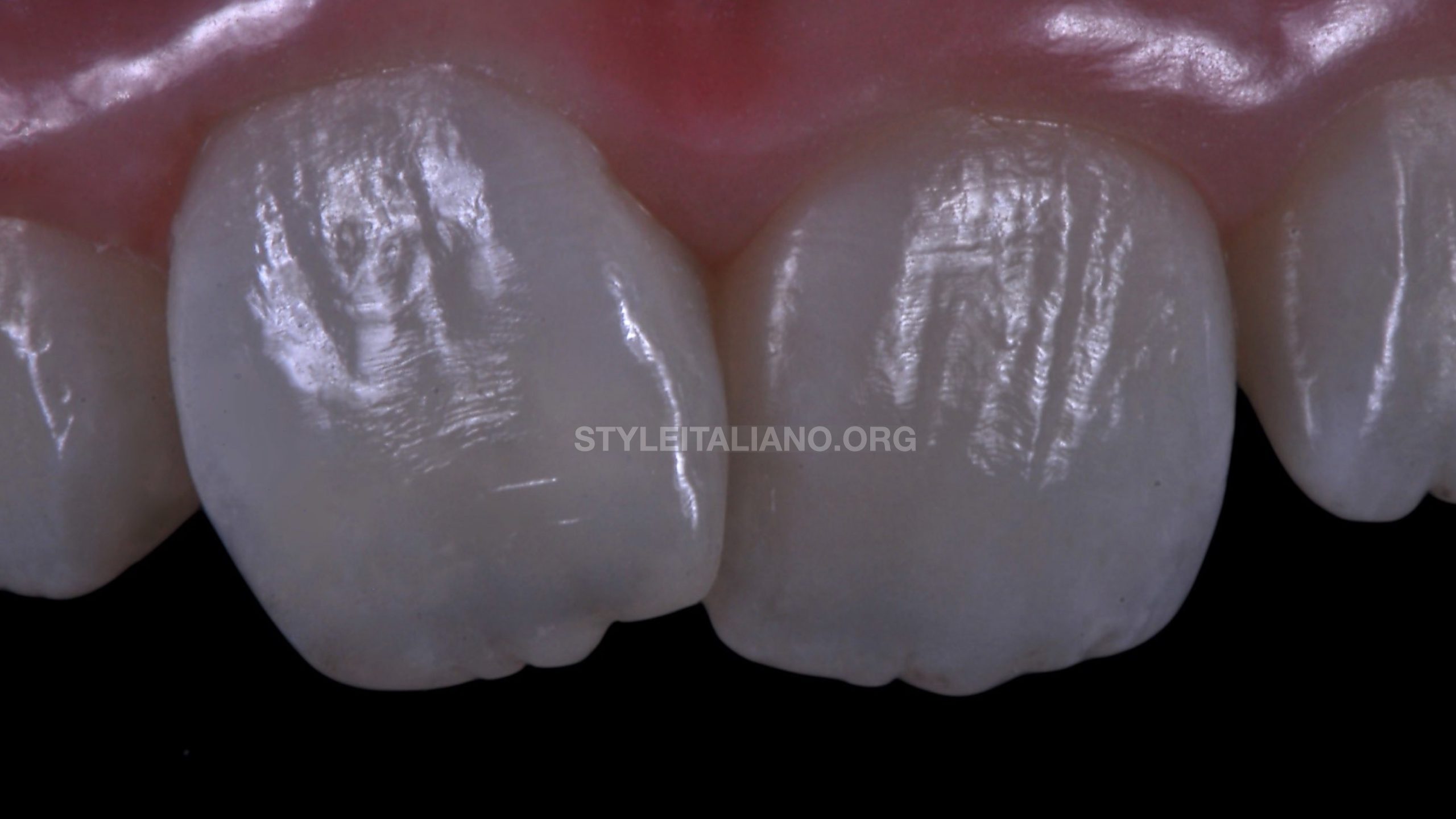
Fig. 7
This is the final result after rehidratation of the tooth and after all the finishing and polishing steps
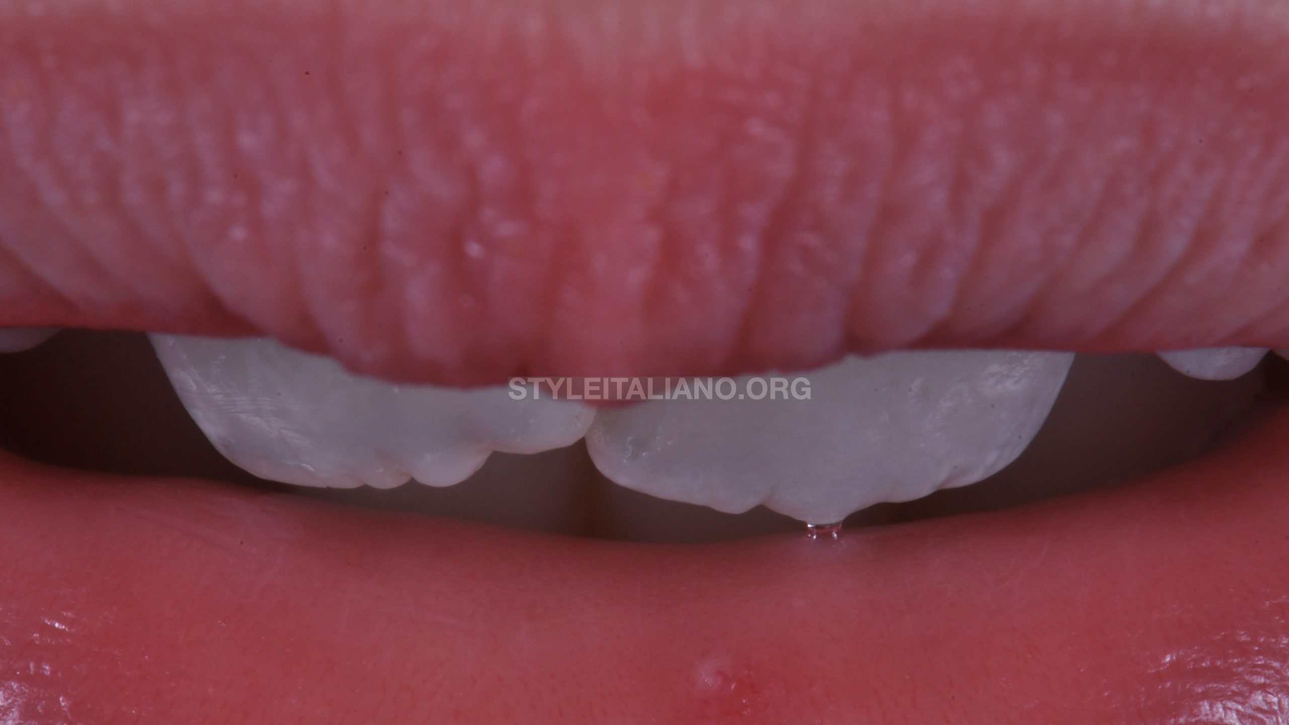
Fig. 8
A detail of the characterizations obtained, essential to give the restoration a natural appearance
Conclusions
A minimally invasive approach is essential for a correct resolution of problems involving anterior teeth, especially in young patients. Our goal must be to restore correct aesthetics and function while preserving the dental structures as much as possible and
With the right knowledge we can easily solve apparently complex clinical cases like this one.
Bibliography
Bakhtiar H, Esmaeili S, Fakhr Tabatabayi S, Ellini MR, Nekoofar MH, Dummer PM. Second-generation Platelet Concentrate (Platelet-rich Fibrin) as a Scaffold in Regenerative Endodontics: A Case Series. J Endod. 2017;43(3):401-8.
Conde MCM, Chisini LA, Sarkis-Onofre R, Schuch HS, Nor JE, Demarco FF. A scoping review of root canal revascularization: relevant aspects for clinical success and tissue formation. Int Endod J. 2017;50(9):860-74.
Dietschi D, Duc O, Krejci I, Sadan A. Biomechanical considerations for the restoration of endodontically treated teeth: a systematic review of the literature. Part II: evaluation of fatigue behavior, interfaces, and in vivo studies. Quintessence Int 2008;39:117—29.
Vanini L. Light and color in anterior composite restorations. Pract Periodontics Aesthet Dent. 1996 Sep;8(7):673-82.
Vanini L, Mangani F, Klimovskaia O. Conservative restoration of anterior teeth. Viterbo (Italy): Editing ACME; 2005.


