CBCT-guided management of external cervical resorption in a mandibular premolar
23/07/2026
Warning: Undefined variable $post in /home/styleendo/htdocs/styleitaliano-endodontics.org/wp-content/plugins/oxygen/component-framework/components/classes/code-block.class.php(133) : eval()'d code on line 2
Warning: Attempt to read property "ID" on null in /home/styleendo/htdocs/styleitaliano-endodontics.org/wp-content/plugins/oxygen/component-framework/components/classes/code-block.class.php(133) : eval()'d code on line 2
External cervical resorption (ECR) is a dynamic external resorptive process initiated by loss of the protective cementum layer, allowing clastic cells from the periodontium to interact with dentine.
Clinically, early ECR may remain asymptomatic and can be discovered only by a localised periodontal defect, altered cervical colour or an incidental radiographic finding. More advanced lesions can present with bleeding on probing, gingival inflammation, pulpitis or periapical disease.
The pericanalar resorption-resistant sheet often protects the pulp in the early stages, but cavitated lesions may eventually approach or communicate with the canal space. For this reason, treatment planning should not be based on clinical inspection alone.
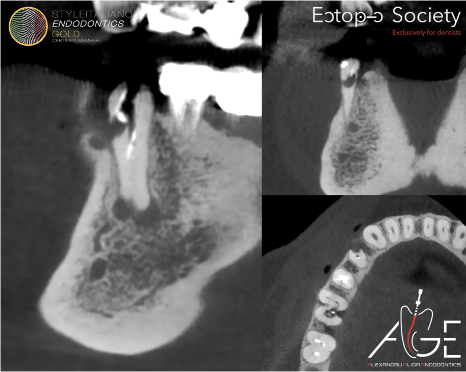
The lesion was interpreted as external cervical resorption and classified as Patel 2Ap.
“2” describes extension into the coronal third of the root, while “A” indicates limited circumferential spread. Although the circumferential extension was favourable, the subgingival portal of entry and close relationship with the canal space (“p”) made simple external restoration alone unsuitable.
In this case, the classification was used not as a label, but as a clinical map: access, debridement, repair and endodontic treatment were planned from the 3D morphology of the defect.
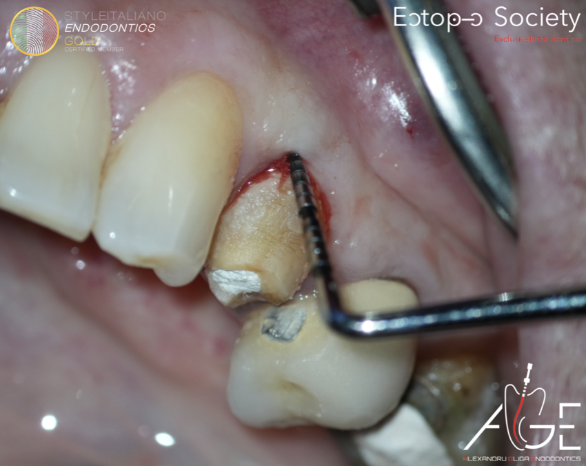
Fig. 1
A 62-year-old female patient was referred for assessment of tooth 44. The patient reported no symptoms. Clinical examination revealed a localised 6 mm probing depth in the mesio-buccal cervical area.
The clinical appearance suggested an accessible cervical defect rather than a generalised periodontal problem. This finding was consistent with a cavitated external cervical resorption lesion and justified 3D radiographic assessment.
The key question was whether the tooth could be managed conservatively while controlling both the external defect and the root canal space.
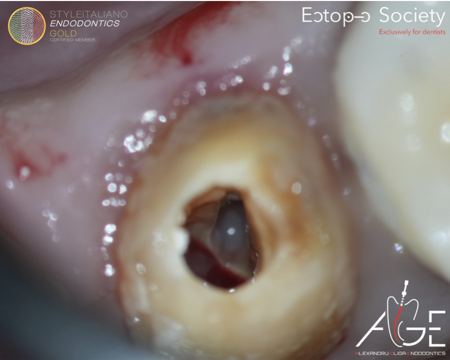
Fig. 2
A one-visit combined approach was selected. The objective was to remove the resorptive tissue, restore the cervical defect and disinfect/obturate the root canal system.
The operative sequence was planned as follows:
1. orthograde endodontic access to inspect the anatomy and protect the canal
surgical exposure of the cervical defect;
2. removal of fibrovascular resorptive tissue;
3. rubber dam isolation;
4. endodontic access and identification of both canals;
5. irrigation with sodium hypochlorite and EDTA;
6. adhesive repair using CLEARFIL™ DC CORE PLUS;
7. warm vertical compaction obturation.
After gaining access through the coronal aspect, fibrovascular tissue was visible in the resorptive area. This finding is typical of a cavitated ECR lesion and may be associated with profuse, localised bleeding.
Removal of this tissue is a diagnostic and therapeutic step. It allows direct inspection of the defect, identifies the sound margins required for repair and clarifies whether the lesion is isolated from, or communicating with, the canal space.
At this stage the treatment strategy remained conservative because the lesion was localised and accessible.
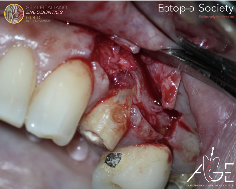
Fig. 3
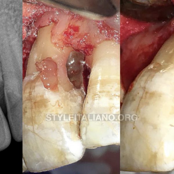
A mucoperiosteal flap was raised to provide controlled access to the mesio-buccal cervical defect. The aim was to visualise the portal of entry and the external component of the lesion without relying on blind instrumentation.
Direct surgical access improves control of debridement and reduces the risk of leaving resorptive tissue behind. It also allows the operator to verify whether the margins are suitable for adhesive repair.
In ECR, exposure and isolation are not separate details; they are part of the biological and restorative prognosis.
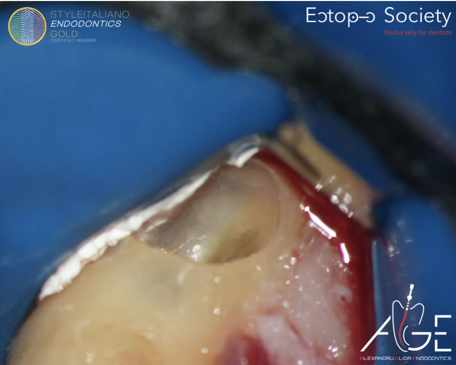
Fig. 4
After surgical access, rubber dam isolation was applied to stabilise the field for endodontic treatment and repair of the defect.
The defect was further inspected under magnification. The fibrovascular tissue was removed, the cavity was cleaned and the restorative margins were evaluated.
This step is critical because the success of the procedure depends not only on canal disinfection, but also on achieving a clean and well-sealed cervical repair.
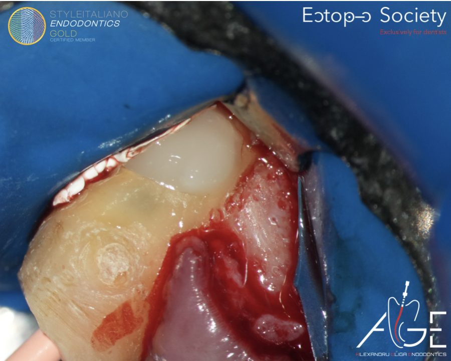
Fig. 5
Once the lesion was debrided and the margins were visible, the cervical defect was restored using CLEARFIL™ DC CORE PLUS (Kuraray Noritake Dental).
In this case, the defect was accessible and had margins that could be controlled surgically and under isolation. The repair was therefore performed with an adhesive restorative approach.
The main objective of the repair was to seal the external cervical communication and re-establish a cleansable cervical contour before completion of the endodontic phase.
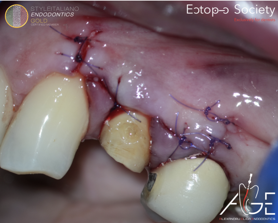
Fig. 6
Following repair of the cervical defect, the flap was repositioned and sutured.
The cervical location of ECR makes periodontal healing a central part of the outcome. A poor contour, residual resorptive tissue or marginal leakage can compromise both soft tissue stability and long-term prognosis.
The immediate surgical objective was stable closure without tension and preservation of the periodontal architecture around the cervical margin.
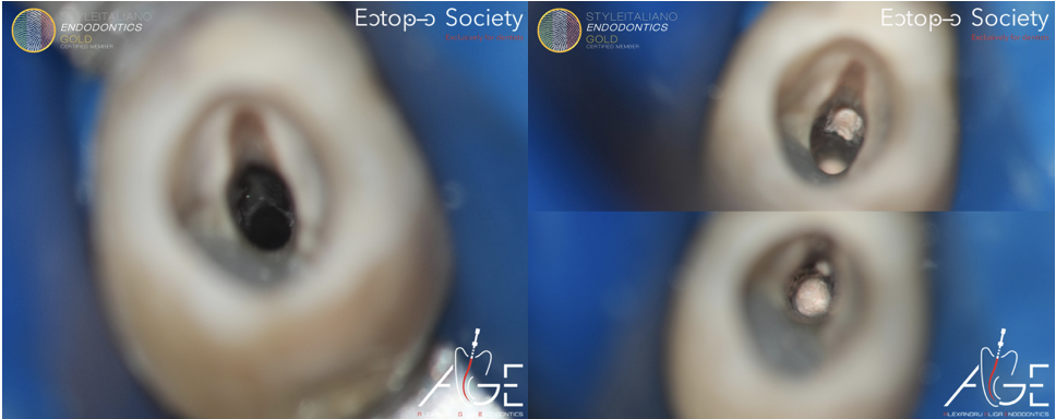
Fig. 7
After cervical management, attention was directed to the root canal system. Tooth 44 presented with a second lingual canal, identified after careful inspection of the chamber floor and canal anatomy.
This finding was clinically important. A missed lingual canal would have compromised disinfection and reduced the value of the cervical repair.
The case therefore had two parallel challenges: the biological control of the resorptive defect and the anatomical control of a two-canal mandibular premolar.
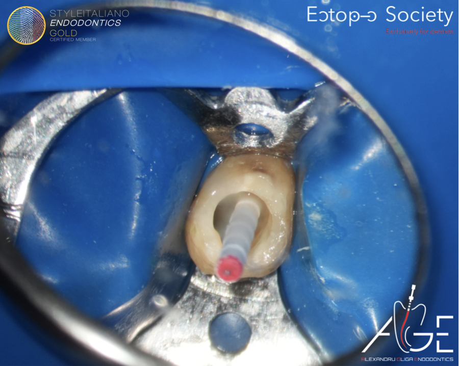
Fig. 8
Root canal treatment was completed in one visit. Both canals were negotiated, prepared and irrigated using sodium hypochlorite and EDTA.
The disinfection strategy aimed to control the canal microbiology while preserving the remaining cervical dentine. This balance is particularly important in ECR because the tooth is already structurally compromised in the cervical area.
After chemomechanical preparation, the canals were ready for obturation.
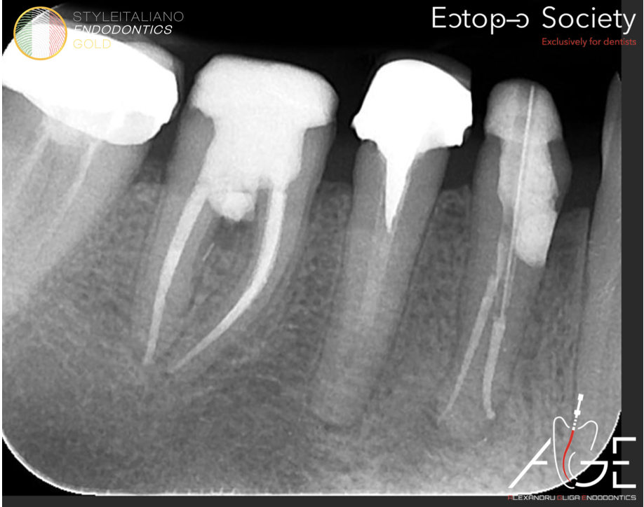
Fig. 9
The canals were obturated using warm vertical compaction. The immediate post-operative periapical radiograph confirmed treatment of both canals and repair of the cervical defect.
Radiographic control is essential after ECR treatment, but it should be interpreted together with the clinical situation. Follow-up must assess symptoms, probing depth, periodontal healing, integrity of the cervical repair and any sign of recurrence.
The final prognosis will depend on both endodontic healing and long-term stability of the cervical restoration.
The ESE Position Statement emphasises that the aims of ECR management are to excavate the resorptive tissue, restore the defect and monitor the tooth for recurrence. Management depends on lesion extent, portal size, accessibility and relationship with the root canal.
This case followed that logic. The lesion was limited enough to be treated, yet complex enough to require a combined strategy. Surgical access provided visibility of the external defect; rubber dam isolation allowed adhesive repair; root canal treatment addressed the internal aspect and the anatomical finding of a second lingual canal.
The result was a conservative treatment of a tooth that could have been misjudged if only the clinical appearance or a two-dimensional radiograph had been considered.
Two findings made this case educational.
First, the fibrovascular tissue inside the lesion confirmed that the cervical defect was active/cavitated and required complete removal before repair.
Second, the second lingual canal changed the endodontic management. The cervical repair could not be considered successful if the canal system remained incompletely treated.
The clinical lesson is therefore simple: in ECR, prognosis is built by sequencing the procedure correctly—plan, visualise, debride, isolate, repair, disinfect and obturate.
Conclusions
External cervical resorption should be assessed three-dimensionally before definitive treatment planning.
The Patel classification is useful because it describes the lesion according to height, circumferential spread and relationship with the canal.
A localised probing defect may be the only clinical sign of a cavitated cervical lesion.
When the lesion is accessible, surgical exposure and isolation allow predictable debridement and repair.
In mandibular premolars, careful inspection for a second lingual canal is mandatory.
Long-term review is required to monitor periodontal healing, restoration integrity and possible recurrence.
Bibliography
1. Patel S, Krastl G, Weiger R, Lambrechts P, Tjäderhane L, Gambarini G, Teng P-H. ESE position statement on root resorption. Int Endod J. 2023;56:792–801.
2. Patel S, Foschi F, Mannocci F, Patel K. External cervical resorption: a three-dimensional classification. Int Endod J. 2018;51:206–214.
3. Patel S, Mavridou AM, Lambrechts P, Saberi N. External cervical resorption—part 1: histopathology, distribution and presentation. Int Endod J. 2018;51:1205–1223.
4. Patel S, Foschi F, Condon R, Pimentel T, Bhuva B. External cervical resorption: part 2—management. Int Endod J. 2018;51:1224–1238.
5. Mavridou AM, Rubbers E, Schryvers A, Maes A, Linssen M, Barendregt DS, et al. A clinical approach strategy for the diagnosis, treatment and evaluation of external cervical resorption. Int Endod J. 2022;55:347–373.
6. Bardini G, Orrù C, Ideo F, Nagendrababu V, Dummer P, Cotti E. Clinical management of external cervical resorption: a systematic review. Aust Endod J. 2023;49:769–787.
7. DeLuca S, Choi A, Pagni S, Alon E. External cervical resorption: relationships between classification, treatment and 1-year outcome with evaluation of the Heithersay and Patel classification systems. J Endod. 2023;49:469–477.
8. Villefrance JS, Kirkevang L-L, Wenzel A, et al. Long-term prognosis for teeth with external cervical resorption based on periapical images and cone beam CT: a clinical study. Int Endod J. 2024;57:1596–1607.