Deep caries due to pre-eruptive intracoronal resorption (PIER) in a recently erupted maxillary second molar: a case report
08/02/2024
Davide Guglielmi
Warning: Undefined variable $post in /home/styleendo/htdocs/styleitaliano-endodontics.org/wp-content/plugins/oxygen/component-framework/components/classes/code-block.class.php(133) : eval()'d code on line 2
Warning: Attempt to read property "ID" on null in /home/styleendo/htdocs/styleitaliano-endodontics.org/wp-content/plugins/oxygen/component-framework/components/classes/code-block.class.php(133) : eval()'d code on line 2
A pre-eruptive intra-coronal resorption (PIER) is a defect located in the dentin of an unerupted tooth, just beneath the dentin-enamel junction.
The depth of the lesion is variable and may also reach the pulp, like in this 13 years old patient.
In the past, these lesions were confused with caries, and were therefore called “hidden” or “pre-eruptive caries”. These defects are usually detected incidentally in routine dental radiographies.
It has been proven that in the pre-eruptive stage the lesions contain soft tissue and inflammatory cells.
The present report describes the clinical management of a case of PEIR on a maxillary second permanent molar in association with a deep caries.
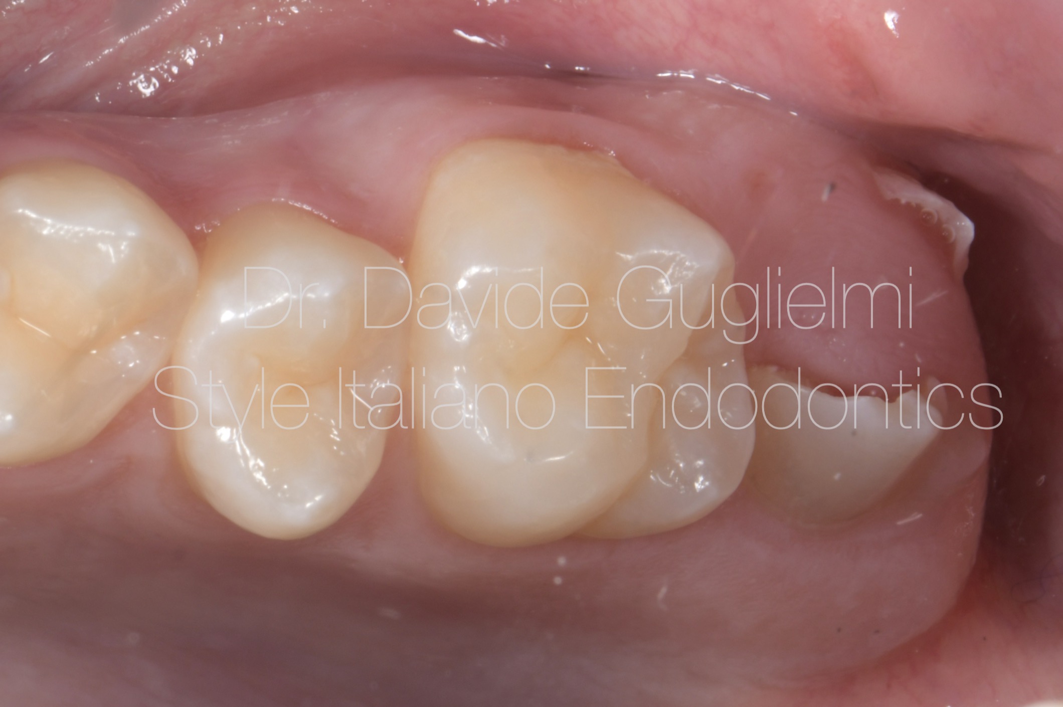
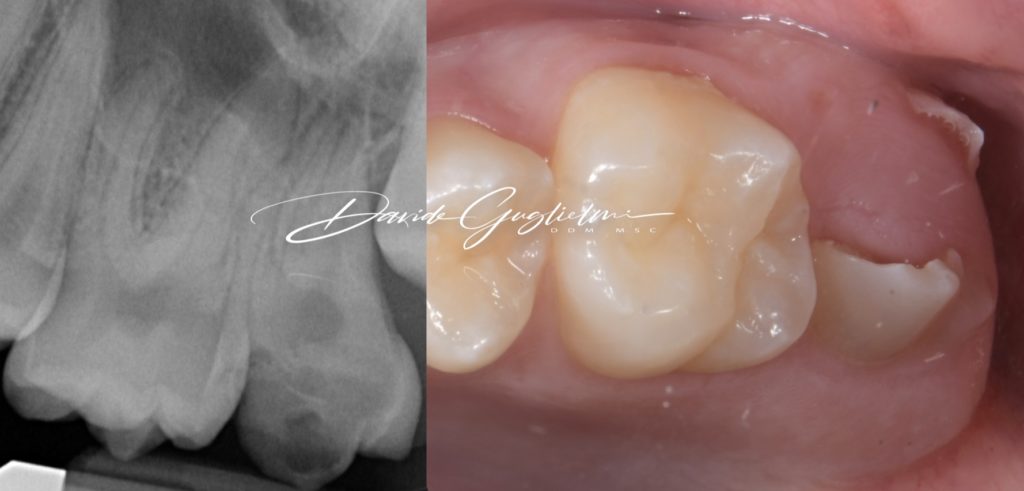
Fig. 1
Occlusal aspect, preoperative view of the asimptomatic partially erupted second permanent maxillary molar of the 13 years old patient.
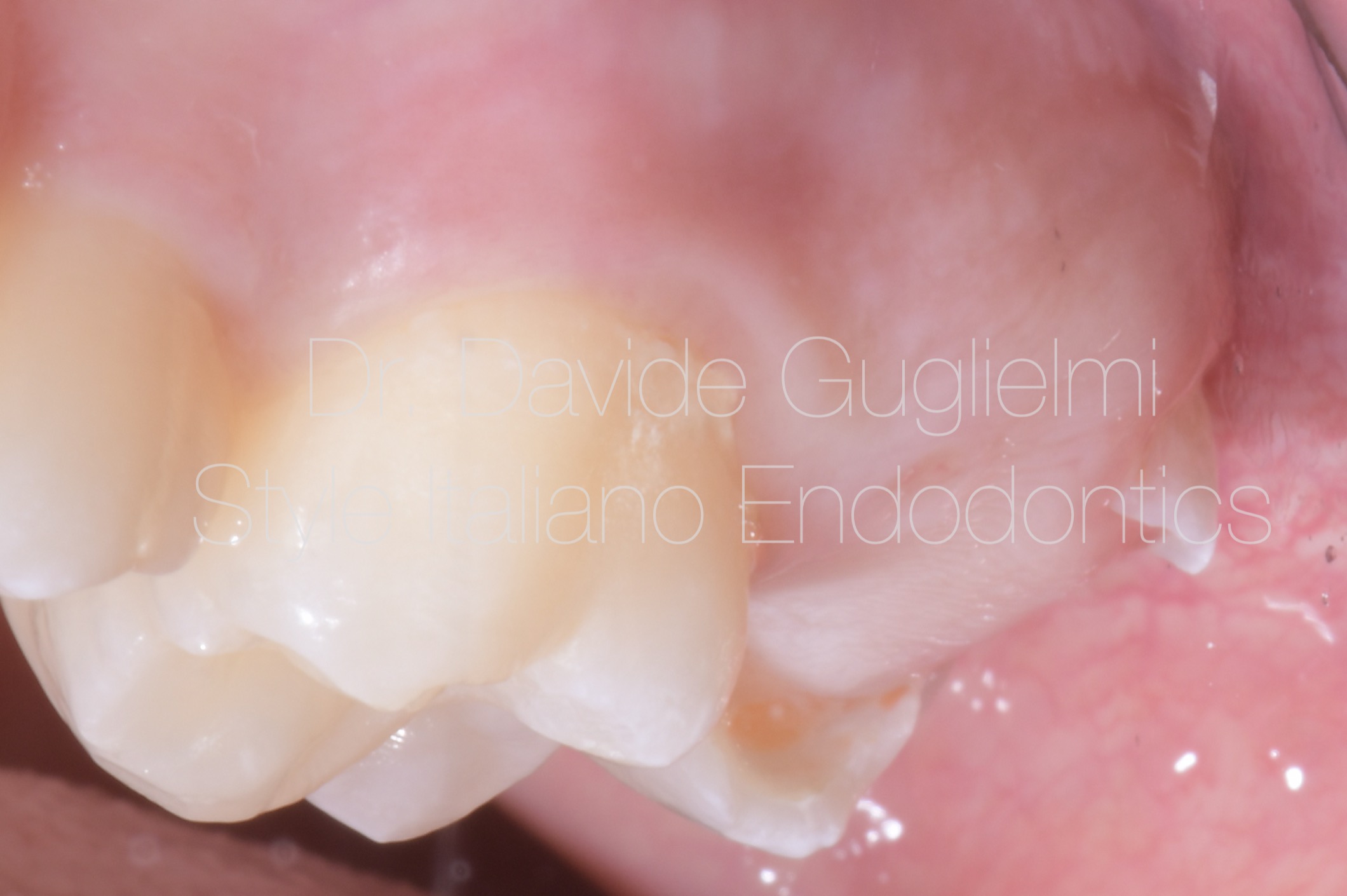
Fig. 2
Buccal aspect, preoperative view of the element 2.6 2.7
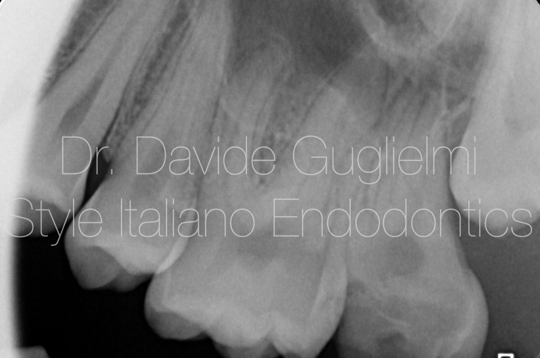
Fig. 3
Preoperative X-ray
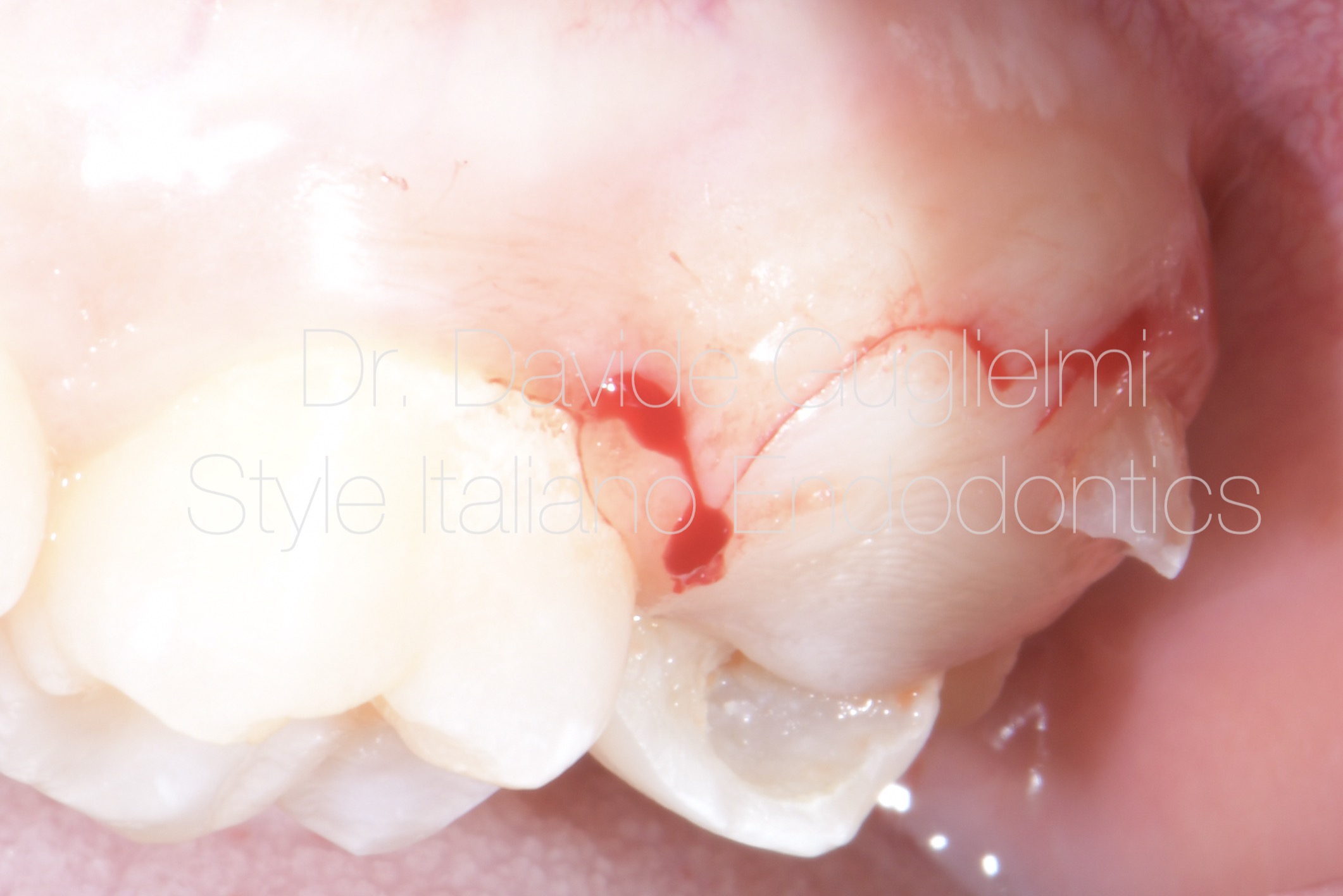
Fig. 4
Flap design
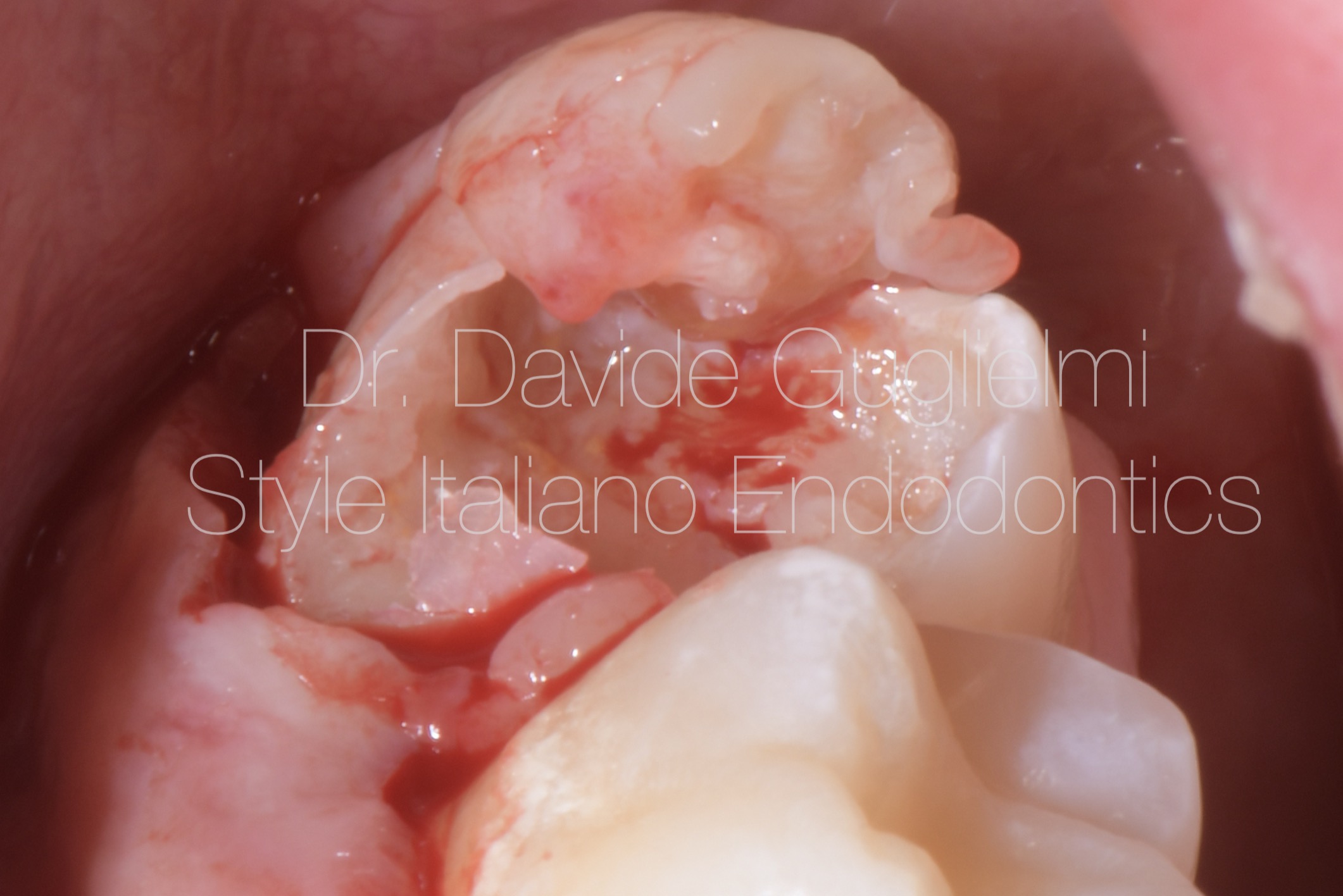
Fig. 5
Flap elevation
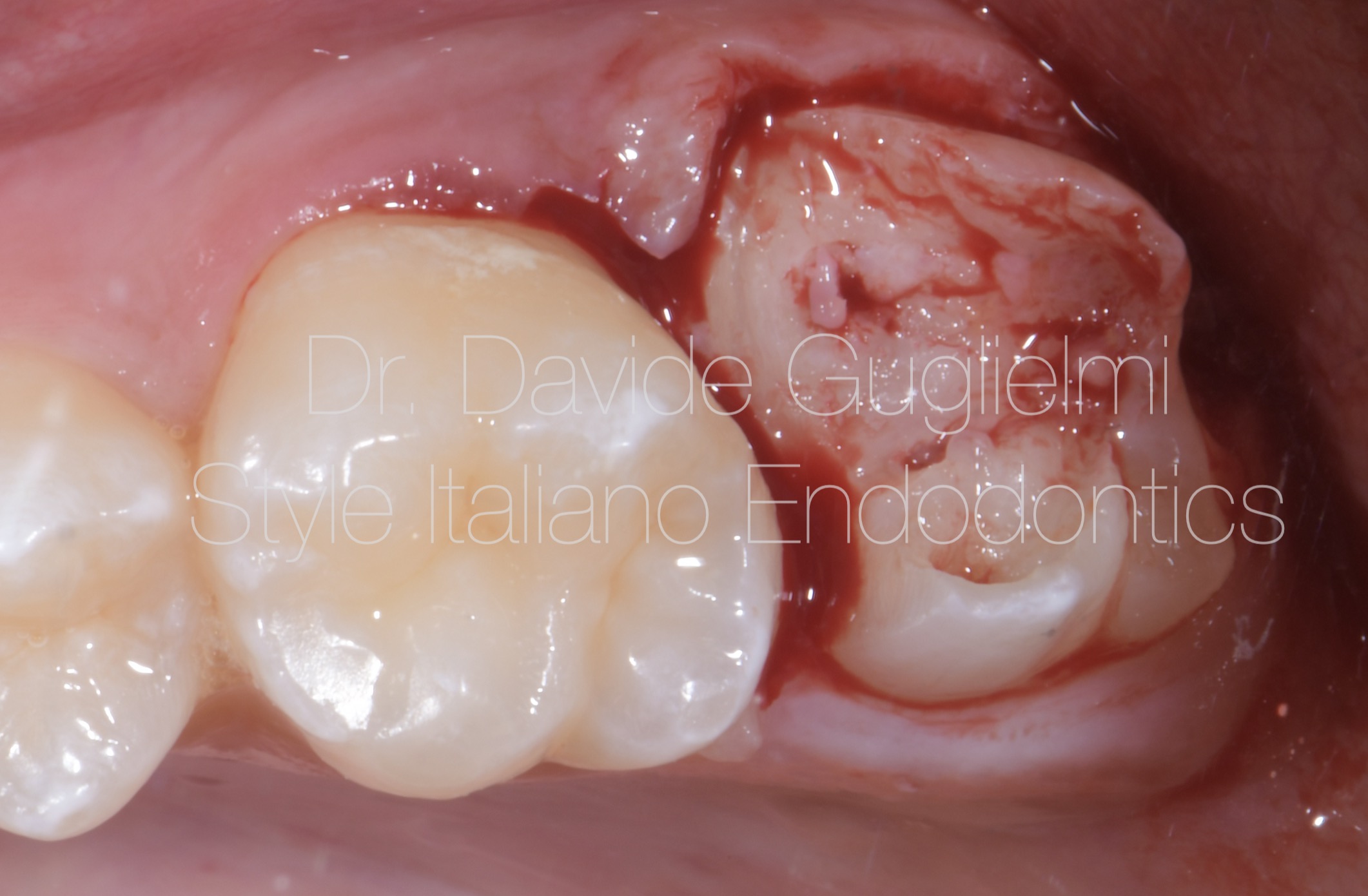
Fig. 6
Secondary flap removal.
The presence of an undefined soft tissue instead of dentin can be easily identified.
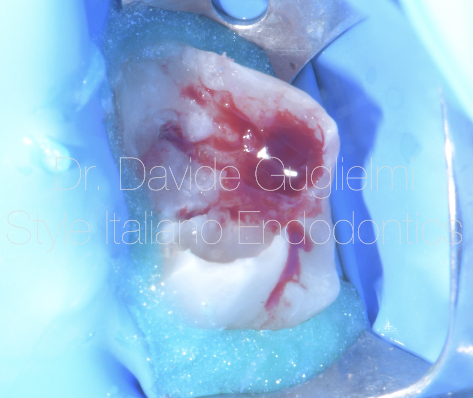
Fig. 7
A full isolated field is obtained by placing both the rubber dam in the classic manner and a liquid dam in the most cervical portion of the tooth.
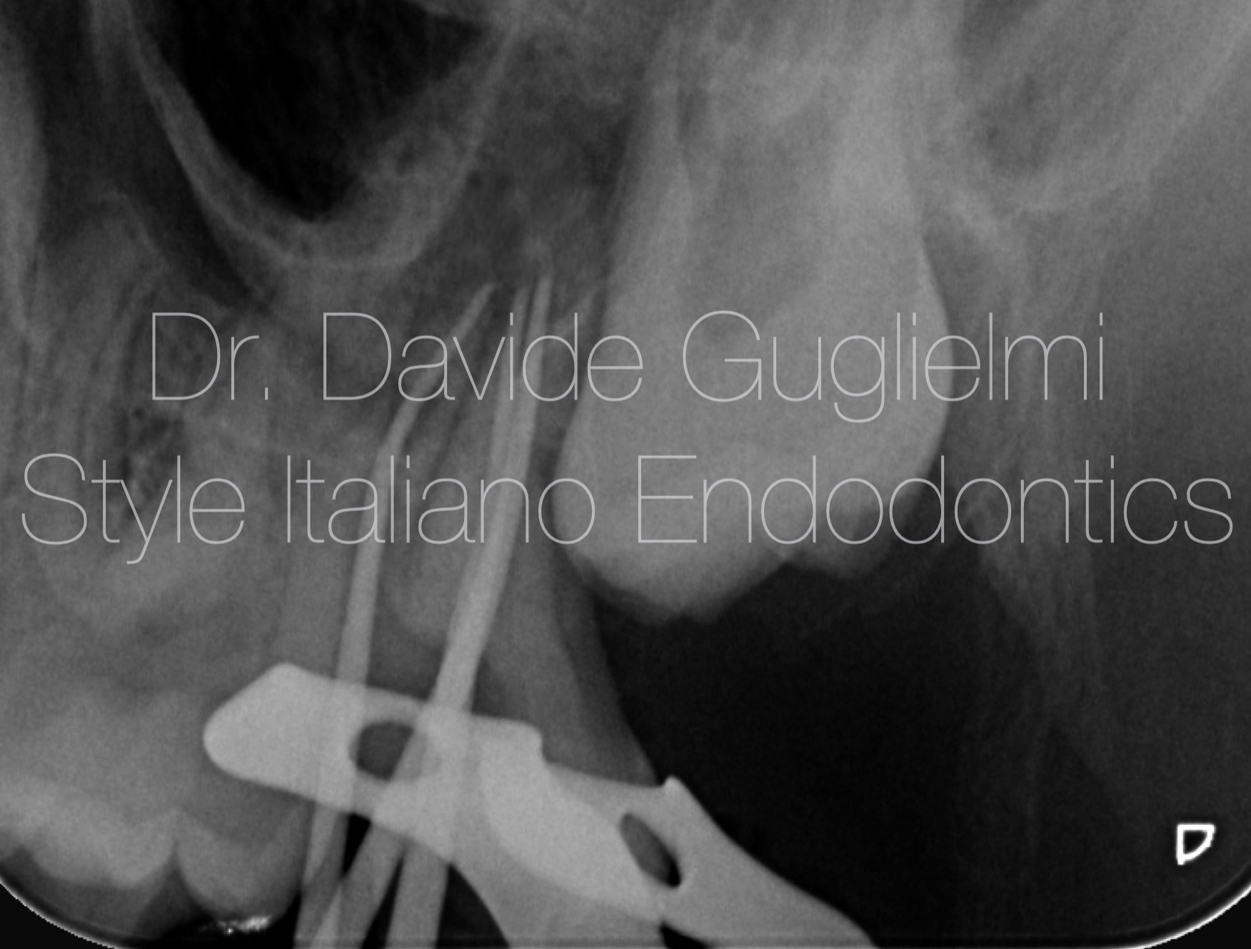
Fig. 8
Master cones X-ray’s been taken during root canal treatment (RCT)
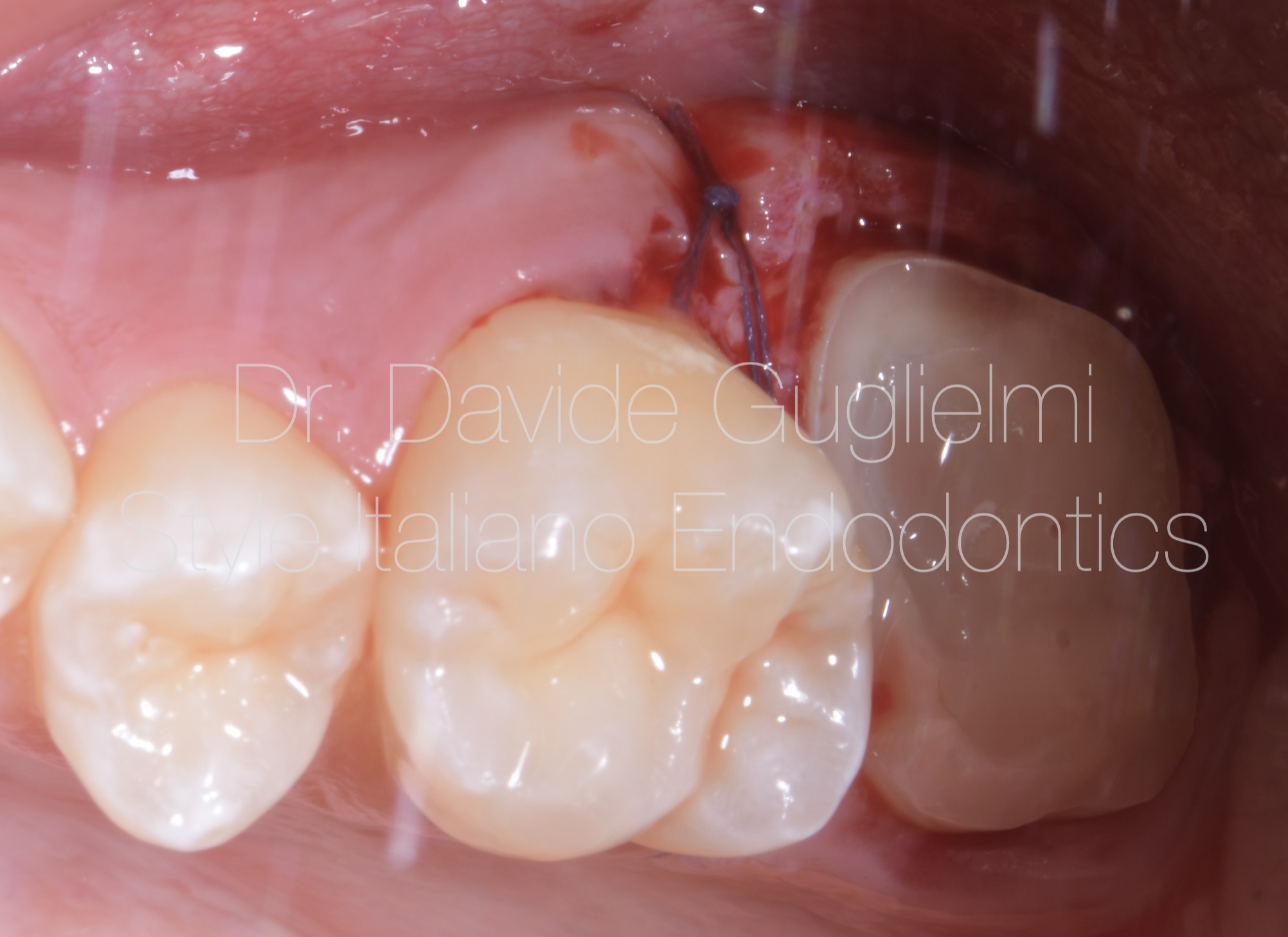
Fig. 9
A resorbible surgical suture, 5/0 (Vycril) is used in order to avoid plaque accumulation in the days following the procedure.
One single vertical mattress stitch is placed on the mesial of the 2.7.
Light and heavy body polyvinylsiloxane impression is taken the same day for the overlay preparation.
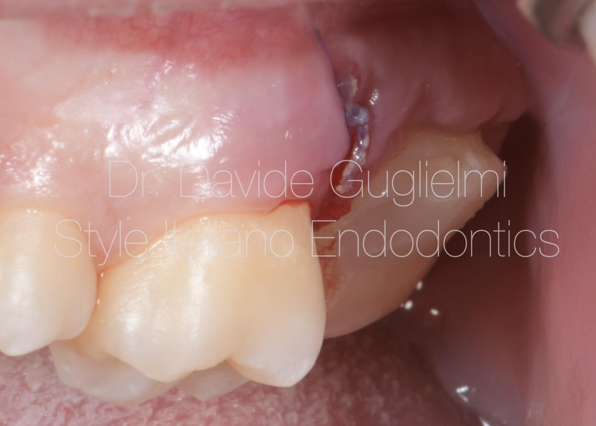
Fig. 10
Buccal aspect of the stitched flap.
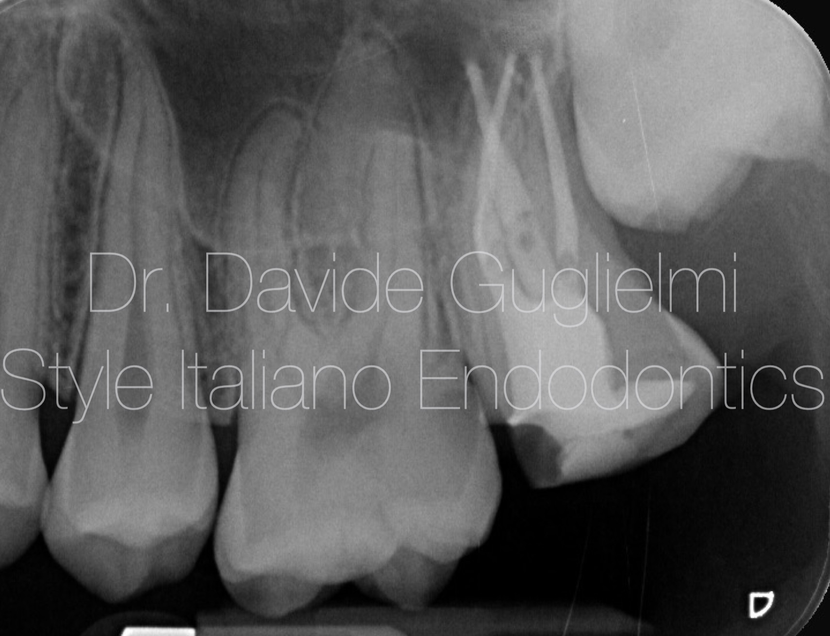
Fig. 11
Post Operative X-ray is taken immediately after the surgical therapy, the RCT (warm vertical obturation) and the build-up reconstruction.
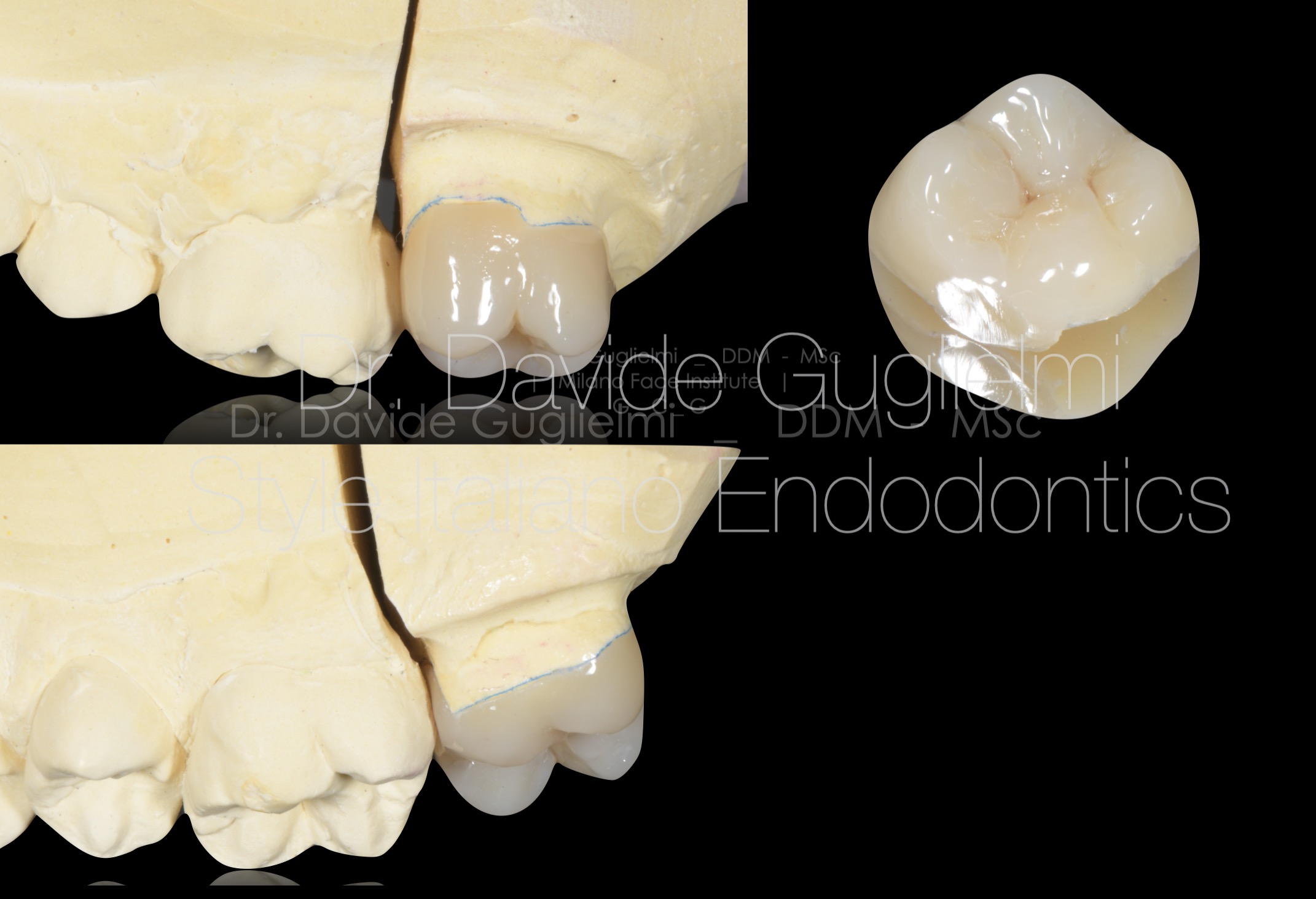
Fig. 12
Three different point of view of the composite overlay.
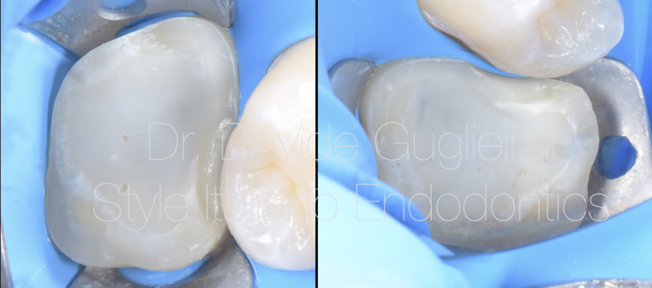
Fig. 13
Occlusal view of the build up and the inter proximal margin.
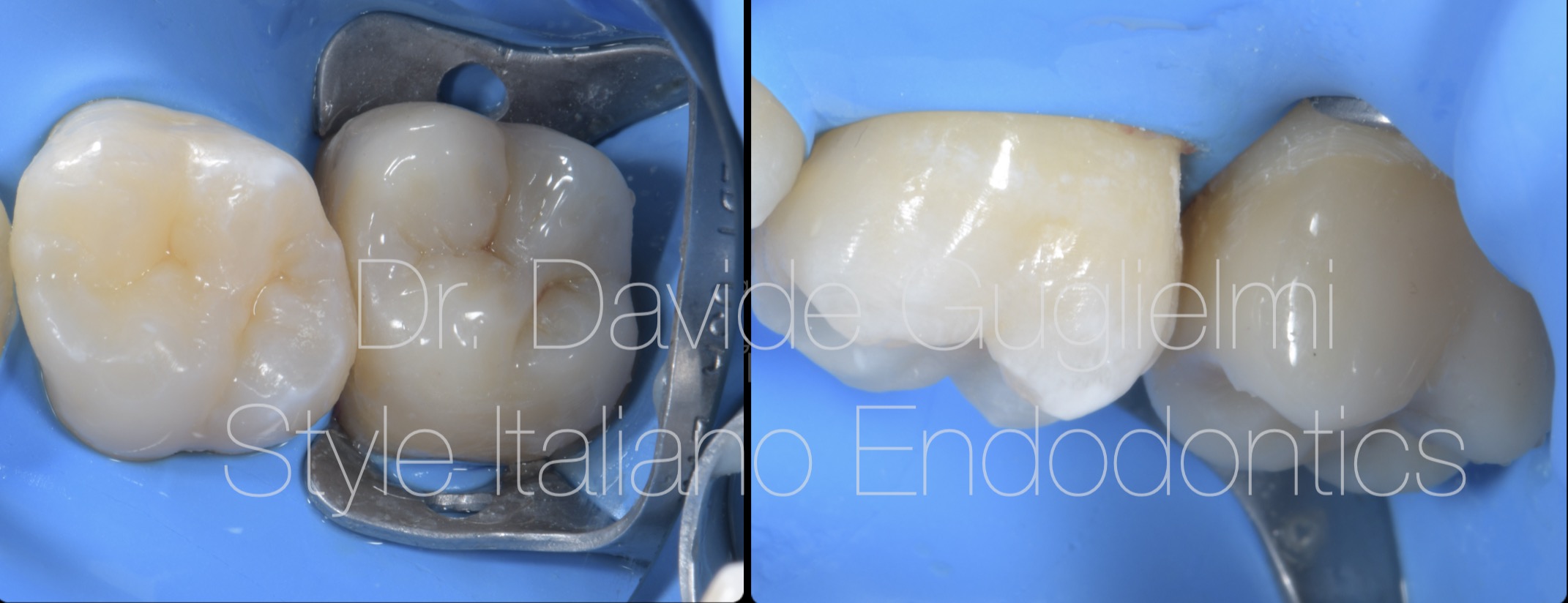
Fig. 14
Occlusal and buccal aspects of the cemented overlay.
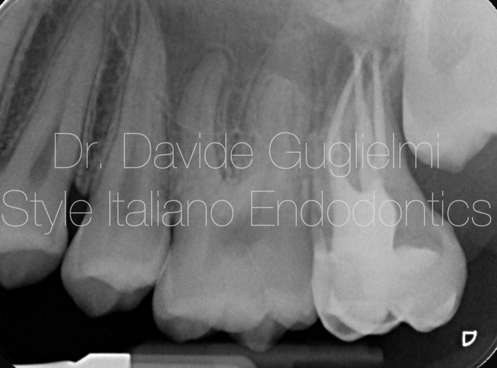
Fig. 15
Post op x-ray
Conclusions
The importance of a correct and prompt diagnosis of a pre eruptive intra-coronal resorption is evident and, if required, can lead to a well-timed treatment in order to prevent pulp involvement.
Bibliography
- Davidovich E, Kreiner B, Peretz B. Treatment of severe pre-eruptive intracoronal resorption of a permanent second molar. Pediatr Dent. 2005 Jan-Feb;27(1):74-7. PMID: 15839399.
- Omar S, Choi J, Nelson B, Shin M, Chen JW. Pre-Eruptive Intracoronal Resorption (PEIR): Literature Review and Case Report. J Calif Dent Assoc. 2015 May;43(5):255-60. PMID: 26798901.
- Kim Seow W. Pre-eruptive intracoronal resorption as an entity of occult caries. Pediatr Dent. 2000; 22:370-375.
- Counihan K.P., O’Connell A.C. Case report: pre-eruptive intra-coronal radiolucencies revisited, Eur Arch Paediatr Dent, 2012 Aug; 13(4):221-6. 3. Kim Seow W, Lu PC, Mcallan LH. Prevalence of pre-eruptive intracoronal dentin defects from panoramic radiographs. Pediatr Dent. 1999; 21:332-339.
- .Sanctis, M. de & Clementini, M. Flap approaches in plastic periodontal and implant surgery: critical elements in design and execution. J Clin Periodontol 41, S108–S122 (2014).
- Ozden B., Acikgoz A. Prevalence and characteristics of intra-coronal resorption in unerupteeth in the permanent dentition: a retrospectivestudy. Oral Radiol 2009, 25:6-13.Veneziani.Posterior indirect adhesive restorations: updated indications and the Morphology Driven Preparation Technique.
- .Veneziani, M. Posterior indirect adhesive restorations: updated indications and the Morphology Driven Preparation Technique. The International Journal of Esthetic Dentistry 12, 204–230 (2017).