THE CURVE CONUNDRUM: ENDODONTIC MANAGEMENT OF A DILACERATED THIRD MOLAR
16/11/2025
Fellow
Warning: Undefined variable $post in /home/styleendo/htdocs/styleitaliano-endodontics.org/wp-content/plugins/oxygen/component-framework/components/classes/code-block.class.php(133) : eval()'d code on line 2
Warning: Attempt to read property "ID" on null in /home/styleendo/htdocs/styleitaliano-endodontics.org/wp-content/plugins/oxygen/component-framework/components/classes/code-block.class.php(133) : eval()'d code on line 2
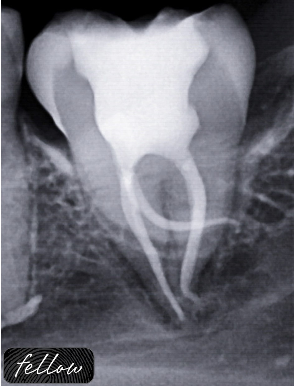
Dilaceration refers to an abnormal angulation or curvature in the root of a tooth, making access to the pulp space difficult and increasing the risk of complications during the procedure. Anatomical proximity to vital structures poses an additional challenge in such cases posing a risk of paresthesia, etc. Such teeth require meticulous attention in terms of treatment planning and often test the limits of modern NiTi instrumentation.
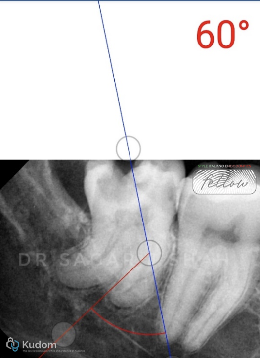
Fig. 1
PREOPERATIVE ASSESSMENT:
Mesial canal curvature- 60 degree

Fig. 2
Distal canal curvature- 30 degree
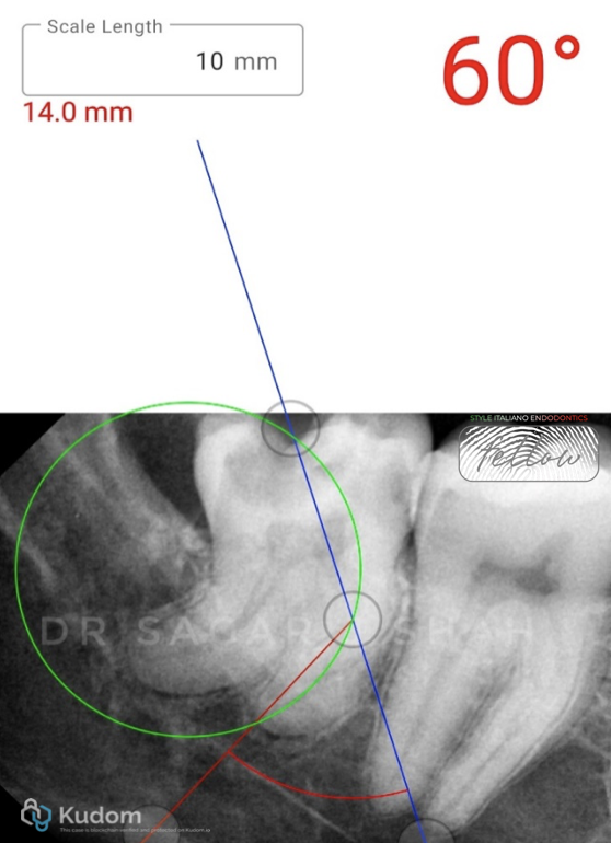
Fig. 3
The radius of curvature- 14 mm
Close proximity to the mandibular canal.
Pulp calcification was seen with the distal root.
AAE DIFFICULTY ASSESSMENT- HIGH
(http://www.aae.org/caseassessment/)
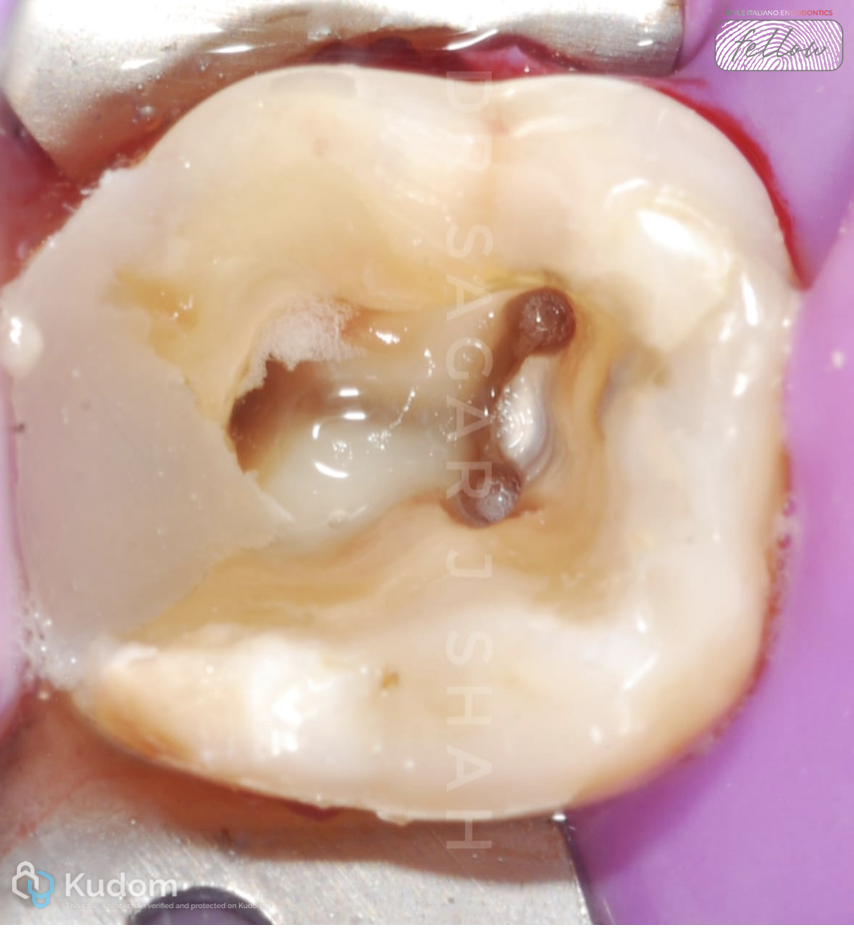
Fig. 4
- ANESTHESIA AND RUBBER DAM APPLICATION
- ACCESS OPENING:
- Performed using SF 12, followed by safe end (EX-24) to achieve complete deroofing.
- Ultrasonic used to refine the access.
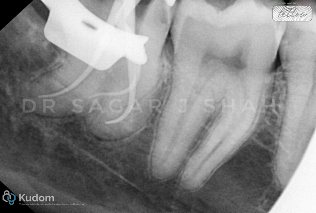
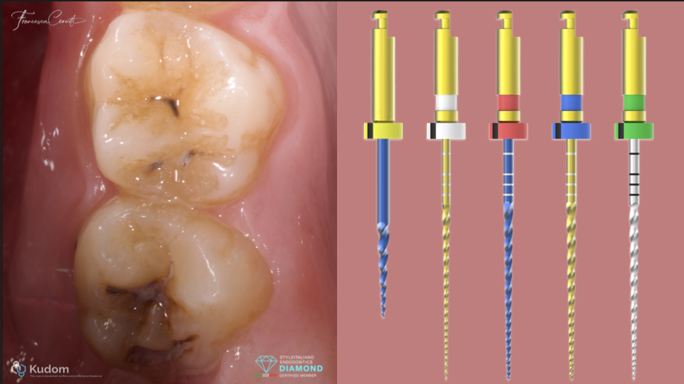
Fig. 5
Classic crown down technique was used with CM wire files for mechanical preparation.
- Preflaring is done with 17/08, 2 mm into the orifice.
- 25/06 was inserted with hand till point of resistance and then the file was activated in a 1 mm stroke with pecking motion apically.
- The sequence was repeated until the file penetrated 13 mm i.e. just above the curvature.
- Working length was then calculated using EAL (Morrita Root ZX 2) using the #8K hand file.
- The glide path was achieved by sequential hand filing with #8K, #10K till they were passive.
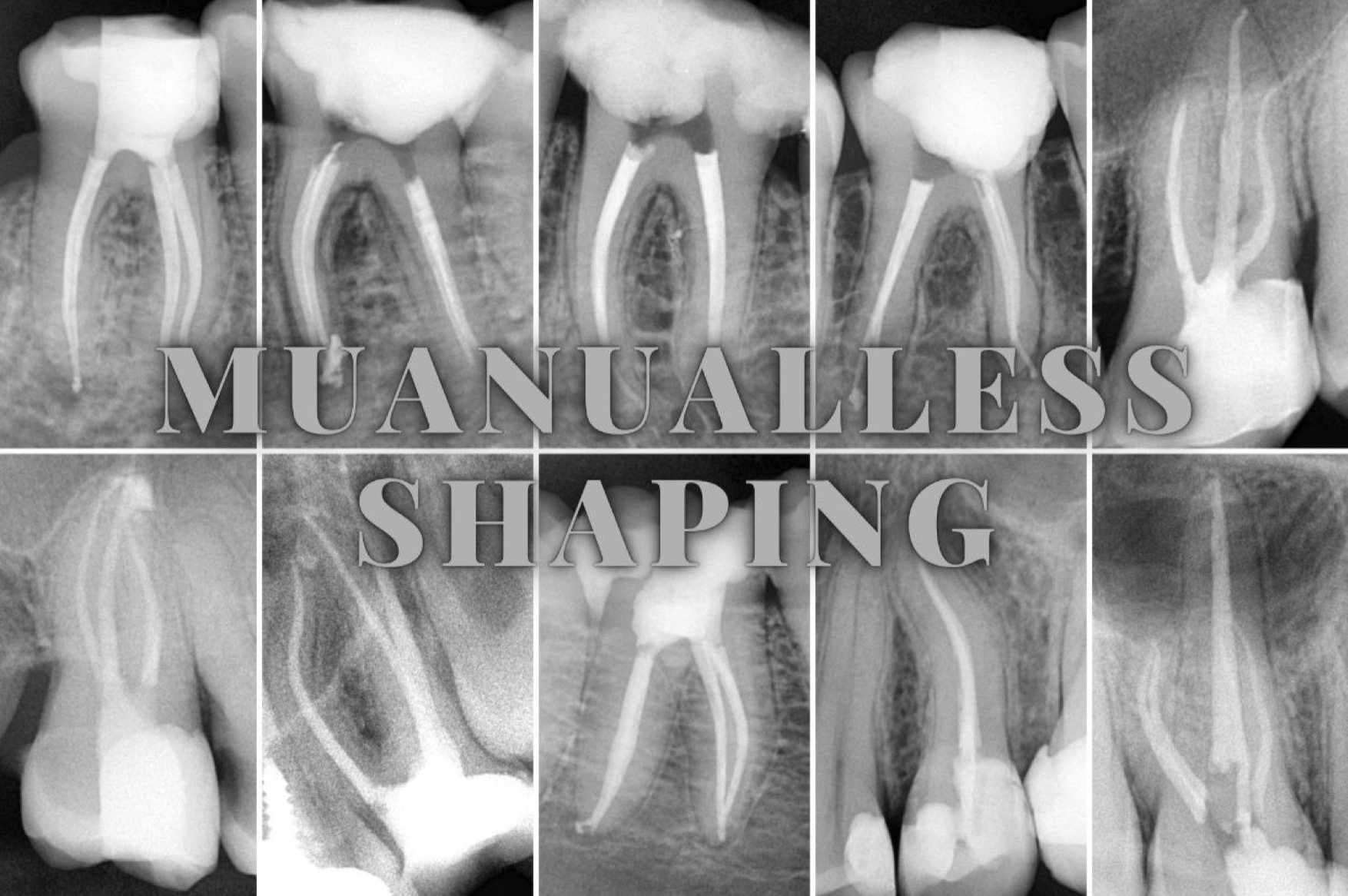
- Shaping was completed with heat treated files to apical size of 20/006 in mesials and 25/06 in distal.
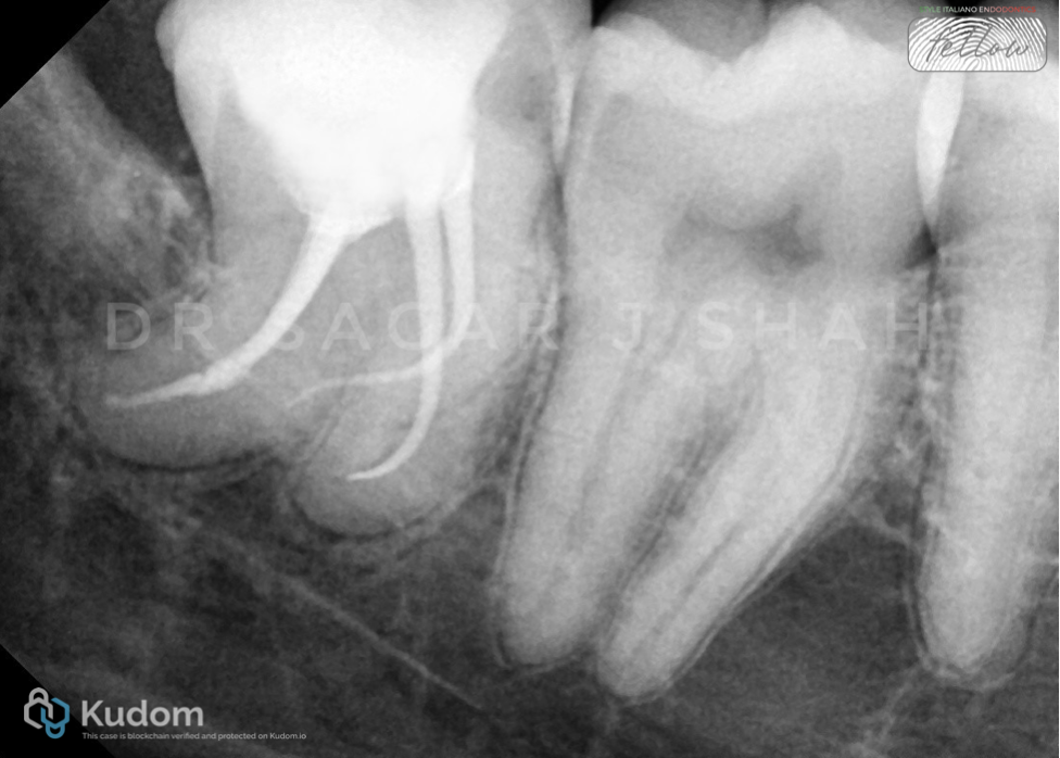
Fig. 6
OBTURATION AND CORE BUILDUP
- Care was taken to keep instrumentation at apical constriction to avoid any mishaps due to close proximity to the mandibular canal
- Cold hydraulic obturation with bioceramic sealer was performed.
- Composite core was done in the same appointment.

Fig. 7
About the author:
Sagar Shah
Conclusions
Successful management of dilacerated teeth requires precise diagnosis, modified access design, use of heat treated NiTi instruments particularly CM wire which shows enhanced cyclic fatigue resistance, Incorporating magnification, appropriate technique, adequate shaping, and efficient irrigation with optimum apical control ensures predictable cleaning and obturation of complex root anatomies specially when in close proximity of vital tooth structures.
Bibliography
- Chaniotis A, Ordinola-Zapata R. Present status and future directions: Management of curved and calcified root canals. Int Endod J. 2022;55(Suppl 3):656-684
- Jafarzadeh H, Abbott PV. Dilaceration: review of an endodontic challenge. J Endod. 2007;33(9):1025-1030
- Çelik B, Çelik ME. Root Dilaceration Using Deep Learning: A Diagnostic Approach. Applied Sciences. 2023; 13(14):8260