Healing of the periapical lesion
30/10/2025
Fellow
Warning: Undefined variable $post in /home/styleendo/htdocs/styleitaliano-endodontics.org/wp-content/plugins/oxygen/component-framework/components/classes/code-block.class.php(133) : eval()'d code on line 2
Warning: Attempt to read property "ID" on null in /home/styleendo/htdocs/styleitaliano-endodontics.org/wp-content/plugins/oxygen/component-framework/components/classes/code-block.class.php(133) : eval()'d code on line 2
The patient reported to our office with a complaint of food lodgement in the lower right back teeth region. During oral examination, proximal caries was found in the lower first molar. There was no pain in the molar tooth. In a radiographic examination there were periapical lesions in both roots. The tooth did not respond to the cold test. A necrotic tooth with a periapical lesion and no pain as symptoms suggest asymptomatic apical periodontitis.

Fig. 1
A preoperative x-ray shows proximal caries extended into the pulp and causing periapical lesion in the mesial and distal roots of the lower first molar. Non-surgical endodontic treatment was planned.
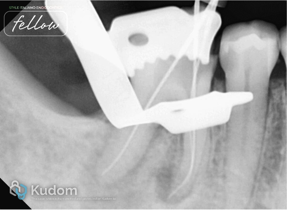
Fig. 2
After rubber dam isolation, caries was removed and deroofing was done. Initial scouting was done with 10 number K-files, and working length was measured. Protaper Gold rotary files were used to enlarge the canal space for better irrigant exchange. The mesial system was prepared till the F1file, and the distal system was prepared till the F2 file in a crown-down manner. Recapitulation was done after every rotary file and in the presence of sodium hypochlorite. Irrigation activation was done with air sonic device. EDTA liquid was use before the final rinsing with sodium hypochlorite.
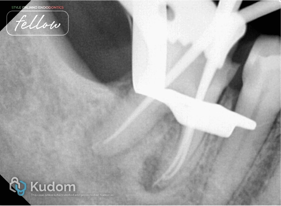
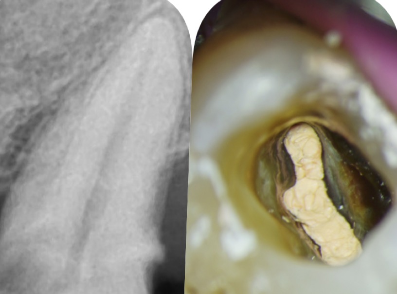
Fig. 3
After irrigation and activation of irrigants, canals were found dry. Non-standardised medium-sized gutta percha cones were checked as master cones.
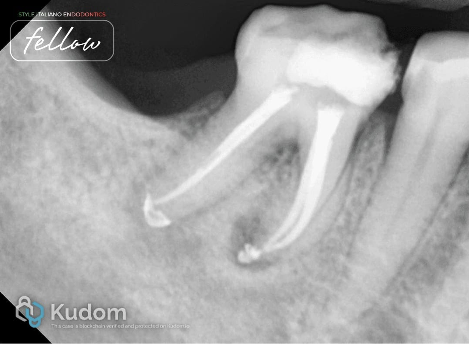
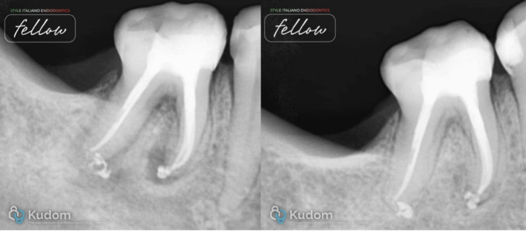
Fig. 4
After-obturation x-ray. Obturation was done with AH Plus sealer and the continuous wave of compaction method.
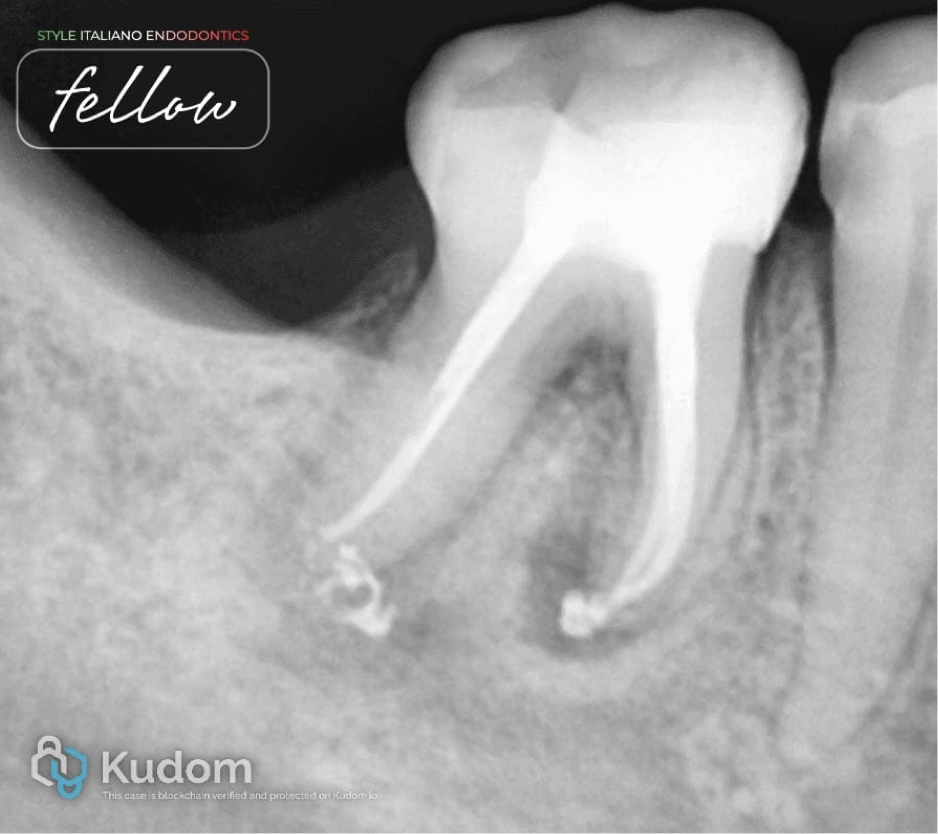
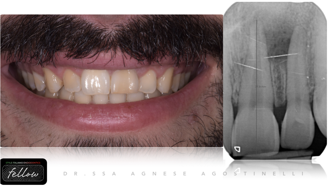
Fig. 5
X-ray after post endodontic restoration with fiber reinforced composite. It shows good corono apical seal.
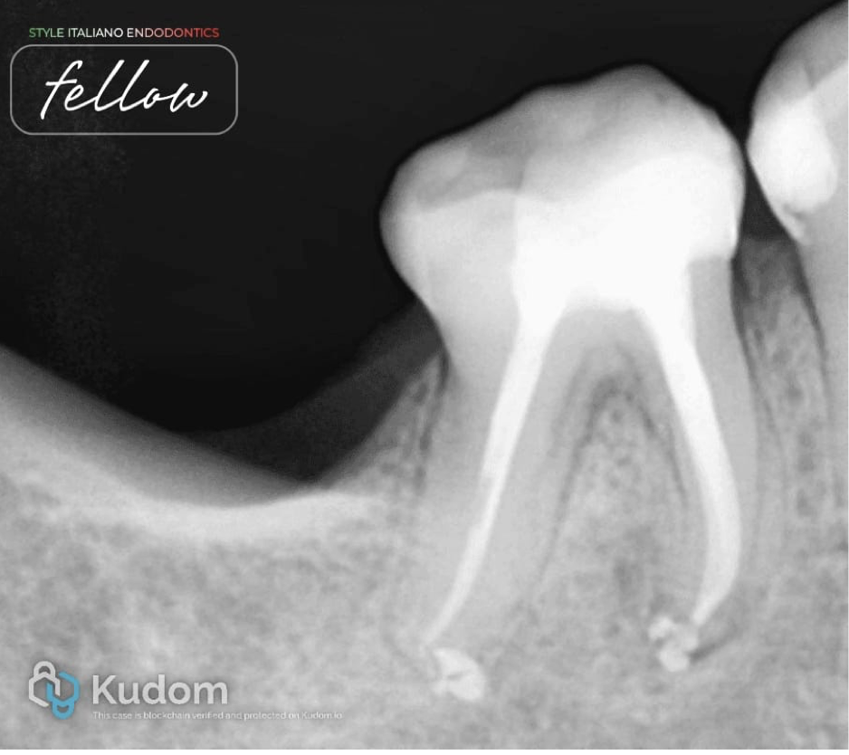
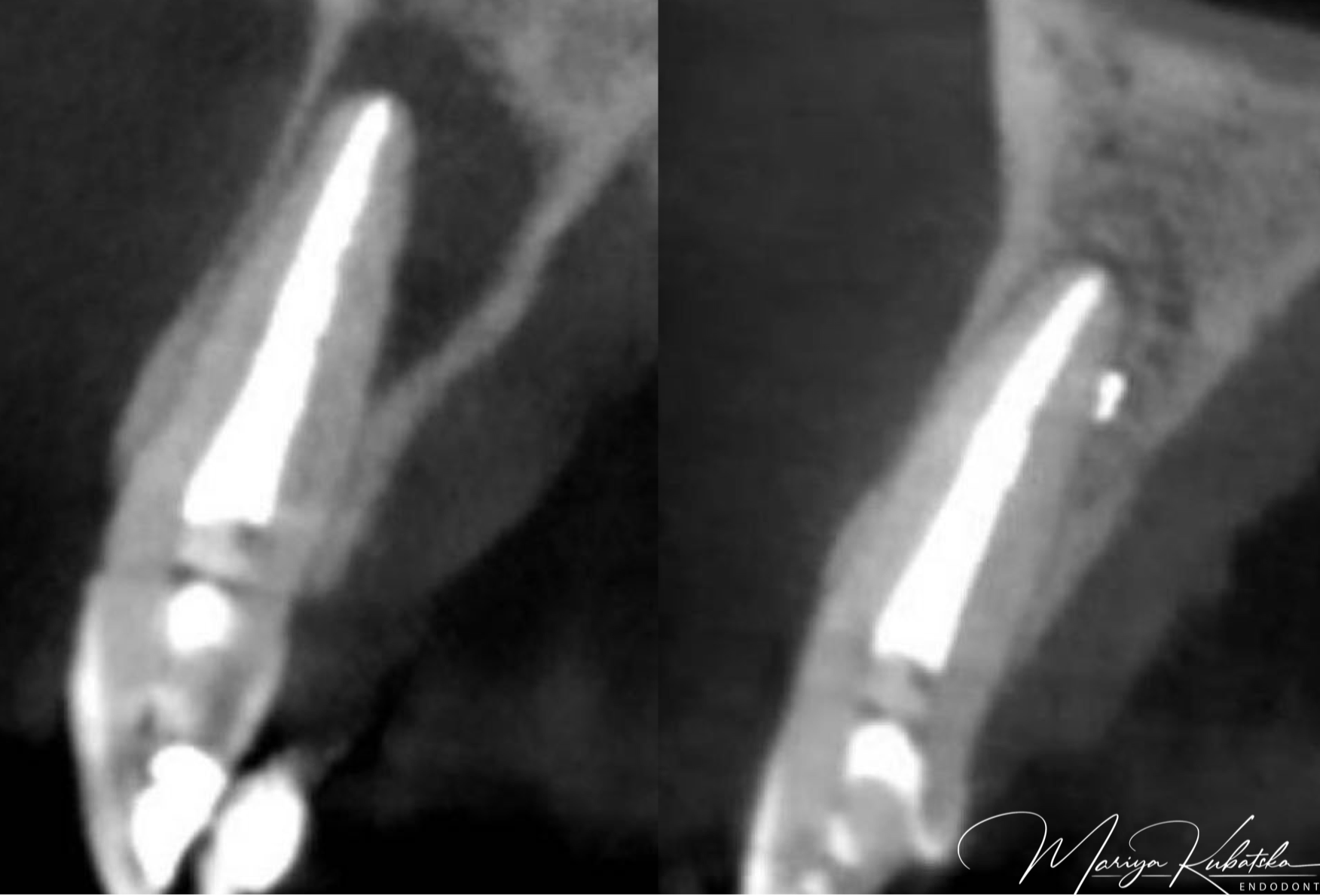
Fig. 6
X-ray taken after completion of the endodontic treatment. It shows healing in both roots. The periapical lesion decreased in size in the mesial and distal root. New bone formation has taken place in the previously lesioned periapical area.

Fig. 7
About the Author
Dr. Parth Padhya
Conclusions
Root canal treatment may suffice for the therapy of asymptomatic apical periodontitis. Removal of the cause of inflammation is usually followed by resorption of the granulomatous tissue and repair with trabeculated bone. There are many factors affecting the healing of the periapical lesion. The goal of the endodontic treatment is to do better cleaning of the canals for disinfection of infected canal spaces. Better cleaning resulted in the healing of the periapical lesion.
Bibliography
1)K.Gulabivala, Yuan Ng : Factors that affect the outcomes of root canal treatment and retreatment- A reframing of the principles, International endodontic journal- volume 56- issue 52, January 2023.
2)Y.-L.Ng, K.Gulabivala : A prospective study of the factors affecting outcomes of nonsurgical rroot treatment: part 1: periapical health, International endodontic journal , volume-44, issue-7/pp.583-609.
3)Bystom A, Happonen RP, Sjogren U, Sundqvist G : Healing of periapical lesion of pulpless teeth after endodontic treatment with controlled asepsis. (1987)Endodontics and dental traumatology 3,58-63.
4)G.M.G.Hommez, C.R.M.Coppens, R.J.G.De Moor : Periapical health related to the quality of coronal restorations and root fillings, International endodontic journal, august 2002/volume 35, issue 8/ pp.680-689.