C-SHAPED MAXILLARY SECOND MOLAR
21/03/2024
Leena Rawal
Warning: Undefined variable $post in /home/styleendo/htdocs/styleitaliano-endodontics.org/wp-content/plugins/oxygen/component-framework/components/classes/code-block.class.php(133) : eval()'d code on line 2
Warning: Attempt to read property "ID" on null in /home/styleendo/htdocs/styleitaliano-endodontics.org/wp-content/plugins/oxygen/component-framework/components/classes/code-block.class.php(133) : eval()'d code on line 2
Root canal treatment of maxillary second molar can be very difficult . Beside the limited accesibility of tooth in mouth with limited mouth opening ,with complex root canal anatomy specially if its a c shaped anatomy with irregular canals and challenge to clean and disinfect the isthmus part specially are the major difficulties of root canal treatment in this particular tooth.
Presenting a case of C Shaped root canal anatomy in maxillary left second molar of a 65 year male came with pain in the same tooth which was diagnosed as irreversible pipits with cold test . We phase all those difficulties mentioned above but with proper equipments and knowledge of the anatomy finally we got satisfactory results we expecting from this case .
Case was completed in single visit and rest details with more pictures are shared in next slides .
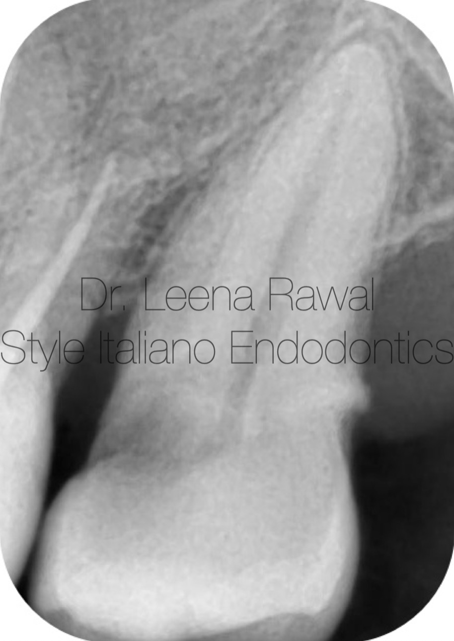
Fig. 1
Pre-operative IOPA of maxillary left second molar showing caries
approaching to pulp ,also showing a positive cold test response
confirms an irreversible pulpitis suggesting a root canal treatment
for the same.
On IOPA root canal external anatomy seems conical .
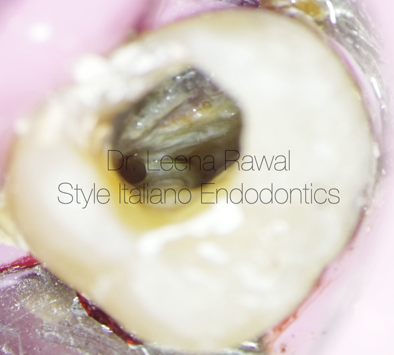
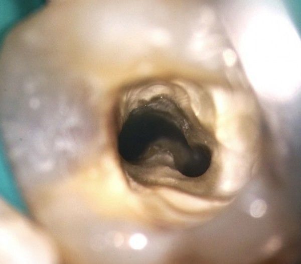
Fig. 2
This image is showing a C shaped root canal anatomy with a
MB orifice and a large oval orifice made by fused distobuccal
& palatal orifices
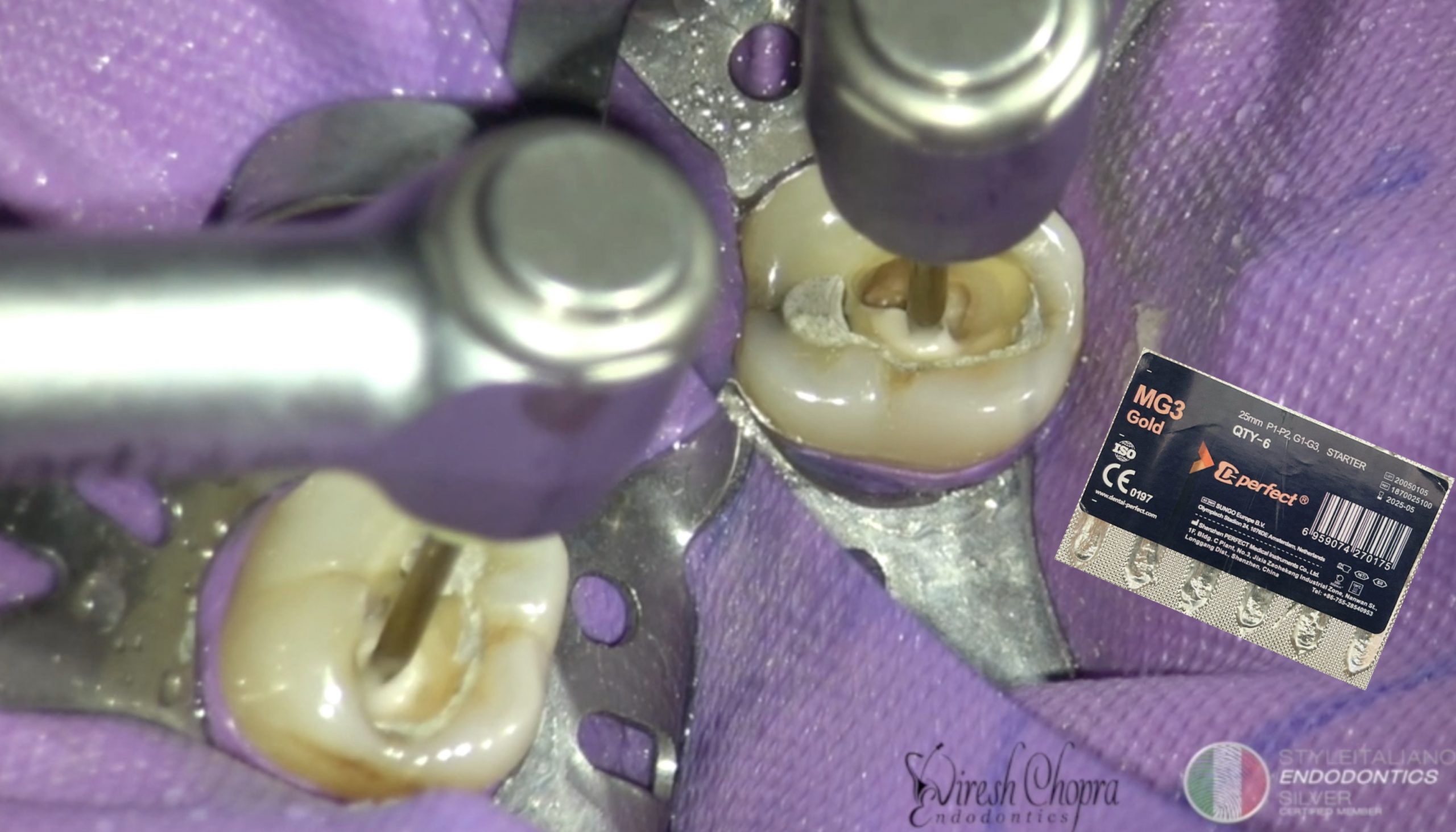
Shaping was done by hyflex CM files .
Irrigation with 5.25% NaOCl ,normal saline & 17% EDTA along
with intracanal heating of NaOCl by fastpack & activation of
irrigants by UltraX.
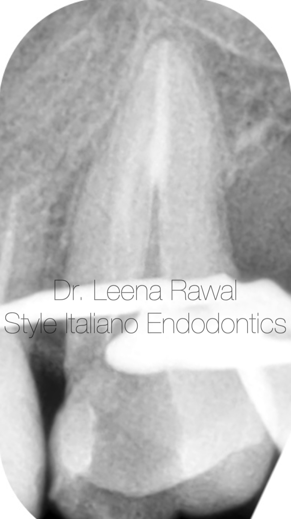
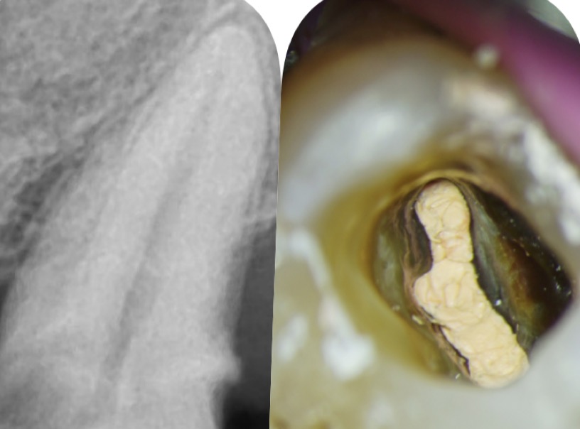
Fig. 3
IOPA showing down-pack procedure done by fastback followed by downtick by fastfill.
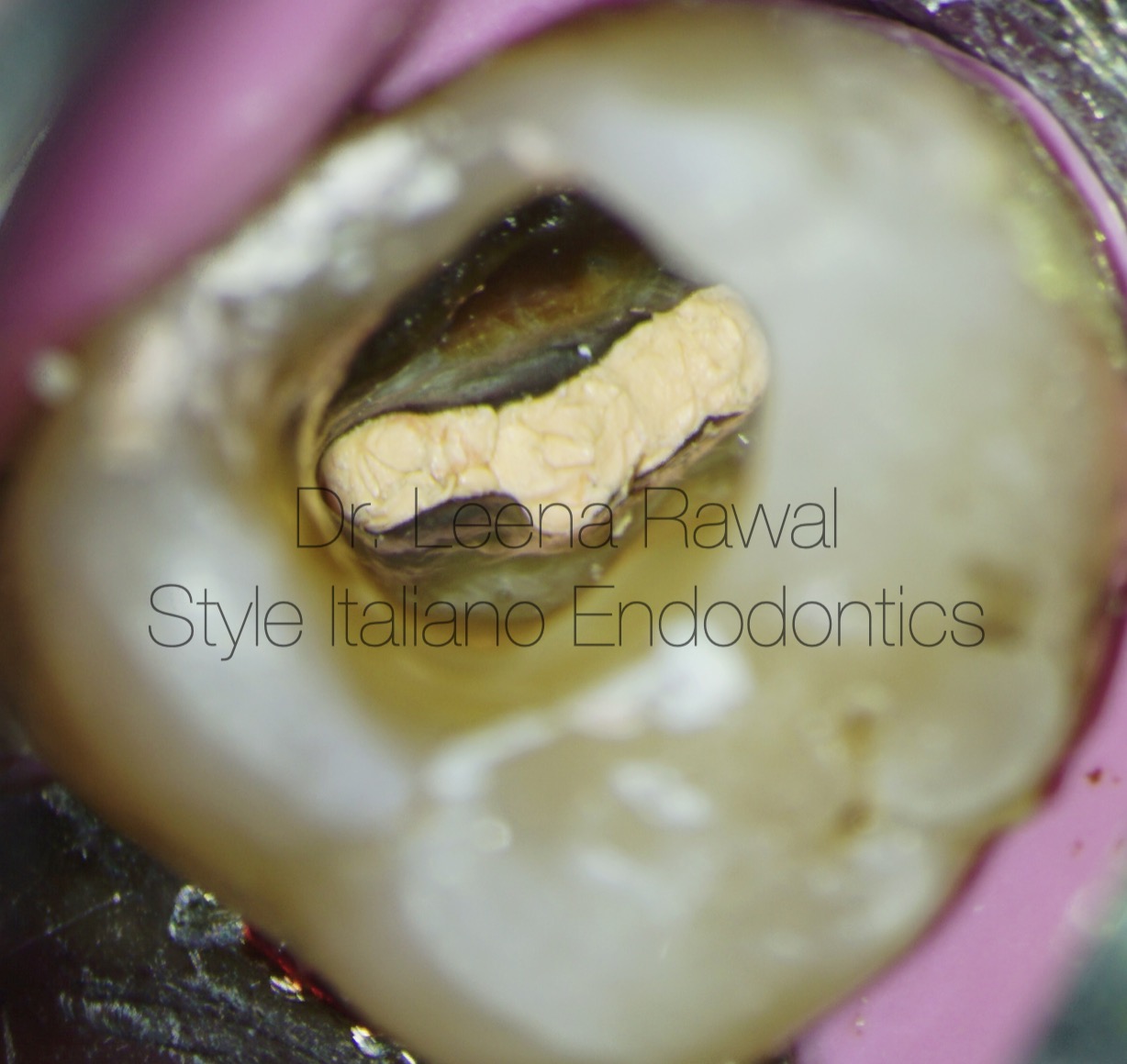
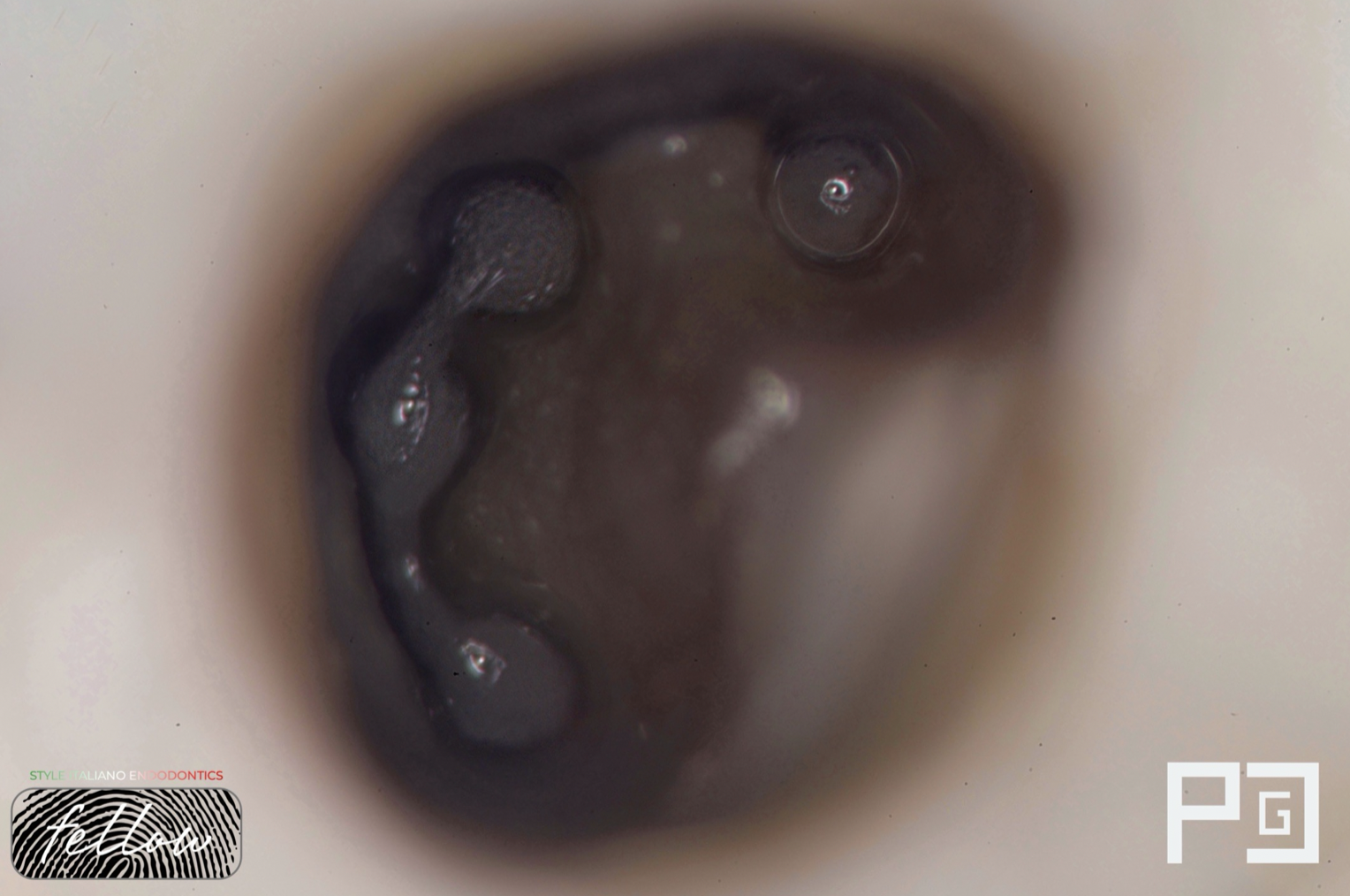
Fig. 4
In this image u can see a beautiful c shaped root canal anatomy
post obturation . Chamber was cleaned by 95% ethanol before
core build with composite was done .
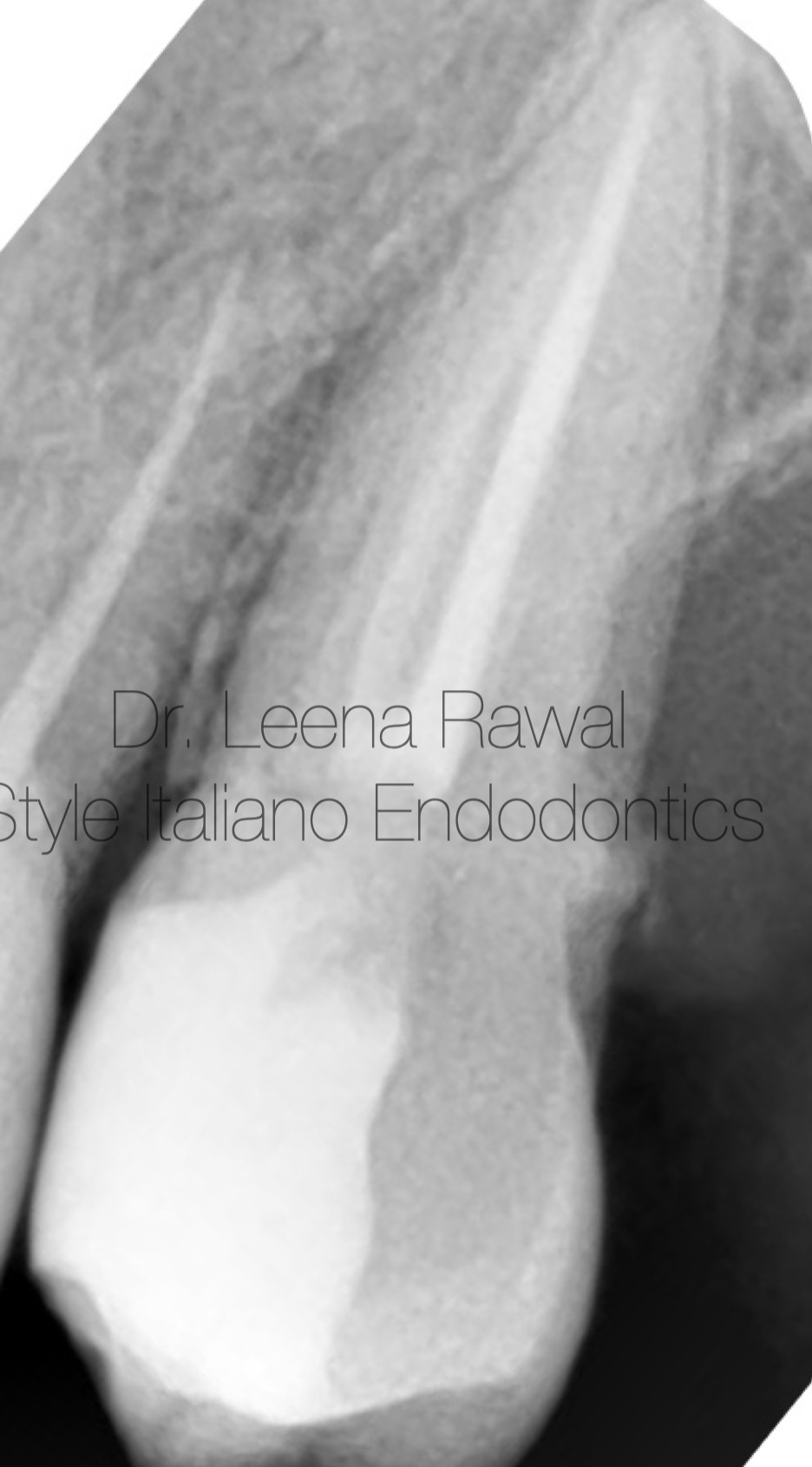
Fig. 5
IOPA post obturation showing core build up with direct composites .
Conclusions
As per this case now we all know that C-shaped canal anatomy is not that common like a mandibular second molar but with a good angulated IOPA readings & by proper isolation with rubber dam, irrigation protocols followed by effective activation of irrigants & last but not the least magnification with an operating microscope can ease to treat such cases very smoothly . After all what we can see we can treat as well.
Bibliography
- Carlsen O ,Alexandersen v , heitmann T ,Jakobsen P. Root canals in one rooted maxillary second molars . Scand J Dent Res. 1992;100:249-56.
- Orban B , Mueller E. The development of bifurcation of multirooted teeth. J Am Dent Assoc. 1929;16:297-319
- Fischlschweiger W , Clausnitzer E. Root formation in molar teeth of the CD -MOUSE. J Ended .1988;14:163-8.
- Jerome CE. C shaped root canal system : Diagnosis,treatment & restoration . Gen Dent. 1994;42:424-7.