Retrieval and retreatment of mandibular molar
06/01/2025
Fellow
Warning: Undefined variable $post in /var/www/vhosts/styleitaliano-endodontics.org/endodontics.styleitaliano.org/wp-content/plugins/oxygen/component-framework/components/classes/code-block.class.php(133) : eval()'d code on line 2
Warning: Attempt to read property "ID" on null in /var/www/vhosts/styleitaliano-endodontics.org/endodontics.styleitaliano.org/wp-content/plugins/oxygen/component-framework/components/classes/code-block.class.php(133) : eval()'d code on line 2
Retrieval along with Retreatment is one of the most challenging procedures in the endodontic therapy,
This article showcases the management of a lower first molar with swparatedinstrumemnt and a periapical lesion.
In Endodontics, we are dealing with a lot of challenges such as anatomy, endodontic mishaps, instrument separation. One of the biggest challenges is an instrument retrieval to enhance the prognosis in retreatments.
When facing a retreatment, the clinician must be aware of retrieval techniques and try to understand the reason of separation to deal with such case in a predictable way.
If we identify the reason we can predictably retreat the case , increasing the success of the endodontic therapy.
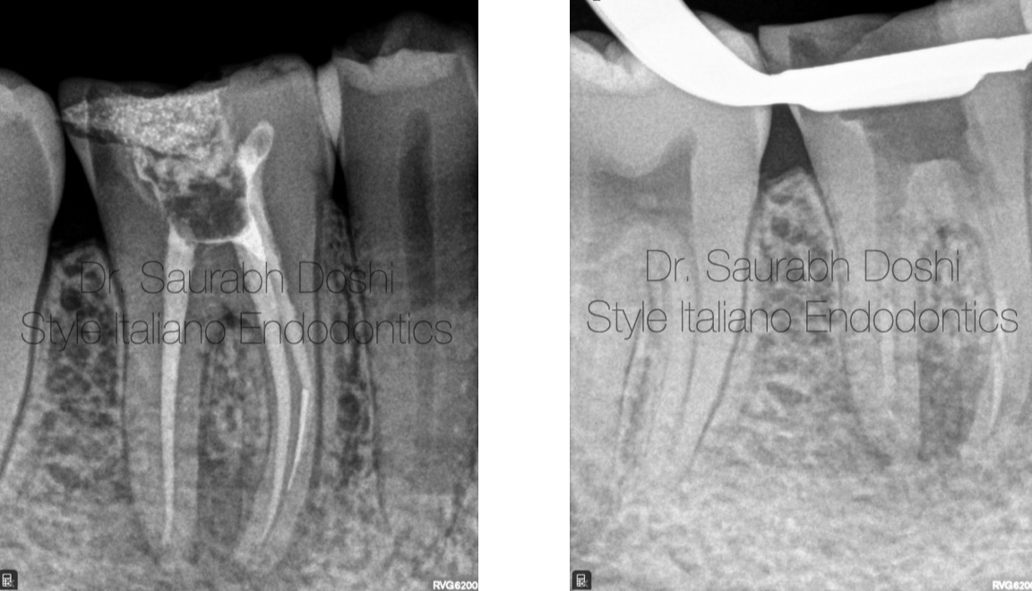
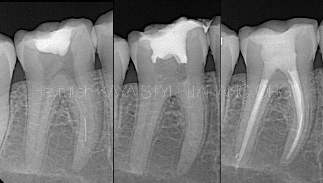
Fig. 1
A patient came with complaining about tenderness and pain on chewing food on the lower first right molar.
Pre-operative radiographs were taken.
Pre-operative radiograph shows :
Short filling on the mesial system with separated insrtuemnt in lingual canal.
Poor Obturation of the mesual canal.
Peri-apical lesions related to each root.
Radiograph suggestive of Removal of gutta percha and instrument was done.
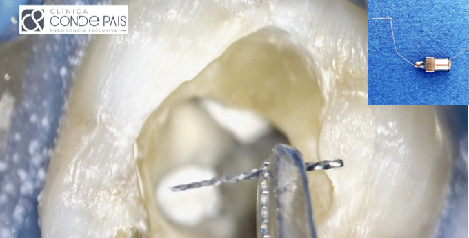
Microscopic photo shows seperatatedinstrument in pulp chamber which was removed using ultrasonic files.

Fig. 2
Radiograph suggestive of Removal of gutta percha and instrument was done.
Microscopic photo shows seperatatedinstrument in pulp chamber which was removed using ultrasonic files.
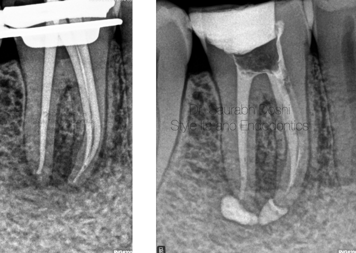
Fig. 3
Root canals were shaped and prepared till 25.06% in mesuial canals and 30.06% in distal canal and master cone selection was done.
Calcium hydroxide intracanal medicament was given for 21 days.
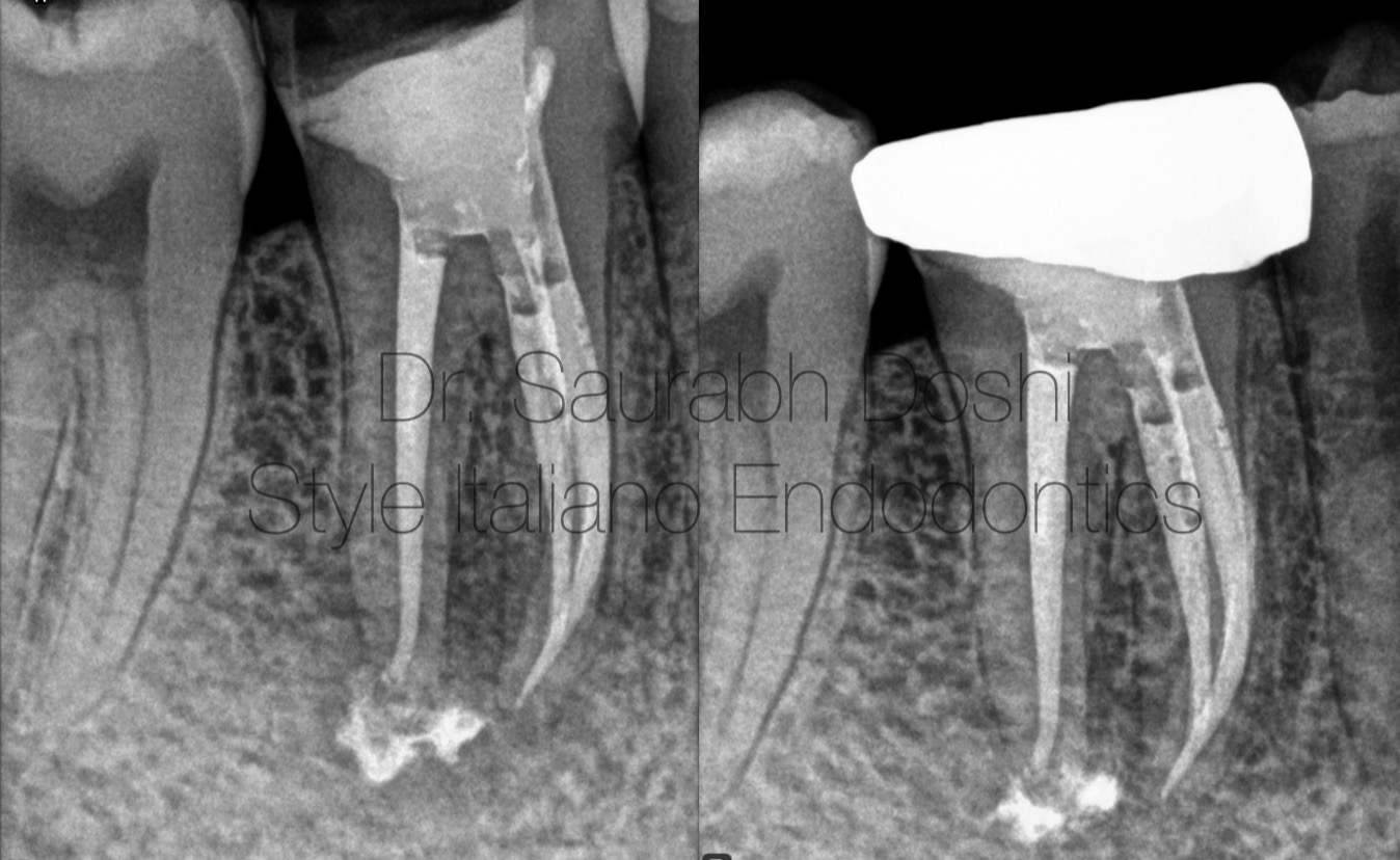
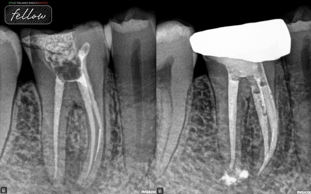
Fig. 4
Root canals were obturated by single cone technique using hydraulic condensation (Ceraseal sealer) and prosthesis cementation was done.
Follow up was done for 3 , 6 , 9 ,12 , 15 ,18 months with periapical area suggestive of healing.

Fig. 5
Dr. Saurabh Doshi
BDS , MDS
Micro-Endodontist and Restorative Dentist.
Proud Diplomate - Indian Board Of Endodontics by Indian Endodontic Society
Diploma in Laser Dentistry ( University of Vienna , Austria)
Fellow of Style Italiano Endodontics Forum.
Author of Book " Root Canal Obturation- Past , Present and Future" Published in International Lambert Academic Publication.
Highly Recommend Endodontist Of The Year At Prestigious Famdent Excellence Awards 2022.
Invited Speaker at National and International Podium including 2nd Style Italiano Endodontics Conference.
Director - Dr. Doshi's Speciality Dental Clinic and Root Canal Centre, Baramati , Maharashtra, India.
Conclusions
Appropriate pre operative assessment of the case, through knowledge of instrument retrieval techniques and root canal filling materials can significantly and predictably increase the outcome of endodontic retreatments.
Bibliography
Shen Y, Peng B, Cheung GS. Factors associated with the removal of fractured NiTi instruments from root canal systems. Oral SurgOral Med Oral Pathol Oral RadiolEndod. 2004;98:605–610.
Madarati AA, Hunter MJ, Dummer PM. Management of intracanal separated instruments. J Endod. 2013 May;39(5):569-81. doi: 10.1016/j.joen.2012.12.033. Epub 2013 Mar 15. PMID: 23611371


